Bilateral mammary duct ectasia induced by sulpiride-associated hyperprolactinemia: A case report

  • Authors:
    • Yizi Cong
    • Haidong Zou
    • Guangdong Qiao
    • Jun Lin
    • Xingmiao Wang
    • Xiaohui Li
    • Yalun Li
    • Shiguang Zhu
  • View Affiliations

  • Published online on: March 12, 2015     https://doi.org/10.3892/ol.2015.3034
  • Pages: 2181-2184
Metrics: Total Views: 0 (Spandidos Publications: | PMC Statistics: )
Total PDF Downloads: 0 (Spandidos Publications: | PMC Statistics: )


Abstract

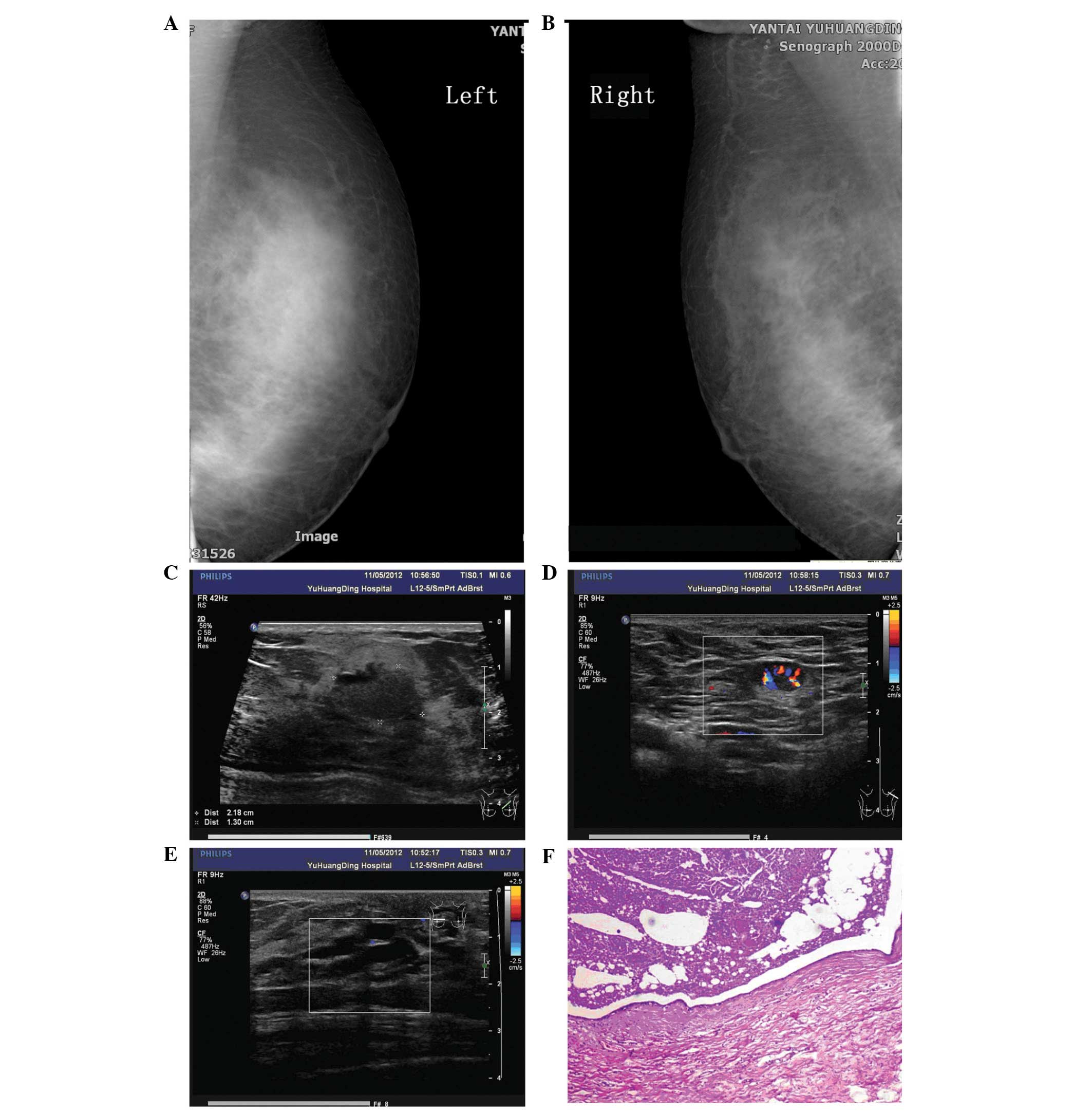
A 32 year old female diagnosed with schizophrenia was treated with sulpiride, trihexyphenidyl and alprazolam for 6 years. A physical examination revealed bilateral nipple retraction and a non-tender mass in the left breast, with little nipple discharge. Tests revealed high levels of carbohydrate antigen 125, serum prolactin and testosterone levels, and ultrasound revealed a number of masses in the bilateral breasts; the largest mass (2.2x1.3 cm) was located in the left breast. A rich blood flow signal was identified around the nodule. The ducts in the bilateral breasts exhibited cystic ectasia. Multiple enlarged lymph nodes were found in the bilateral axillae. Mammography revealed thickened breast tissue without an evident mass, and calcification. A segmental mastectomy was performed and subsequent histological examination revealed multiple dilated ducts, the largest of which contained eosinophilic material. The pathological diagnosis was of breast duct dilatation. Bacterial culture and drug sensitivity analysis of the secretions from the cystic cavity revealed no bacterial growth, and an acid fast bacillus stain was negative. Extravasation of the surgical wound occurred 1 month later, and Staphylococcus epidermidis was observed using a bacterial culture. This was treated with moxifloxacin for 1 week. It was suggested that the patient should switch to a prolactin sparing antipsychotic in view of the hyperprolactinemia, however, the patient refused. After a clinical follow-up of 16 months, the wound had healed well and no palpable mass was found in the breast.
View Figures
View References

Related Articles

Journal Cover

May-2015
Volume 9 Issue 5

Print ISSN: 1792-1074
Online ISSN:1792-1082

Sign up for eToc alerts

Recommend to Library

Copy and paste a formatted citation
x
Spandidos Publications style
Cong Y, Zou H, Qiao G, Lin J, Wang X, Li X, Li Y and Zhu S: Bilateral mammary duct ectasia induced by sulpiride-associated hyperprolactinemia: A case report. Oncol Lett 9: 2181-2184, 2015
APA
Cong, Y., Zou, H., Qiao, G., Lin, J., Wang, X., Li, X. ... Zhu, S. (2015). Bilateral mammary duct ectasia induced by sulpiride-associated hyperprolactinemia: A case report. Oncology Letters, 9, 2181-2184. https://doi.org/10.3892/ol.2015.3034
MLA
Cong, Y., Zou, H., Qiao, G., Lin, J., Wang, X., Li, X., Li, Y., Zhu, S."Bilateral mammary duct ectasia induced by sulpiride-associated hyperprolactinemia: A case report". Oncology Letters 9.5 (2015): 2181-2184.
Chicago
Cong, Y., Zou, H., Qiao, G., Lin, J., Wang, X., Li, X., Li, Y., Zhu, S."Bilateral mammary duct ectasia induced by sulpiride-associated hyperprolactinemia: A case report". Oncology Letters 9, no. 5 (2015): 2181-2184. https://doi.org/10.3892/ol.2015.3034