Introduction
Primary tumors arising from the mesentery are rare.
The incidence of primary lesions among mesentery neoplasms is ∼1%;
for instance, in a previously-conducted review of a large series of
patients with mesenteric abnormalities detected using CT, only one
case was determined to be a primary tumor (1). Perivascular epithelioid cell tumors
(PEComas) are a recently-defined family of rare tumors composed of
distinctive perivascular epithelioid cells (PECs). To the best of
our knowledge, only seven cases of mesenteric PEComa have been
described in the English literature to date (2–5), including
five female patients, one adult male patient and one young boy.
Considering the small number of reported cases, the clinical and
imaging features of the tumor have yet to be adequately determined.
The present study describes the case of a 48-year-old female
patient with mesenteric malignant PEComa. The aim of the current
study was to accumulate information regarding PEComas to improve
the diagnostic specificity of the disease.
Case report
A 48 year-old female patient presented to
Yuhuangding Hospital (Yantai, China) in March 2008 with a lower
abdominal mass that had been gradually increasing in size. The
patient initially noticed a fist-sized, painless mass ∼3 months
prior to admittance, but no history of abdominal pain, melena, body
weight loss or a change in bowel habits was noted. During an
abdominal examination, the patient experienced pain upon palpation
and a nontender mass was identified in the lower abdomen. No
abnormalities were identified during standard blood tests and chest
X-rays. The levels of the carcinoembryonic antigen and carbohydrate
antigen (CA) 19-9 tumor markers were within the normal range;
however, the level of CA125 was markedly increased (137.2 U/ml;
normal range, <35 U/ml). The colonoscopy results were normal;
however, ultrasonography of the abdomen identified a highly
vascularized heterogeneous mass located in the lower abdomen.
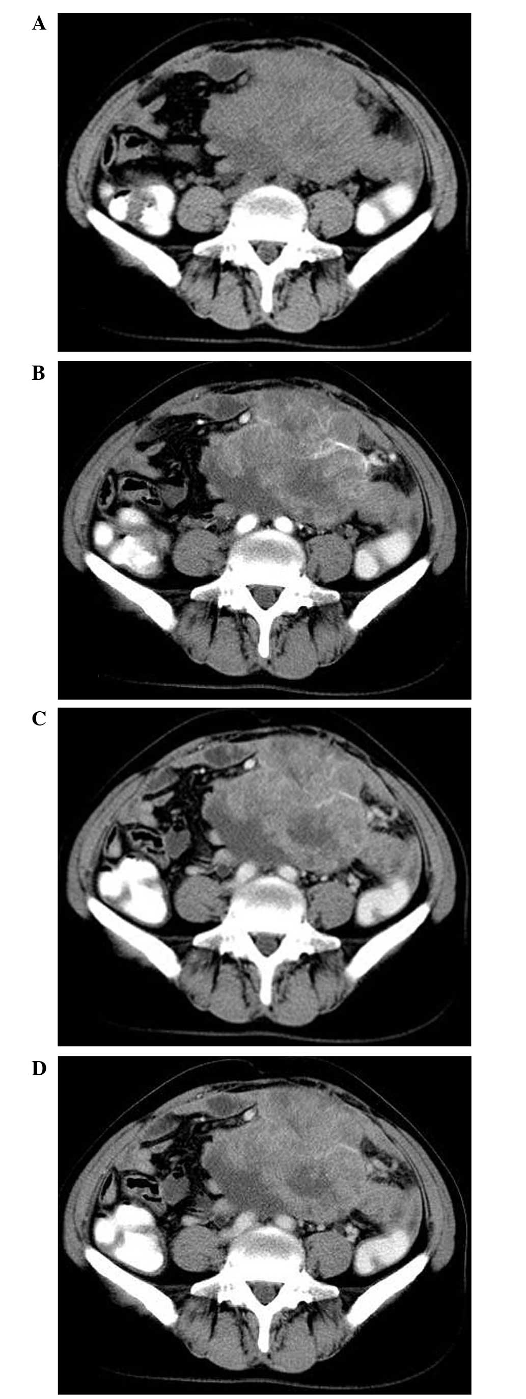
Abdominal plain computed tomography (CT) scanning revealed a large,
partially ill-defined and heterogeneous mass with a size of
12.5×8.5 cm occupying the lower abdomen (Fig. 1A). Upon contrast-enhanced CT imaging,
the tumor displayed nonhomogeneous contrast-enhancement with
hypodense areas, indicating myxoid change, hemorrhage or necrosis.
Furthermore, multiple tumor vessels were observed in the tumor
during arterial phase imaging (Fig.
1B-D) and no significant fat component was visible. Therefore,
the CT findings indicated a malignant tumor, possibly of
mesenchymal origin, such as leiomyosarcoma. Consequently, a
diagnostic exploratory laparotomy was performed. During surgery, a
large lobulated tumor measuring 14 cm maximally was identified,
arising in the mesentery and resulting in constriction and
distortion of the small intestine. The tumor was successfully
resected by a combined resection of 30 cm of the small intestine
and end-to-end anastomosis of the small intestine. The external
surface of the tumor was lobulated and irregular, while sections
through the tumor revealed the presence of solid, grayish-yellow
tissue with areas of fresh and old hemorrhage, as well as
necrosis.
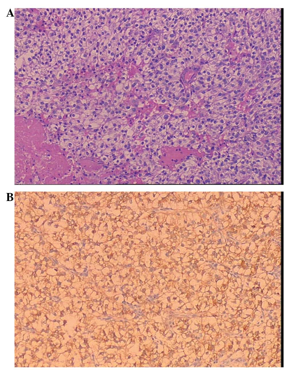
Histologically, the tumor was composed of
epithelioid cells with clear to lightly eosinophilic cytoplasm that
grew in a sheet-like pattern and were arranged in a radial fashion
around blood vessels. Furthermore, the tumor cells exhibited
striking nuclear atypia and elevated mitotic activity (Fig. 2A), observed using hematoxylin and
eosin staining (Baihao Biological Technology Co., Ltd.; Tianjin,
China). Upon immunohistochemical staining using EnVision™ (Dako,
Carpinteria, CA, USA), the tumor cells were strongly positive for
human melanoma black (HMB)-45 (Fig.
2B), but negative for melan A, actin, desmin, cytokeratin and
vimentin. The histological and immunohistochemical features were
consistent with a malignant form of PEComa. Therefore, following
surgery, the patient underwent adjuvant chemotherapy with 200 mg/dl
oxaliplatin per month for three cycles. Subsequently, the serum
CA125 levels decreased to the normal range. At a follow-up 60
months after diagnosis, the patient demonstrated no evidence of
disease. The study was approved by the ethics committee of Shandong
Medical Imaging Research Institute, Shandong University, (Jinan,
China) and Written informed consent was obtained from the
patient.
Discussion
PEComa is a type of mesenchymal neoplasm composed of
histologically and immunohistochemically distinctive PECs (6). In 1992, Bonetti et al (7) initially described PECs, and in 1996,
Zamboni et al (8) used the
term PEComa to describe this rare family of lesions. At present,
the PEComa family includes conventional angiomyolipomas (AMLs),
clear cell sugar tumors, lymphangioleiomyomatosis and PEComa-not
otherwise specified, which is a group of rare, morphologically and
immunophenotypically similar tumors.
PEComas have been identified in almost every site in
the body, including the liver, kidney, falciform ligament,
gynecological regions, small and large bowel, retroperitoneum,
abdominal wall, extremities and neck (2,9). They
typically occur in middle-aged patients, with predominance in
female individuals (female to male ratio, 6:1) (2).
PEComas arising from the mesentery are rare, and
currently there is no uniform criteria for diagnosis. They are
predominantly composed of nests and sheets of epithelioid cells;
however, spindle cells with clear to granular eosinophilic
cytoplasm are also observed in certain cases, demonstrating focal
invasion of the blood vessel walls. PEComas typically exhibit
positive immunohistochemical staining for the two melanocytic
markers, HMB-45 and melan-A, and the smooth muscle markers, actin
and desmin (3,4,10,11). Additionally, more recently, cathepsin
K has been identified as a potentially more powerful marker than
the aforementioned commonly used markers (12). However, immunohistochemical staining
is not definite and not all previously diagnosed PEComas were
positive for all of these markers. In the present study,
histologically, the tumor morphology was typical; therefore based
on the characteristic PECs and the unique phenotypic feature of
HMB-45 expression, a diagnosis of PEComa was determined.
The clinical behavior of PEComa is typically benign,
however, aggressive behavior is occasionally displayed. Prediction
of malignant PEComa behavior based on microscopic attributes is
currently limited due to the lack of clear criteria. However, in
2005, Folpe et al (2)
described the following six histological features, which are
indicative of high-risk PEComa: Large tumor size (median diameter,
>5 cm), infiltrative pattern, high nuclear grade and
cellularity, high mitotic rate (>1/50 high power fields),
necrosis and vascular invasion. PEComas with ≥2 of the
aforementioned high-risk features should be considered malignant.
In addition, Folpe et al (2)
stated that specific cases with no high-risk features may still
exhibit aggressive behavior. Therefore, according to the
aforementioned criteria, the tumor observed in the current patient
may be classified as malignant.
Numerous cases of PEComa (particularly classic AML)
may occur sporadically or in association with the tuberous
sclerosis complex (TSC) (13). These
tumors appear to be associated with specific genetic alterations of
the TSC, including the loss of TSC1 (chromosome 9q34) or TSC2
(chromosome 16p13.3) genes (14).
However, the current patient did not have a personal or a family
history of TSC.
PEComas arising in the mesentery are rare. Including
the present case, to the best of our knowledge, only eight
mesenteric PEComas have been reported thus far (2–5), including
six in female patients, one in an adult male patient and one in a
young boy. The reported tumor size is variable, with a maximum size
range of 4–27 cm. Common clinical symptoms of mesenteric PEComas
are abdominal pain, abdominal distention and a palpable mass.
However, such tumors are typically asymptomatic and tend to grow to
a large size prior to diagnosis (as also observed in the current
case) since the mobility of the mesentery permits the tumors to
occupy an anatomically large space without exhibiting adverse
effects. During follow-up, no recurrence occurred in five patients;
however, the remaining three patients, who exhibited ≥3 high-risk
features, developed local recurrence.
Preoperative imaging examinations, including
abdominal ultrasonography and CT/magnetic resonance imaging (MRI)
scans, are useful tools for the identification and diagnosis of the
origin and extension of a mesenteric mass. However, the
aforementioned imaging modalities are not sufficiently sensitive to
enable the diagnosis of PEComa, with the exception of classic AML
with macroscopic fat, since PEComas demonstrate a wide spectrum of
imaging findings; thus, their imaging characteristics are
nonspecific (11,15,16).
However, Tan et al (15)
identified that PEComas quickly enhanced in the arterial and venous
phases of enhanced CT and MRI imaging. In the present study, the
tumor was diagnosed as a malignant tumor by performing a CT scan.
The tumor presented as a large, lobulated, heterogeneous soft
tissue mass and displayed nonhomogeneous contrast-enhancement with
multiple tumor vessels visualized on arterial phase imaging.
Malignant PEComas arising in the mesentery should be distinguished
from other mesenteric neoplasms, such as leiomyosarcoma, malignant
fibrous histiocytoma, fibrosarcoma, liposarcoma and metastatic
carcinomas. However, due to similar imaging appearances,
preoperative diagnosis is difficult using radiological criteria
alone. Therefore, diagnosis can only be confirmed following
histological analysis of the tumor.
Currently, the most effective treatment strategy for
PEComas is surgical resection. In addition, adjuvant therapy is
recommended for all patients with malignant features; however, the
role of adjuvant therapy remains unclear due to the rarity of the
disease. The current patient received adjuvant chemotherapy with
oxaliplatin following tumor resection, resulting in effective
control of disease progression and the prevention of local
recurrence. Wagner et al (17)
have previously identified that inhibition of mammalian target of
rapamycin complex 1, which is pathologically activated by loss of
the TSC1/TSC2 tumor suppressor complex, is a rational mechanistic
target for the development of novel PEComa treatment strategies.
Furthermore, a long-term periodic follow-up is required in all
patients presenting with PEComas.
In conclusion, PEComas are a family of rare
mesenchymal neoplasms that should preoperatively be considered in
the differential diagnosis of lesions arising in the mesentery.
However, preoperative imaging examinations are not sufficiently
sensitive to enable diagnosis of PEComas, with the exception of
classic AMLs with macroscopic fat.
References
|
1
|
Whitley NO, Bohlman ME and Baker LP: CT
patterns of mesenteric disease. J Comput Assist Tomogr. 6:490–496.
1982. View Article : Google Scholar : PubMed/NCBI
|
|
2
|
Folpe AL, Mentzel T, Lehr HA, Fisher C,
Balzer BL and Weiss SW: Perivascular epithelioid cell neoplasms of
soft tissue and gynecologic origin: a clinicopathologic study of 26
cases and review of the literature. Am J Surg Pathol. 29:1558–1575.
2005. View Article : Google Scholar : PubMed/NCBI
|
|
3
|
Chen IY, Yang SF, Chen FM and Chai CY:
Abdominopelvic perivascular epithelioid cell tumor with overt
malignancy: a case report. Kaohsiung J Med Sci. 21:277–281. 2005.
View Article : Google Scholar : PubMed/NCBI
|
|
4
|
Gross E, Vernea F, Weintraub M and
Koplewitz BZ: Perivascular epithelioid cell tumor of the ascending
colon mesentery in a child: case report and review of the
literature. J Pediatr Surg. 45:830–833. 2010. View Article : Google Scholar : PubMed/NCBI
|
|
5
|
Lai CL, Hsu KF, Yu JC, et al: Malignant
perivascular epithelioid cell tumor of the mesentery: A case report
and literature review. Onkologie. 35:114–117. 2012. View Article : Google Scholar : PubMed/NCBI
|
|
6
|
Folpe AL: Neoplasms with perivascular
epithelioid cell differentiation (PEComas)Pathology and Genetics of
Tumours of Soft Tissue and Bone. Fletcher CDM, Unni KK and Mertens
F: IARC Press; Lyon: pp. 221–222. 2002
|
|
7
|
Bonetti F, Pea M, Martignoni G and Zamboni
G: PEC and sugar. Am J Surg Pathol. 16:307–308. 1992. View Article : Google Scholar : PubMed/NCBI
|
|
8
|
Zamboni G, Pea M, Martignoni G, et al:
Clear cell ‘sugar’ tumor of the pancreas. A novel member of the
family of lesions characterized by the presence of perivascular
cells. Am J Surg Pathol. 20:722–730. 1996. View Article : Google Scholar : PubMed/NCBI
|
|
9
|
Weiss SW and Goldblum JR: Perivascular
epithelioid cell family of tumorsEnzinger and Weiss's Soft Tissue
Tumors. 5th. Weiss SW and Goldblum JR: Mosby Elsevier;
Philadelphia, PA: pp. 1138–1156. 2008
|
|
10
|
Hornick JL and Fletcher CD: PEComa: what
do we know so far? Histopathology. 48:75–82. 2006. View Article : Google Scholar : PubMed/NCBI
|
|
11
|
Prasad SR, Sahani DV, Mino-Kenudson M, et
al: Neoplasms of the perivascular epithelioid cell involving the
abdomen and the pelvis: cross-sectional imaging findings. J Comput
Assist Tomogr. 31:688–696. 2007. View Article : Google Scholar : PubMed/NCBI
|
|
12
|
Rao Q, Cheng L, Xia QY, et al: Cathepsin K
expression in a wide spectrum of perivascular epithelioid cell
neoplasms (PEComas): a clinicopathological study emphasizing
extrarenal PEComas. Histopathology. 62:642–650. 2013. View Article : Google Scholar : PubMed/NCBI
|
|
13
|
Casper KA, Donnelly LF, Chen B and Bissler
JJ: Tuberous sclerosis complex: renal imaging findings. Radiology.
225:451–456. 2002. View Article : Google Scholar : PubMed/NCBI
|
|
14
|
Folpe AL and Kwiatkowski DJ: Perivascular
epithelioid cell neoplasms: pathology and pathogenesis. Hum Pathol.
41:1–15. 2010. View Article : Google Scholar : PubMed/NCBI
|
|
15
|
Tan Y, Zhang H and Xiao EH: Perivascular
epithelioid cell tumour: dynamic CT, MRI and clinicopathological
characteristics – analysis of 32 cases and review of the
literature. Clin Radiol. 68:555–561. 2013. View Article : Google Scholar : PubMed/NCBI
|
|
16
|
Baez JC, Landry JM, Saltzman JR, Qian X,
Zinner MJ and Mortele KJ: Pancreatic PEComa (sugar tumor): MDCT and
EUS features. JOP Pancreas. 10:679–682. 2009.
|
|
17
|
Wagner AJ, Malinowska-Kolodziej I, Morgan
JA, et al: Clinical activity of mTOR inhibition with sirolimus in
malignant perivascular epithelioid cell tumors: targeting the
pathogenic activation of mTORC1 in tumors. J Clin Oncol.
28:835–840. 2010. View Article : Google Scholar : PubMed/NCBI
|