Spandidos Publications Logo
  • About
    • About Spandidos
    • Aims and Scopes
    • Abstracting and Indexing
    • Editorial Policies
    • Reprints and Permissions
    • Job Opportunities
    • Terms and Conditions
    • Contact
  • Journals
    • All Journals
    • Oncology Letters
      • Oncology Letters
      • Information for Authors
      • Editorial Policies
      • Editorial Board
      • Aims and Scope
      • Abstracting and Indexing
      • Bibliographic Information
      • Archive
    • International Journal of Oncology
      • International Journal of Oncology
      • Information for Authors
      • Editorial Policies
      • Editorial Board
      • Aims and Scope
      • Abstracting and Indexing
      • Bibliographic Information
      • Archive
    • Molecular and Clinical Oncology
      • Molecular and Clinical Oncology
      • Information for Authors
      • Editorial Policies
      • Editorial Board
      • Aims and Scope
      • Abstracting and Indexing
      • Bibliographic Information
      • Archive
    • Experimental and Therapeutic Medicine
      • Experimental and Therapeutic Medicine
      • Information for Authors
      • Editorial Policies
      • Editorial Board
      • Aims and Scope
      • Abstracting and Indexing
      • Bibliographic Information
      • Archive
    • International Journal of Molecular Medicine
      • International Journal of Molecular Medicine
      • Information for Authors
      • Editorial Policies
      • Editorial Board
      • Aims and Scope
      • Abstracting and Indexing
      • Bibliographic Information
      • Archive
    • Biomedical Reports
      • Biomedical Reports
      • Information for Authors
      • Editorial Policies
      • Editorial Board
      • Aims and Scope
      • Abstracting and Indexing
      • Bibliographic Information
      • Archive
    • Oncology Reports
      • Oncology Reports
      • Information for Authors
      • Editorial Policies
      • Editorial Board
      • Aims and Scope
      • Abstracting and Indexing
      • Bibliographic Information
      • Archive
    • Molecular Medicine Reports
      • Molecular Medicine Reports
      • Information for Authors
      • Editorial Policies
      • Editorial Board
      • Aims and Scope
      • Abstracting and Indexing
      • Bibliographic Information
      • Archive
    • World Academy of Sciences Journal
      • World Academy of Sciences Journal
      • Information for Authors
      • Editorial Policies
      • Editorial Board
      • Aims and Scope
      • Abstracting and Indexing
      • Bibliographic Information
      • Archive
    • International Journal of Functional Nutrition
      • International Journal of Functional Nutrition
      • Information for Authors
      • Editorial Policies
      • Editorial Board
      • Aims and Scope
      • Abstracting and Indexing
      • Bibliographic Information
      • Archive
    • International Journal of Epigenetics
      • International Journal of Epigenetics
      • Information for Authors
      • Editorial Policies
      • Editorial Board
      • Aims and Scope
      • Abstracting and Indexing
      • Bibliographic Information
      • Archive
    • Medicine International
      • Medicine International
      • Information for Authors
      • Editorial Policies
      • Editorial Board
      • Aims and Scope
      • Abstracting and Indexing
      • Bibliographic Information
      • Archive
  • Articles
  • Information
    • Information for Authors
    • Information for Reviewers
    • Information for Librarians
    • Information for Advertisers
    • Conferences
  • Language Editing
Spandidos Publications Logo
  • About
    • About Spandidos
    • Aims and Scopes
    • Abstracting and Indexing
    • Editorial Policies
    • Reprints and Permissions
    • Job Opportunities
    • Terms and Conditions
    • Contact
  • Journals
    • All Journals
    • Biomedical Reports
      • Information for Authors
      • Editorial Policies
      • Editorial Board
      • Aims and Scope
      • Abstracting and Indexing
      • Bibliographic Information
      • Archive
    • Experimental and Therapeutic Medicine
      • Information for Authors
      • Editorial Policies
      • Editorial Board
      • Aims and Scope
      • Abstracting and Indexing
      • Bibliographic Information
      • Archive
    • International Journal of Epigenetics
      • Information for Authors
      • Editorial Policies
      • Editorial Board
      • Aims and Scope
      • Abstracting and Indexing
      • Bibliographic Information
      • Archive
    • International Journal of Functional Nutrition
      • Information for Authors
      • Editorial Policies
      • Editorial Board
      • Aims and Scope
      • Abstracting and Indexing
      • Bibliographic Information
      • Archive
    • International Journal of Molecular Medicine
      • Information for Authors
      • Editorial Policies
      • Editorial Board
      • Aims and Scope
      • Abstracting and Indexing
      • Bibliographic Information
      • Archive
    • International Journal of Oncology
      • Information for Authors
      • Editorial Policies
      • Editorial Board
      • Aims and Scope
      • Abstracting and Indexing
      • Bibliographic Information
      • Archive
    • Medicine International
      • Information for Authors
      • Editorial Policies
      • Editorial Board
      • Aims and Scope
      • Abstracting and Indexing
      • Bibliographic Information
      • Archive
    • Molecular and Clinical Oncology
      • Information for Authors
      • Editorial Policies
      • Editorial Board
      • Aims and Scope
      • Abstracting and Indexing
      • Bibliographic Information
      • Archive
    • Molecular Medicine Reports
      • Information for Authors
      • Editorial Policies
      • Editorial Board
      • Aims and Scope
      • Abstracting and Indexing
      • Bibliographic Information
      • Archive
    • Oncology Letters
      • Information for Authors
      • Editorial Policies
      • Editorial Board
      • Aims and Scope
      • Abstracting and Indexing
      • Bibliographic Information
      • Archive
    • Oncology Reports
      • Information for Authors
      • Editorial Policies
      • Editorial Board
      • Aims and Scope
      • Abstracting and Indexing
      • Bibliographic Information
      • Archive
    • World Academy of Sciences Journal
      • Information for Authors
      • Editorial Policies
      • Editorial Board
      • Aims and Scope
      • Abstracting and Indexing
      • Bibliographic Information
      • Archive
  • Articles
  • Information
    • For Authors
    • For Reviewers
    • For Librarians
    • For Advertisers
    • Conferences
  • Language Editing
Login Register Submit
  • This site uses cookies
  • You can change your cookie settings at any time by following the instructions in our Cookie Policy. To find out more, you may read our Privacy Policy.

    I agree
Search articles by DOI, keyword, author or affiliation
Search
Advanced Search
presentation
Oncology Letters
Join Editorial Board Propose a Special Issue
Print ISSN: 1792-1074 Online ISSN: 1792-1082
Journal Cover
July-2015 Volume 10 Issue 1

Full Size Image

Sign up for eToc alerts
Recommend to Library

Journals

International Journal of Molecular Medicine

International Journal of Molecular Medicine

International Journal of Molecular Medicine is an international journal devoted to molecular mechanisms of human disease.

International Journal of Oncology

International Journal of Oncology

International Journal of Oncology is an international journal devoted to oncology research and cancer treatment.

Molecular Medicine Reports

Molecular Medicine Reports

Covers molecular medicine topics such as pharmacology, pathology, genetics, neuroscience, infectious diseases, molecular cardiology, and molecular surgery.

Oncology Reports

Oncology Reports

Oncology Reports is an international journal devoted to fundamental and applied research in Oncology.

Experimental and Therapeutic Medicine

Experimental and Therapeutic Medicine

Experimental and Therapeutic Medicine is an international journal devoted to laboratory and clinical medicine.

Oncology Letters

Oncology Letters

Oncology Letters is an international journal devoted to Experimental and Clinical Oncology.

Biomedical Reports

Biomedical Reports

Explores a wide range of biological and medical fields, including pharmacology, genetics, microbiology, neuroscience, and molecular cardiology.

Molecular and Clinical Oncology

Molecular and Clinical Oncology

International journal addressing all aspects of oncology research, from tumorigenesis and oncogenes to chemotherapy and metastasis.

World Academy of Sciences Journal

World Academy of Sciences Journal

Multidisciplinary open-access journal spanning biochemistry, genetics, neuroscience, environmental health, and synthetic biology.

International Journal of Functional Nutrition

International Journal of Functional Nutrition

Open-access journal combining biochemistry, pharmacology, immunology, and genetics to advance health through functional nutrition.

International Journal of Epigenetics

International Journal of Epigenetics

Publishes open-access research on using epigenetics to advance understanding and treatment of human disease.

Medicine International

Medicine International

An International Open Access Journal Devoted to General Medicine.

Journal Cover
July-2015 Volume 10 Issue 1

Full Size Image

Sign up for eToc alerts
Recommend to Library

  • Article
  • Citations
    • Cite This Article
    • Download Citation
    • Create Citation Alert
    • Remove Citation Alert
    • Cited By
  • Similar Articles
    • Related Articles (in Spandidos Publications)
    • Similar Articles (Google Scholar)
    • Similar Articles (PubMed)
  • Download PDF
  • Download XML
  • View XML
Case Report

Nasopharyngeal actinomycosis on 18F-fluorodeoxyglucose positron emission tomography/computed tomography: A case report

  • Authors:
    • Yu‑Hsuan Lin
    • Chin Hu
    • Hao‑Wen Chuang
    • Ming‑Yee Lin
  • View Affiliations / Copyright

    Affiliations: Department of Otolaryngology, Head and Neck Surgery, Kaohsiung Veterans General Hospital, Kaohsiung 813, Taiwan, R.O.C., Department of Nuclear Medicine, Kaohsiung Veterans General Hospital, Kaohsiung 813, Taiwan, R.O.C., Department of Pathology, Kaohsiung Veterans General Hospital, Kaohsiung 813, Taiwan, R.O.C.
  • Pages: 260-262
    |
    Published online on: April 28, 2015
       https://doi.org/10.3892/ol.2015.3157
  • Expand metrics +
Metrics: Total Views: 0 (Spandidos Publications: | PMC Statistics: )
Metrics: Total PDF Downloads: 0 (Spandidos Publications: | PMC Statistics: )
Cited By (CrossRef): 0 citations Loading Articles...

This article is mentioned in:


Abstract

Actinomycosis is a chronic disease characterized by slow progression, abscess formation, tissue fibrosis and draining sinuses. Occurrence originating from the nasopharynx is extremely low. The present study described the case of a 46-year-old otherwise healthy female, with no remarkable history of mucosal injury or teeth rottenness, who presented with an asymptomatic nasopharyngeal mass that was detected incidentally by 18F‑fluorodeoxyglucose positron emission tomography/computed tomography. A nasopharyngoscopy revealed an unclear demarcated granular mass. The patient then underwent a biopsy. Based on the obtained clinical images, microbiological results and histological findings, a diagnosis of actinomycosis was established. The patient experienced an uneventful eradication of the disease after two months of oral antibiotic treatment with amoxicillin. In conclusion, these findings indicate that actinomycosis should be included in the differential diagnosis of nasopharyngeal neoplasms.

Introduction

Actinomycosis is a rare, chronic and indolent progressive granulomatous infection that is typically caused by the bacteria, Actinomyces israelii (1–3). Classically, actinomycosis presents in three clinical forms: Cervicofacial, thoracic and abdominopelvic (1,2). Cervicofacial actinomycosis accounts for around 60% of cases, with the mandible being the most frequently affected anatomic site (1–3). Though the clinical manifestations vary, an abscess formation with subsequent draining sinus and fistula formation is most commonly observed (1–3). An adequate course of antibiotics is the cornerstone of treatment (1–3). Surgical debridement is necessary in certain circumstances (1). The prognosis is positive, providing that early diagnosis and treatment occurs (3). Sustained observation is mandatory for detection of recurrence. Occurrence of nasopharyngeal actinomycosis is rare, and infection can occur without prior mucosal injury or an immunocompromised status (1). Therefore, it is advisable to always maintain a high level of clinical suspicion when treating patients with a persistent nasopharyngeal mass that presents with vague symptoms. The present study described a case of this rare disease entity. The clinical course, microbiologic findings and images were also presented.

Case report

An otherwise healthy 46-year-old Asian female 
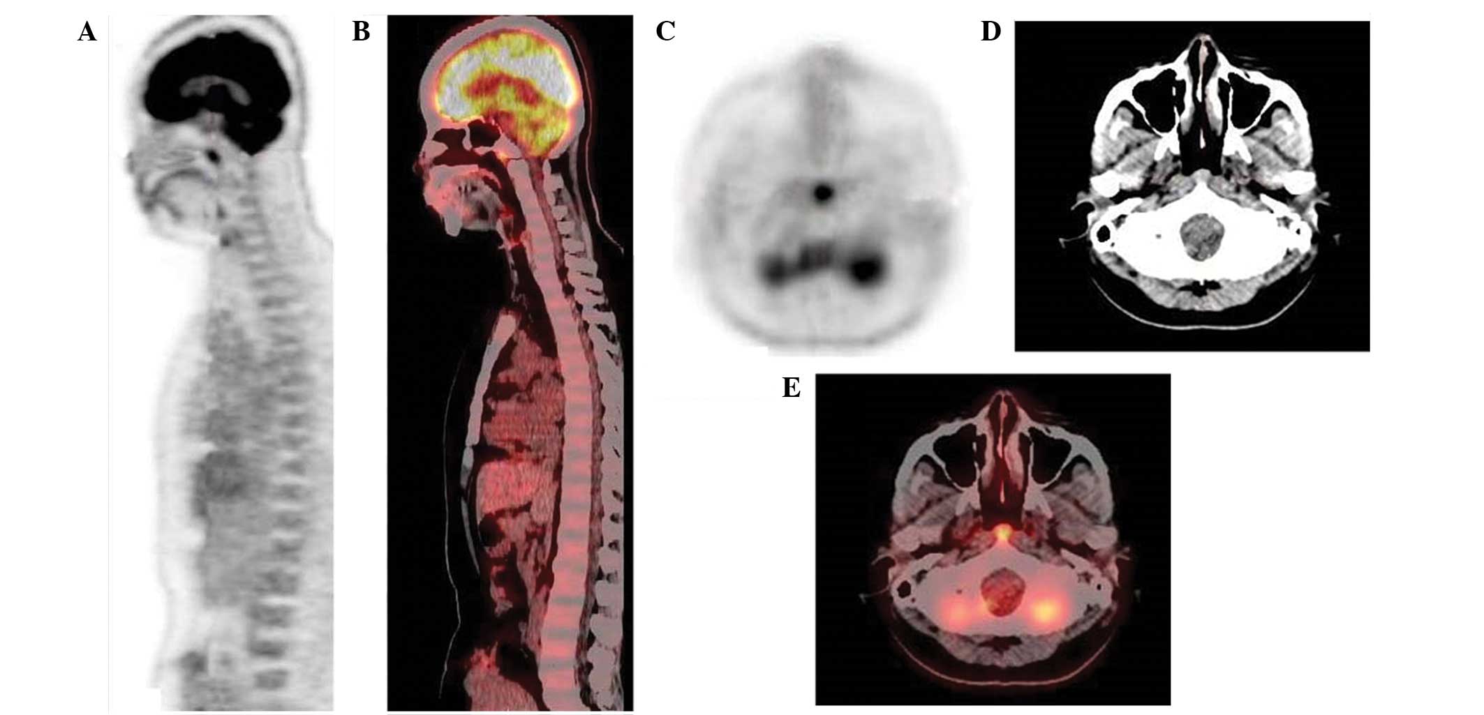
underwent a 18F-fluorodeoxyglucose (18F-FDG) positron emission tomography (PET)/computed tomography (CT) whole-body cancer screening at the Kaohsiung Veterans General Hospital (Kaohsiung, Taiwan). Intense, focal 18F-FDG uptake (standard uptake value, 6.2) was identified over the nasopharynx (Fig. 1). The patient was transferred to the otolaryngology outpatient department following a suspected nasopharyngeal malignancy in March 2013. A detailed history identified no other symptoms affecting the four general areas of compliant: aural, nasal, neck, or miscellaneous accounted for by cranial nerve involvement. In addition, the medical history of the patient, in relation to maxillofacial trauma or dental 
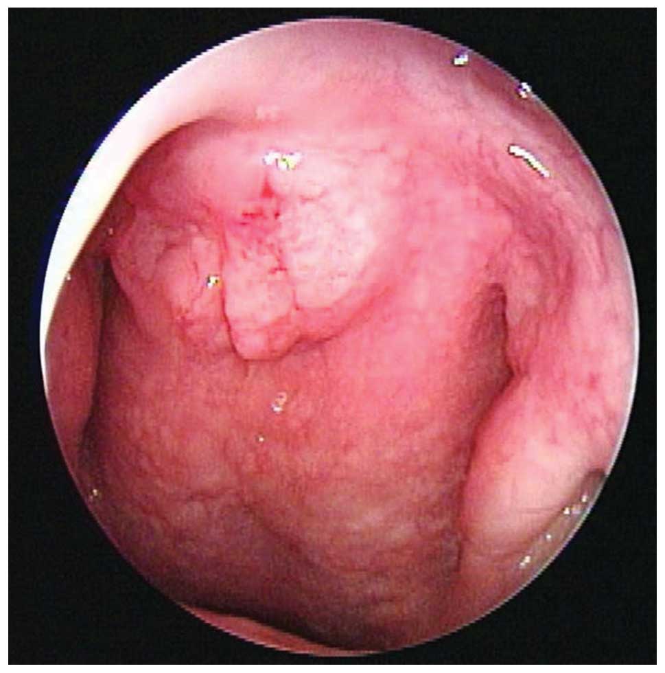
manipulation, was unremarkable. Direct nasopharyngoscopy with a rigid telescope revealed an unclearly demarcated granular mass originating from the roof of the nasopharynx (Fig. 2). A biopsy was performed and a sample of the fluid from the mass was sent for comprehensive microbiology and pathology analysis. The results revealed that the sample 
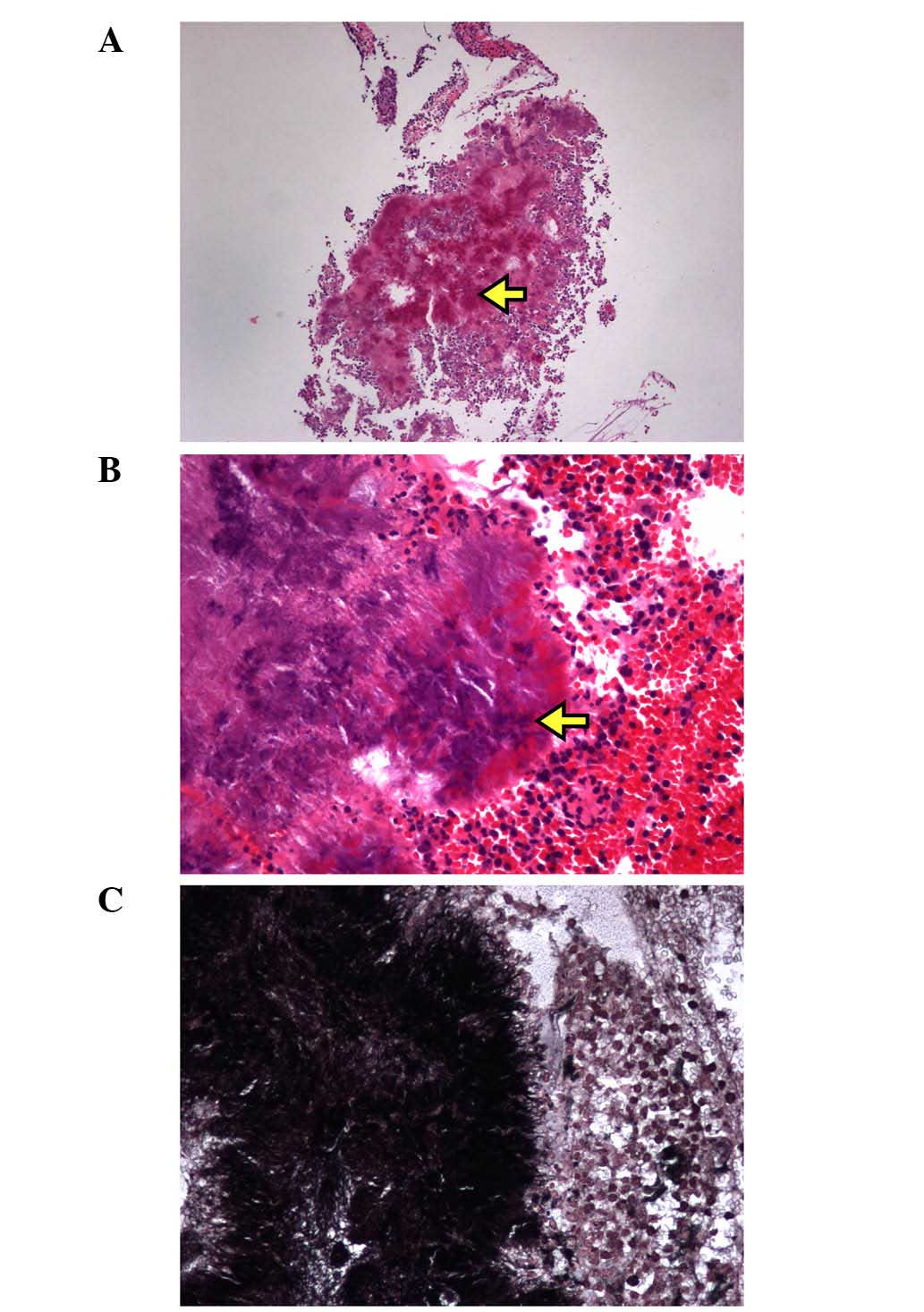
contained Gram-positive filamentous rods, but was negative for acid-fast staining. Despite this, the culture medium failed to grow any colonies. Microscopic analysis at low magnification identified cauliflower-shaped sulfur granules in association with acute and chronic inflammation (Fig. 3A). Higher-power microscopic examination revealed that these granules were surrounded by a rosette of clubbed filaments (Fig. 3B). Furthermore, Grocott-Gomori methenamine silver staining revealed the presence of filamentous rods (Fig. 3C). Based on these findings, the patient was diagnosed with nasopharyngeal actinomycosis. After two months of oral antibiotic treatment with 500 mg amoxicillin four times per day, recovery was uneventful, with no evidence of recurrence over the following 17 months. Written informed consent was obtained from the patient prior to publication of the study, and the study was approved by the Ethics Committee of the Institutional Review Board of Kaohsiung Veterans General Hospital (Kaohsiung, Taiwan).

Figure 1.

Coronal (A) PET and (B) fused PET/CT scans, as well as axial (C) PET, (D) CT and (E) fused PET/CT scans revealing a focal hot lesion (maximum standard uptake value, 6.2) located at the roof of the nasopharynx and measuring ~1 cm in size. PET, positron emission tomography; CT, computed tomography.

Figure 2.

Nasopharyngoscopic examination revealing a granular mass originating from the nasopharynx.

Figure 3.

(A) Pictomicrograph from the nasopharyngeal biopsy revealing typical sulfur granules (arrow; H&E staining; magnification, ×100). (B) The central core of the sulfur granules contained organized aggregates of filaments with club-like eosinophilic structures, referred to as the Splendore-Hoeppli phenomenon (arrow; H&E staining; magnification, ×400). (C) Grocott-Gomori methenamine silver staining of the specimen (magnification, ×400). H&E, hematoxylin and eosin.

Discussion

Human actinomycosis was first described in 1878 by James Israel (4). Actinomycosis is a rare anaerobic bacterial infection, typically caused by Actinomyces israelii (1,2). The members of the pathogenic Actinomyces species do not exist freely in nature, but are commensals that normally inhabit the oropharynx (particularly the tonsillar crypts and the gingivodental crevices) (4), the abdominopelvic region or the female genitourinary organs (1). Antecedent tissue injury with coinfection by other pathogens that act in a synergistic manner may lead to infection at any site in the body. Orocervicofacial actinomycosis is the most common form of the disease, accounting for up to 60% of all cases (2). Lesions are frequently located at the angle of the jaw or in the submandibular region (1). Common presenting features include an acute painful abscess or chronic indolent soft-tissue swelling, from which sinus tracts can develop over time. At present, the clinical diagnosis of actinomycosis is challenging, and therefore the disease has been referred to as a ‘masquerader of head and neck’ disease (1–3).

Diagnosis is established most accurately by isolating the Actinomyces species in cultures of clinical specimens (1–3). However, previous studies have reported that <50% of cases highly suspected to suffer from actinomycosis lead to the growth of the organism in cultures (1–3). This is considered to be due to the requirement of strict anaerobic culturing conditions, previous antibiotic use or the overgrowth of a concomitant organism (1–9). Clinicians must be knowledgeable when submitting specimens in order to optimize the recovery of these fastidious anaerobic bacteria. Under microscopic examination, colonies of the organism form typical sulfur granules and present as round or oval basophilic masses on hematoxylin-eosin slides, the centres of which contain organized aggregates of filaments with club-like eosinophilic structures that are referred to as the Splendore-Hoeppli phenomenon (1–3). A Gram-stained smear of the specimen may exhibit beaded, branched Gram-positive filamentous rods (1–3). The utilization of specific stains, including Grocott-Gomori methenamine silver stain, MacCallum-Goodpasture stain, p-aminosalicylic acid or Brown-Brenn stain, may aid in the visualization of the bacilli (1). A specific differential consideration is a diagnosis of nocardiosis (9), which is positive for acid-fast staining. Therefore, the combination of clinical images, microbiological results and histological findings should enable a successful diagnosis of actinomycosis (1–3).

The treatment of actinomycosis involves prolonged antibiotic treatment and/or surgical resection (1–9). Surgery may be reserved for certain circumstances, such as the excision of the sinus tract, resection of necrotic tissue, sequestration of bone and drainage of abscesses (1). Although surgical intervention promotes recovery, it is not curative by itself. High-dose penicillin (18–24 million units per day) administered over a prolonged period is the standard form of therapy (1–3). Doxycycline, clindamycin and erythromycin may be used as effective alternative regimens, particularly for patients who are allergic to penicillin (1–3). However, previous data suggested that not all cases warrant long-term antibiotics. Sharkawy (2) and Oostman and Smego (3) stratified cases into mild and complicated infections, and concluded that the modern approach to treatment can be individualized depending on the site of infection, severity of disease and the patient's response to treatment. In addition, Daamen and Johnson (5) and Chiang et al (8) successfully treated patients with nasopharyngeal actinomycosis using a four-week administration of oral antibiotics.

To the best of our knowledge, nasopharyngeal actinomycosis resulting in a draining fistula has not been previously reported. Nasopharyngeal actinomycosis frequently presents as a non-tender virulent granuloma that is capable of expanding into contiguous tissue without regard for facial or anatomical barriers. In terms of its mass effect, it may cause unilateral otitis media with effusion (6), nasal airway obstruction (5) and carotid occlusion (7).

Since one of the hallmarks of nasopharyngeal carcinoma is its marked racial/ethnic and geographic distribution in Southeast Asia, recommendations for workup have changed over time as technologies have improved. PET scanning, albeit not routinely recommended, is not only capable of revealing the location of tumors, but also provides valuable information concerning metabolic tumor volume. Unlike in malignant disease, positive FDG uptake depends on the presence and activity of inflammatory leukocytes (10). In the present study, it was hypothesized that acute inflammation with direct actinomycetes invasion was the main cause of increased uptake of 18F-FDG.

In conclusion, although the occurrence of nasopharyngeal actinomycosis is extremely low, it is advisable to always maintain a high level of clinical suspicion when treating patients with a persistent nasopharyngeal mass that presents with only vague symptoms. Healthy individuals may be infected without any predisposing factors. A presumptive diagnosis is established by the presence of sulfur granules containing Gram-positive filamentous rods, but a negative result for acid-fast staining (1). The prompt initiation of an appropriate therapy is crucial for the eradication of this insidious disease. In addition, prolonged observation and follow-up are mandatory in order to detect recurrence. The present study fills a breach in the clinical literature regarding this unique disease entity. Future studies should aim to provide further innovative information regarding the clinical evaluation and management of nasopharyngeal actinomycosis.

References

1 

Wong VK, Turmezei TD and Weston VC: Actinomycosis. BMJ. 343:d60992011. View Article : Google Scholar

2 

Sharkawy AA: Cervicofacial actinomycosis and mandibular osteomyelitis. Infect Dis Clin North Am. 21:543–556. 2007. View Article : Google Scholar : PubMed/NCBI

3 

Oostman O and Smego RA: Cervicofacial actinomycosis: Diagnosis and management. Curr Infect Dis Rep. 7:170–174. 2005. View Article : Google Scholar : PubMed/NCBI

4 

Melgarejo Moreno P, Hellin Meseguer D, Marco Garrido A, Galindo Ortego X, Ruiz Macia JA and Hostalet F: A correlation between age and Actinomyces in the adenotonsillar tissue of children. B-ENT. 2:95–97. 2006.

5 

Daamen N and Johnson JT: Nasopharyngeal actinomycosis: a rare cause of nasal airway obstruction. Laryngoscope. 114:1403–1405. 2004. View Article : Google Scholar : PubMed/NCBI

6 

Ono T, Yoshida Y, Izumaru S and Nakashima T: A case of nasopharyngeal actinomycosis leading to otitis media with effusion. Auris Nasus Larynx. 33:451–454. 2006. View Article : Google Scholar : PubMed/NCBI

7 

Kalra V and Malhotra A: Actinomycosis of the nasopharynx causing carotid occlusion. Clin Neuroradiol. 23:129–131. 2013. View Article : Google Scholar : PubMed/NCBI

8 

Chiang CW, Chang YL and Lou PJ: Actinomycosis imitating nasopharyngeal carcinoma. Ann Otol Rhinol Laryngol. 109:605–607. 2000. View Article : Google Scholar : PubMed/NCBI

9 

Kolb CM, Ostrander S, Ramsey MJ, Burgos RM and Belnap C: Pathology quiz case 1. Actinomycosis osteomyelitis of the temporal bone. Arch Otolaryngol Head Neck Surg. 138:203–205. 2012. View Article : Google Scholar : PubMed/NCBI

10 

Rini JN and Palestro CJ: Imaging of infection and inflammation with 18F-FDG-labeled leukocytes. Q J Nucl Med Mol Imaging. 50:143–146. 2006.PubMed/NCBI

Related Articles

  • Abstract
  • View
  • Download
  • Twitter
Copy and paste a formatted citation
Spandidos Publications style
Lin YH, Hu C, Chuang HW and Lin MY: Nasopharyngeal actinomycosis on 18F-fluorodeoxyglucose positron emission tomography/computed tomography: A case report. Oncol Lett 10: 260-262, 2015.
APA
Lin, Y., Hu, C., Chuang, H., & Lin, M. (2015). Nasopharyngeal actinomycosis on 18F-fluorodeoxyglucose positron emission tomography/computed tomography: A case report. Oncology Letters, 10, 260-262. https://doi.org/10.3892/ol.2015.3157
MLA
Lin, Y., Hu, C., Chuang, H., Lin, M."Nasopharyngeal actinomycosis on 18F-fluorodeoxyglucose positron emission tomography/computed tomography: A case report". Oncology Letters 10.1 (2015): 260-262.
Chicago
Lin, Y., Hu, C., Chuang, H., Lin, M."Nasopharyngeal actinomycosis on 18F-fluorodeoxyglucose positron emission tomography/computed tomography: A case report". Oncology Letters 10, no. 1 (2015): 260-262. https://doi.org/10.3892/ol.2015.3157
Copy and paste a formatted citation
x
Spandidos Publications style
Lin YH, Hu C, Chuang HW and Lin MY: Nasopharyngeal actinomycosis on 18F-fluorodeoxyglucose positron emission tomography/computed tomography: A case report. Oncol Lett 10: 260-262, 2015.
APA
Lin, Y., Hu, C., Chuang, H., & Lin, M. (2015). Nasopharyngeal actinomycosis on 18F-fluorodeoxyglucose positron emission tomography/computed tomography: A case report. Oncology Letters, 10, 260-262. https://doi.org/10.3892/ol.2015.3157
MLA
Lin, Y., Hu, C., Chuang, H., Lin, M."Nasopharyngeal actinomycosis on 18F-fluorodeoxyglucose positron emission tomography/computed tomography: A case report". Oncology Letters 10.1 (2015): 260-262.
Chicago
Lin, Y., Hu, C., Chuang, H., Lin, M."Nasopharyngeal actinomycosis on 18F-fluorodeoxyglucose positron emission tomography/computed tomography: A case report". Oncology Letters 10, no. 1 (2015): 260-262. https://doi.org/10.3892/ol.2015.3157
Follow us
  • Twitter
  • LinkedIn
  • Facebook
About
  • Spandidos Publications
  • Careers
  • Cookie Policy
  • Privacy Policy
How can we help?
  • Help
  • Live Chat
  • Contact
  • Email to our Support Team