Spandidos Publications Logo
  • About
    • About Spandidos
    • Aims and Scopes
    • Abstracting and Indexing
    • Editorial Policies
    • Reprints and Permissions
    • Job Opportunities
    • Terms and Conditions
    • Contact
  • Journals
    • All Journals
    • Oncology Letters
      • Oncology Letters
      • Information for Authors
      • Editorial Policies
      • Editorial Board
      • Aims and Scope
      • Abstracting and Indexing
      • Bibliographic Information
      • Archive
    • International Journal of Oncology
      • International Journal of Oncology
      • Information for Authors
      • Editorial Policies
      • Editorial Board
      • Aims and Scope
      • Abstracting and Indexing
      • Bibliographic Information
      • Archive
    • Molecular and Clinical Oncology
      • Molecular and Clinical Oncology
      • Information for Authors
      • Editorial Policies
      • Editorial Board
      • Aims and Scope
      • Abstracting and Indexing
      • Bibliographic Information
      • Archive
    • Experimental and Therapeutic Medicine
      • Experimental and Therapeutic Medicine
      • Information for Authors
      • Editorial Policies
      • Editorial Board
      • Aims and Scope
      • Abstracting and Indexing
      • Bibliographic Information
      • Archive
    • International Journal of Molecular Medicine
      • International Journal of Molecular Medicine
      • Information for Authors
      • Editorial Policies
      • Editorial Board
      • Aims and Scope
      • Abstracting and Indexing
      • Bibliographic Information
      • Archive
    • Biomedical Reports
      • Biomedical Reports
      • Information for Authors
      • Editorial Policies
      • Editorial Board
      • Aims and Scope
      • Abstracting and Indexing
      • Bibliographic Information
      • Archive
    • Oncology Reports
      • Oncology Reports
      • Information for Authors
      • Editorial Policies
      • Editorial Board
      • Aims and Scope
      • Abstracting and Indexing
      • Bibliographic Information
      • Archive
    • Molecular Medicine Reports
      • Molecular Medicine Reports
      • Information for Authors
      • Editorial Policies
      • Editorial Board
      • Aims and Scope
      • Abstracting and Indexing
      • Bibliographic Information
      • Archive
    • World Academy of Sciences Journal
      • World Academy of Sciences Journal
      • Information for Authors
      • Editorial Policies
      • Editorial Board
      • Aims and Scope
      • Abstracting and Indexing
      • Bibliographic Information
      • Archive
    • International Journal of Functional Nutrition
      • International Journal of Functional Nutrition
      • Information for Authors
      • Editorial Policies
      • Editorial Board
      • Aims and Scope
      • Abstracting and Indexing
      • Bibliographic Information
      • Archive
    • International Journal of Epigenetics
      • International Journal of Epigenetics
      • Information for Authors
      • Editorial Policies
      • Editorial Board
      • Aims and Scope
      • Abstracting and Indexing
      • Bibliographic Information
      • Archive
    • Medicine International
      • Medicine International
      • Information for Authors
      • Editorial Policies
      • Editorial Board
      • Aims and Scope
      • Abstracting and Indexing
      • Bibliographic Information
      • Archive
  • Articles
  • Information
    • Information for Authors
    • Information for Reviewers
    • Information for Librarians
    • Information for Advertisers
    • Conferences
  • Language Editing
Spandidos Publications Logo
  • About
    • About Spandidos
    • Aims and Scopes
    • Abstracting and Indexing
    • Editorial Policies
    • Reprints and Permissions
    • Job Opportunities
    • Terms and Conditions
    • Contact
  • Journals
    • All Journals
    • Biomedical Reports
      • Information for Authors
      • Editorial Policies
      • Editorial Board
      • Aims and Scope
      • Abstracting and Indexing
      • Bibliographic Information
      • Archive
    • Experimental and Therapeutic Medicine
      • Information for Authors
      • Editorial Policies
      • Editorial Board
      • Aims and Scope
      • Abstracting and Indexing
      • Bibliographic Information
      • Archive
    • International Journal of Epigenetics
      • Information for Authors
      • Editorial Policies
      • Editorial Board
      • Aims and Scope
      • Abstracting and Indexing
      • Bibliographic Information
      • Archive
    • International Journal of Functional Nutrition
      • Information for Authors
      • Editorial Policies
      • Editorial Board
      • Aims and Scope
      • Abstracting and Indexing
      • Bibliographic Information
      • Archive
    • International Journal of Molecular Medicine
      • Information for Authors
      • Editorial Policies
      • Editorial Board
      • Aims and Scope
      • Abstracting and Indexing
      • Bibliographic Information
      • Archive
    • International Journal of Oncology
      • Information for Authors
      • Editorial Policies
      • Editorial Board
      • Aims and Scope
      • Abstracting and Indexing
      • Bibliographic Information
      • Archive
    • Medicine International
      • Information for Authors
      • Editorial Policies
      • Editorial Board
      • Aims and Scope
      • Abstracting and Indexing
      • Bibliographic Information
      • Archive
    • Molecular and Clinical Oncology
      • Information for Authors
      • Editorial Policies
      • Editorial Board
      • Aims and Scope
      • Abstracting and Indexing
      • Bibliographic Information
      • Archive
    • Molecular Medicine Reports
      • Information for Authors
      • Editorial Policies
      • Editorial Board
      • Aims and Scope
      • Abstracting and Indexing
      • Bibliographic Information
      • Archive
    • Oncology Letters
      • Information for Authors
      • Editorial Policies
      • Editorial Board
      • Aims and Scope
      • Abstracting and Indexing
      • Bibliographic Information
      • Archive
    • Oncology Reports
      • Information for Authors
      • Editorial Policies
      • Editorial Board
      • Aims and Scope
      • Abstracting and Indexing
      • Bibliographic Information
      • Archive
    • World Academy of Sciences Journal
      • Information for Authors
      • Editorial Policies
      • Editorial Board
      • Aims and Scope
      • Abstracting and Indexing
      • Bibliographic Information
      • Archive
  • Articles
  • Information
    • For Authors
    • For Reviewers
    • For Librarians
    • For Advertisers
    • Conferences
  • Language Editing
Login Register Submit
  • This site uses cookies
  • You can change your cookie settings at any time by following the instructions in our Cookie Policy. To find out more, you may read our Privacy Policy.

    I agree
Search articles by DOI, keyword, author or affiliation
Search
Advanced Search
presentation
Oncology Letters
Join Editorial Board Propose a Special Issue
Print ISSN: 1792-1074 Online ISSN: 1792-1082
Journal Cover
July-2015 Volume 10 Issue 1

Full Size Image

Sign up for eToc alerts
Recommend to Library

Journals

International Journal of Molecular Medicine

International Journal of Molecular Medicine

International Journal of Molecular Medicine is an international journal devoted to molecular mechanisms of human disease.

International Journal of Oncology

International Journal of Oncology

International Journal of Oncology is an international journal devoted to oncology research and cancer treatment.

Molecular Medicine Reports

Molecular Medicine Reports

Covers molecular medicine topics such as pharmacology, pathology, genetics, neuroscience, infectious diseases, molecular cardiology, and molecular surgery.

Oncology Reports

Oncology Reports

Oncology Reports is an international journal devoted to fundamental and applied research in Oncology.

Experimental and Therapeutic Medicine

Experimental and Therapeutic Medicine

Experimental and Therapeutic Medicine is an international journal devoted to laboratory and clinical medicine.

Oncology Letters

Oncology Letters

Oncology Letters is an international journal devoted to Experimental and Clinical Oncology.

Biomedical Reports

Biomedical Reports

Explores a wide range of biological and medical fields, including pharmacology, genetics, microbiology, neuroscience, and molecular cardiology.

Molecular and Clinical Oncology

Molecular and Clinical Oncology

International journal addressing all aspects of oncology research, from tumorigenesis and oncogenes to chemotherapy and metastasis.

World Academy of Sciences Journal

World Academy of Sciences Journal

Multidisciplinary open-access journal spanning biochemistry, genetics, neuroscience, environmental health, and synthetic biology.

International Journal of Functional Nutrition

International Journal of Functional Nutrition

Open-access journal combining biochemistry, pharmacology, immunology, and genetics to advance health through functional nutrition.

International Journal of Epigenetics

International Journal of Epigenetics

Publishes open-access research on using epigenetics to advance understanding and treatment of human disease.

Medicine International

Medicine International

An International Open Access Journal Devoted to General Medicine.

Journal Cover
July-2015 Volume 10 Issue 1

Full Size Image

Sign up for eToc alerts
Recommend to Library

  • Article
  • Citations
    • Cite This Article
    • Download Citation
    • Create Citation Alert
    • Remove Citation Alert
    • Cited By
  • Similar Articles
    • Related Articles (in Spandidos Publications)
    • Similar Articles (Google Scholar)
    • Similar Articles (PubMed)
  • Download PDF
  • Download XML
  • View XML
Case Report

Primary glioblastoma of the cerebellar vermis: A case report

  • Authors:
    • Xiaolin Jing
    • Guohua Shen
    • Minggang Su
    • Rong Tian
    • Zhiyun Jia
  • View Affiliations / Copyright

    Affiliations: Department of Nuclear Medicine, West China Hospital, Sichuan University, Chengdu, Sichuan 610041, P.R. China
  • Pages: 402-404
    |
    Published online on: May 6, 2015
       https://doi.org/10.3892/ol.2015.3188
  • Expand metrics +
Metrics: Total Views: 0 (Spandidos Publications: | PMC Statistics: )
Metrics: Total PDF Downloads: 0 (Spandidos Publications: | PMC Statistics: )
Cited By (CrossRef): 0 citations Loading Articles...

This article is mentioned in:


Abstract

Cerebellar glioblastoma is a rare adult tumor. The accurate diagnosis of cerebellar glioblastoma is important for establishing a suitable therapeutic schedule. However, it is occasionally difficult to diagnosis these tumors. Clinical presentation, computed tomography (CT) and magnetic resonance imaging can provide useful information, but they may not lead to a definitive diagnosis. Positron emission tomography/computed tomography (PET/CT) may provide a novel way of forming a differential diagnoses. The lesions of glioblastoma multiforme (GBM) rarely occur in the cerebellum, with prior studies reporting that only 0.4‑3.4% of all GBM tumors occur here. In the current study, a case of primary cerebellar glioblastoma is presented and the physiopathology, clinical presentation, diagnosis, differential diagnosis, treatment and general outcome of this disease are discussed. A 61‑year‑old female presented with nausea, vomiting, balance problems and cerebellar signs. Cranial magnetic resonance imaging (MRI) and positron emission tomography/computed tomography (PET/CT) examination demonstrated one regular contour of a mass lesion in the cerebellar vermis. Following surgery, glioblastoma was histologically confirmed. The outcome of the patient was favorable after 18 months of follow‑up. Cerebellar GBM should be considered in the differential diagnosis of a cerebellar mass lesion, and PET/CT may provide a novel identification method for different cerebellar mass lesions.

Introduction

Glioblastoma multiforme (GBM) is the most frequently occurring malignant primary brain tumor in adults. These tumors generally develop in the fifth and sixth decades of life (1,2). However, these lesions rarely occur in the cerebellum, with prior studies reporting that only 0.4–3.4% of all GBM tumors occur in this location (3–5). Hypertension, impaired balance and gait disturbance are typical clinical manifestations (1). The best treatment for cerebellar glioblastoma is removal of as much of the tumors as possible but keeping surgical morbidity to a minimum (2). The prognosis of cerebellar glioblastoma is similar to that of anaplasic astrocytoma (1). In the current study, a case of primary cerebellar glioblastoma is presented, and the physiopathology, clinical presentation, diagnosis, differential diagnosis, treatment and general outcome of this disease is discussed.

Case report

A 61-year-old female with no other medical history was referred to West China Hospital of Sichuan University (Chengdu, China) and presented with nausea, vomiting and balance problems that had persisted for >1 month. A neurological examination demonstrated cerebellar signs, including positive bilateral finger-nose and knee-shin tests. Tumor marker analysis and other 
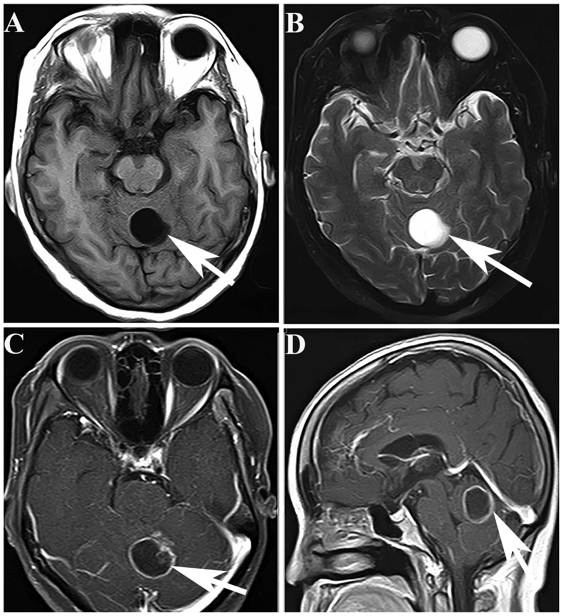
blood tests were negative. Cranial magnetic resonance imaging (MRI) showed one regular contour of a mass lesion in the cerebellar vermis. The mass was present as a hypointense and hyperintense lesion in the cerebellar vermis in T1- and T2-weighted images, respectively (Fig. 1A and B). The mass had well-defined borders and large areas of central necrosis. T1-weighted magnetic enhanced imaging showed a well-defined, heterogeneously ring-enhancing lesion, with one additional enhanced node (Fig. 1C and D). The provisional diagnosis was of a metastatic lesion of the cerebellum. The patient 
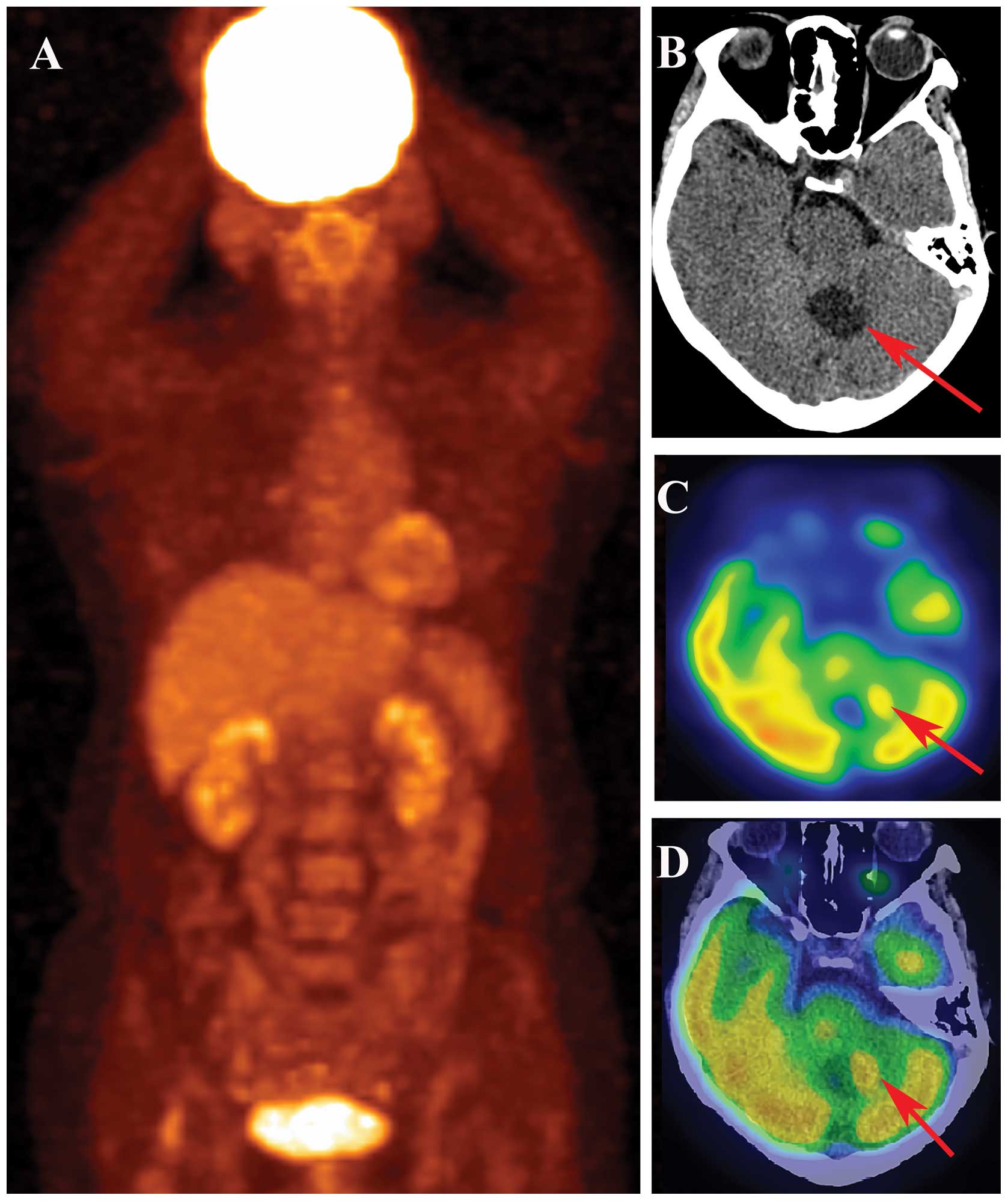
signed the written informed consent and then underwent a positron emission tomography/computed tomography (PET/CT) examination. There was no abnormal glucose uptake on coronal PET imaging (Fig. 2A). Cerebral CT showed one hypodense lesion with a nodule alongside the cerebellar vermis (Fig. 2B). In PET imaging, the nodule was observed to have much higher glucose uptake than other tissues (Fig. 2C and D). Therefore, the patient was diagnosed with primary brain tumors.

Figure 1.

Cranial magnetic resonance imaging showing one lesion in the cerebellar vermis. (A) T1-weighted image showing a well-defined hypointense tumor in the vermis of the cerebellum (arrow). (B) T2-weighted image showing a well-distributed hyperintense lesion without evident surrounding edema (arrow). (C and D) T1-weighted magnetic resonance enhanced images showing a well-defined, ring-enhancing lesion with one additional enhanced nodule (arrow).

Figure 2.

(A) Coronal positron emission tomography (PET) image showing no abnormal glucose uptake. (B) Computed tomography scan showing a round cystic mass with more hypodense expression than normal brain tissue. In addition, there is a hyperdense nodule alongside the mass (arrow). (C and D) In the PET and PET/CT images, the nodule (arrow) shows much higher glucose uptake compared with the surrounding brain tissue.

The patient underwent a cerebellar lesion resection at West China Hospital of Sichuan University (Chengdu, China) and 
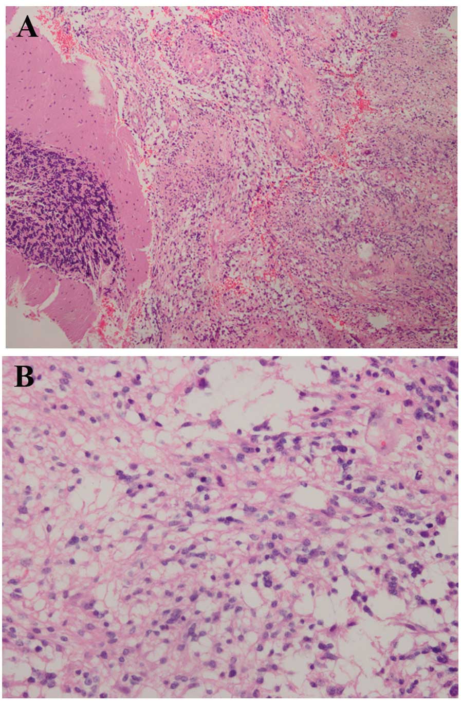
the pathology of the lesion diagnosed a glioblastoma (class IV; Fig. 3). Following the surgery, histopathological examination revealed a cellular tumor, which was consistent with glioblastoma that met the World Health Organization criteria of grade 4 astrocytoma. The appearance of the tumor was consistent with necrosis, pseudo-palisading and vascular hyperplasia. Photomicrograph of tumor sections demonstrated an intense cellular density and nuclear pleomorphism, which was characterized by marked anisokaryosis and larger nuclei than is normal. The cells had an abundant and eosinophilic cytoplasm and umerous mitoses were observed (Fig. 3).

Figure 3.

Tumor sections showing an intense cellular density and nuclear pleomorphism. (A) The tumor cells are characterized by marked anisokaryosis; the nuclei are larger than normal. The cells have an abundant and eosinophilic cytoplasm. Numerous mitoses are observed. Hematoxylin and eosin stain; (A) ×100 and (B) ×400 magnification.

The patient had modest brain edema following the surgery for a few days, but improved immediately after diuretics were administered. The neurologic signs improved and the clinical presentation disappeared. The patient did not receive radiotherapy or chemotherapy after the surgery. There was no sign of tumor recurrence after 18 months.

Discussion

Cerebellar GBM in adults is rare, and thus comprises only a small proportion of all GBM tumors of the brain. GBM of the cerebellum can be observed in all age groups. A study has shown that the male-to-female ratio for this disease is 2:1, and that ~70% of the tumors occur in adults (average age, 46.7 years), while 30% occur in children (average age, 10.4 years) (6). GBM is a stage 4 tumor according to the World Health Organization classification (7). The prognosis of this entity is uncertain, and factors associated with prognosis have been unclear. According to the case study of a 41-year-old male with a cerebellar glioblastoma developing in the cerebellar hemisphere who was treated 35 years previously by radiotherapy, radiation may be a cause for inducing GBM (8): The patient had medulloblastoma (MB) in the cerebellar vermis, and postoperatively he received a total of 40 Gy radiations to the whole brain and 30.5 Gy to the spine without chemotherapy (8).

Patients with GBM typically present with increased hypertension, impaired balance and gait disturbance (1). Upon examination, patients exhibit cerebellar signs, as occurred in the present case. The symptoms of dizziness, mental confusion and neck pain can also be present (9). A diagnosis of cerebellar GBM is not usually pre-operatively suspected, although certain CT and MRI characteristics may indicate it (6,9). Cerebellar metastases, hemangioblastomas, brain abscesses and anaplastic astrocytomas are common differential diagnoses in adults. Brain abscesses often have symptoms of infection. Abscess walls are mostly smooth without nodules. Enhanced CT and MRI scans show incomplete ring-like reinforcements, which are thin and uniform. Hemangioblastomas always occur singly, and >90% occur in the cerebellar hemisphere. The CT and MRI indications are similar to those in the present case. The pathology of a lesion can only be confirmed after surgery. Brain metastases always have necrosis within them and show greater than one lesion with marked peripheral edema (1,2,4,9). The history of a primary tumor is important. In fact, to identify these diseases, a number of different examinations are required, and they may take a long time to complete. At the West China Hospital of Sichuan University, patients are provided with PET/CT examinations according to whether lesions take up glucose or not, so that this disease can be identified. However, reports of signs of different brain tumors in PET/CT scans are rare. As PET/CT scans become increasingly common, they may become a novel method for identifying brain tumors.

Cerebellar glioblastoma treatment is usually palliative, consisting of surgery, radiotherapy and chemotherapy (1,2). Decisions regarding surgery depend on factors such as patient age, performance, diseased region in the brain and resectability of the tumor (1,2,4,6). Subsequent to surgery, patients can be treated with radiotherapy and chemotherapy. However, GBM is the highest graded astrocytoma and the recurrence rate following surgery is high. Moreover, the prognosis of patients with glioblastoma is poor, with a 2-year survival rate of 10% and <5% of the patients surviving for a long period of time. The mean survival time for patients with cerebellar glioblastoma after the beginning of symptomatology has been reported to be 12–19 months (6,10).

The present study reports a rare case of cerebellar GBM. PET/CT examination may provide a novel method for diagnosing GBM and providing differential diagnoses for metastases, abscesses and hemangioblastomas in the cerebellum.

Acknowledgements

This study was supported by the National Natural Science Foundation (grant nos. 81271532 and 30900378).

References

1 

El El Maaqili MR, Hossini A, El Fatemi N, et al: Primary glioblastoma of the cerebellum in a 19-year-old woman: a case report. J Med Case Rep. 6:3292012. View Article : Google Scholar : PubMed/NCBI

2 

Grahovac G, Tomac D, Lambasa S, et al: Cerebellar glioblastomas: Pathophysiology, clinical presentation and management. Acta Neurochir. 151:653–657. 2009. View Article : Google Scholar : PubMed/NCBI

3 

Babu R, Sharma R, Karikari IO, et al: Outcome and prognostic factors in adult cerebellar glioblastoma. J Clin Neurosci. 20:1117–1121. 2013. View Article : Google Scholar : PubMed/NCBI

4 

Kuroiwa T, Numaguchi Y, Rothman MI, et al: Posterior fossa glioblastoma multiforme: MR findings. AJNR Am J Neuroradiol. 16:583–589. 1995.PubMed/NCBI

5 

Roth JG and Elvidge AR: Glioblastoma multiforme: A clinical survey. J Neurosurg. 17:736–750. 1960. View Article : Google Scholar : PubMed/NCBI

6 

Mattos JP, Marenco HA, Campos JM, et al: Cerebellar glioblastoma multiforme in an adult. Arq Neuropsiquiatr. 64:132–135. 2006. View Article : Google Scholar : PubMed/NCBI

7 

Louis DN, Ohgaki H, Wiestler OD, et al: The 2007 WHO classification of tumours of the central nervous system. Acta Neuropathol. 114:97–109. 2007. View Article : Google Scholar : PubMed/NCBI

8 

Hamasaki K, Nakamura H, Ueda Y, et al: Radiation-induced glioblastoma occurring 35 years after radiation therapy for medulloblastoma: case report. Brain Tumor Pathol. 27:39–43. 2010. View Article : Google Scholar : PubMed/NCBI

9 

Demir MK, Hakan T, Akinci O and Berkman Z: Primary cerebellar glioblastoma multiforme. Diagn Interv Radiol. 11:83–86. 2005.PubMed/NCBI

10 

Kulkarni AV, Becker LE, Jay V, et al: Primary cerebellar glioblastomas multiforme in children Report of four cases. J Neurosurg. 90:546–550. 1999. View Article : Google Scholar : PubMed/NCBI

Related Articles

  • Abstract
  • View
  • Download
  • Twitter
Copy and paste a formatted citation
Spandidos Publications style
Jing X, Shen G, Su M, Tian R and Jia Z: Primary glioblastoma of the cerebellar vermis: A case report. Oncol Lett 10: 402-404, 2015.
APA
Jing, X., Shen, G., Su, M., Tian, R., & Jia, Z. (2015). Primary glioblastoma of the cerebellar vermis: A case report. Oncology Letters, 10, 402-404. https://doi.org/10.3892/ol.2015.3188
MLA
Jing, X., Shen, G., Su, M., Tian, R., Jia, Z."Primary glioblastoma of the cerebellar vermis: A case report". Oncology Letters 10.1 (2015): 402-404.
Chicago
Jing, X., Shen, G., Su, M., Tian, R., Jia, Z."Primary glioblastoma of the cerebellar vermis: A case report". Oncology Letters 10, no. 1 (2015): 402-404. https://doi.org/10.3892/ol.2015.3188
Copy and paste a formatted citation
x
Spandidos Publications style
Jing X, Shen G, Su M, Tian R and Jia Z: Primary glioblastoma of the cerebellar vermis: A case report. Oncol Lett 10: 402-404, 2015.
APA
Jing, X., Shen, G., Su, M., Tian, R., & Jia, Z. (2015). Primary glioblastoma of the cerebellar vermis: A case report. Oncology Letters, 10, 402-404. https://doi.org/10.3892/ol.2015.3188
MLA
Jing, X., Shen, G., Su, M., Tian, R., Jia, Z."Primary glioblastoma of the cerebellar vermis: A case report". Oncology Letters 10.1 (2015): 402-404.
Chicago
Jing, X., Shen, G., Su, M., Tian, R., Jia, Z."Primary glioblastoma of the cerebellar vermis: A case report". Oncology Letters 10, no. 1 (2015): 402-404. https://doi.org/10.3892/ol.2015.3188
Follow us
  • Twitter
  • LinkedIn
  • Facebook
About
  • Spandidos Publications
  • Careers
  • Cookie Policy
  • Privacy Policy
How can we help?
  • Help
  • Live Chat
  • Contact
  • Email to our Support Team