Spandidos Publications Logo
  • About
    • About Spandidos
    • Aims and Scopes
    • Abstracting and Indexing
    • Editorial Policies
    • Reprints and Permissions
    • Job Opportunities
    • Terms and Conditions
    • Contact
  • Journals
    • All Journals
    • Oncology Letters
      • Oncology Letters
      • Information for Authors
      • Editorial Policies
      • Editorial Board
      • Aims and Scope
      • Abstracting and Indexing
      • Bibliographic Information
      • Archive
    • International Journal of Oncology
      • International Journal of Oncology
      • Information for Authors
      • Editorial Policies
      • Editorial Board
      • Aims and Scope
      • Abstracting and Indexing
      • Bibliographic Information
      • Archive
    • Molecular and Clinical Oncology
      • Molecular and Clinical Oncology
      • Information for Authors
      • Editorial Policies
      • Editorial Board
      • Aims and Scope
      • Abstracting and Indexing
      • Bibliographic Information
      • Archive
    • Experimental and Therapeutic Medicine
      • Experimental and Therapeutic Medicine
      • Information for Authors
      • Editorial Policies
      • Editorial Board
      • Aims and Scope
      • Abstracting and Indexing
      • Bibliographic Information
      • Archive
    • International Journal of Molecular Medicine
      • International Journal of Molecular Medicine
      • Information for Authors
      • Editorial Policies
      • Editorial Board
      • Aims and Scope
      • Abstracting and Indexing
      • Bibliographic Information
      • Archive
    • Biomedical Reports
      • Biomedical Reports
      • Information for Authors
      • Editorial Policies
      • Editorial Board
      • Aims and Scope
      • Abstracting and Indexing
      • Bibliographic Information
      • Archive
    • Oncology Reports
      • Oncology Reports
      • Information for Authors
      • Editorial Policies
      • Editorial Board
      • Aims and Scope
      • Abstracting and Indexing
      • Bibliographic Information
      • Archive
    • Molecular Medicine Reports
      • Molecular Medicine Reports
      • Information for Authors
      • Editorial Policies
      • Editorial Board
      • Aims and Scope
      • Abstracting and Indexing
      • Bibliographic Information
      • Archive
    • World Academy of Sciences Journal
      • World Academy of Sciences Journal
      • Information for Authors
      • Editorial Policies
      • Editorial Board
      • Aims and Scope
      • Abstracting and Indexing
      • Bibliographic Information
      • Archive
    • International Journal of Functional Nutrition
      • International Journal of Functional Nutrition
      • Information for Authors
      • Editorial Policies
      • Editorial Board
      • Aims and Scope
      • Abstracting and Indexing
      • Bibliographic Information
      • Archive
    • International Journal of Epigenetics
      • International Journal of Epigenetics
      • Information for Authors
      • Editorial Policies
      • Editorial Board
      • Aims and Scope
      • Abstracting and Indexing
      • Bibliographic Information
      • Archive
    • Medicine International
      • Medicine International
      • Information for Authors
      • Editorial Policies
      • Editorial Board
      • Aims and Scope
      • Abstracting and Indexing
      • Bibliographic Information
      • Archive
  • Articles
  • Information
    • Information for Authors
    • Information for Reviewers
    • Information for Librarians
    • Information for Advertisers
    • Conferences
  • Language Editing
Spandidos Publications Logo
  • About
    • About Spandidos
    • Aims and Scopes
    • Abstracting and Indexing
    • Editorial Policies
    • Reprints and Permissions
    • Job Opportunities
    • Terms and Conditions
    • Contact
  • Journals
    • All Journals
    • Biomedical Reports
      • Information for Authors
      • Editorial Policies
      • Editorial Board
      • Aims and Scope
      • Abstracting and Indexing
      • Bibliographic Information
      • Archive
    • Experimental and Therapeutic Medicine
      • Information for Authors
      • Editorial Policies
      • Editorial Board
      • Aims and Scope
      • Abstracting and Indexing
      • Bibliographic Information
      • Archive
    • International Journal of Epigenetics
      • Information for Authors
      • Editorial Policies
      • Editorial Board
      • Aims and Scope
      • Abstracting and Indexing
      • Bibliographic Information
      • Archive
    • International Journal of Functional Nutrition
      • Information for Authors
      • Editorial Policies
      • Editorial Board
      • Aims and Scope
      • Abstracting and Indexing
      • Bibliographic Information
      • Archive
    • International Journal of Molecular Medicine
      • Information for Authors
      • Editorial Policies
      • Editorial Board
      • Aims and Scope
      • Abstracting and Indexing
      • Bibliographic Information
      • Archive
    • International Journal of Oncology
      • Information for Authors
      • Editorial Policies
      • Editorial Board
      • Aims and Scope
      • Abstracting and Indexing
      • Bibliographic Information
      • Archive
    • Medicine International
      • Information for Authors
      • Editorial Policies
      • Editorial Board
      • Aims and Scope
      • Abstracting and Indexing
      • Bibliographic Information
      • Archive
    • Molecular and Clinical Oncology
      • Information for Authors
      • Editorial Policies
      • Editorial Board
      • Aims and Scope
      • Abstracting and Indexing
      • Bibliographic Information
      • Archive
    • Molecular Medicine Reports
      • Information for Authors
      • Editorial Policies
      • Editorial Board
      • Aims and Scope
      • Abstracting and Indexing
      • Bibliographic Information
      • Archive
    • Oncology Letters
      • Information for Authors
      • Editorial Policies
      • Editorial Board
      • Aims and Scope
      • Abstracting and Indexing
      • Bibliographic Information
      • Archive
    • Oncology Reports
      • Information for Authors
      • Editorial Policies
      • Editorial Board
      • Aims and Scope
      • Abstracting and Indexing
      • Bibliographic Information
      • Archive
    • World Academy of Sciences Journal
      • Information for Authors
      • Editorial Policies
      • Editorial Board
      • Aims and Scope
      • Abstracting and Indexing
      • Bibliographic Information
      • Archive
  • Articles
  • Information
    • For Authors
    • For Reviewers
    • For Librarians
    • For Advertisers
    • Conferences
  • Language Editing
Login Register Submit
  • This site uses cookies
  • You can change your cookie settings at any time by following the instructions in our Cookie Policy. To find out more, you may read our Privacy Policy.

    I agree
Search articles by DOI, keyword, author or affiliation
Search
Advanced Search
presentation
Oncology Letters
Join Editorial Board Propose a Special Issue
Print ISSN: 1792-1074 Online ISSN: 1792-1082
Journal Cover
July-2015 Volume 10 Issue 1

Full Size Image

Sign up for eToc alerts
Recommend to Library

Journals

International Journal of Molecular Medicine

International Journal of Molecular Medicine

International Journal of Molecular Medicine is an international journal devoted to molecular mechanisms of human disease.

International Journal of Oncology

International Journal of Oncology

International Journal of Oncology is an international journal devoted to oncology research and cancer treatment.

Molecular Medicine Reports

Molecular Medicine Reports

Covers molecular medicine topics such as pharmacology, pathology, genetics, neuroscience, infectious diseases, molecular cardiology, and molecular surgery.

Oncology Reports

Oncology Reports

Oncology Reports is an international journal devoted to fundamental and applied research in Oncology.

Experimental and Therapeutic Medicine

Experimental and Therapeutic Medicine

Experimental and Therapeutic Medicine is an international journal devoted to laboratory and clinical medicine.

Oncology Letters

Oncology Letters

Oncology Letters is an international journal devoted to Experimental and Clinical Oncology.

Biomedical Reports

Biomedical Reports

Explores a wide range of biological and medical fields, including pharmacology, genetics, microbiology, neuroscience, and molecular cardiology.

Molecular and Clinical Oncology

Molecular and Clinical Oncology

International journal addressing all aspects of oncology research, from tumorigenesis and oncogenes to chemotherapy and metastasis.

World Academy of Sciences Journal

World Academy of Sciences Journal

Multidisciplinary open-access journal spanning biochemistry, genetics, neuroscience, environmental health, and synthetic biology.

International Journal of Functional Nutrition

International Journal of Functional Nutrition

Open-access journal combining biochemistry, pharmacology, immunology, and genetics to advance health through functional nutrition.

International Journal of Epigenetics

International Journal of Epigenetics

Publishes open-access research on using epigenetics to advance understanding and treatment of human disease.

Medicine International

Medicine International

An International Open Access Journal Devoted to General Medicine.

Journal Cover
July-2015 Volume 10 Issue 1

Full Size Image

Sign up for eToc alerts
Recommend to Library

  • Article
  • Citations
    • Cite This Article
    • Download Citation
    • Create Citation Alert
    • Remove Citation Alert
    • Cited By
  • Similar Articles
    • Related Articles (in Spandidos Publications)
    • Similar Articles (Google Scholar)
    • Similar Articles (PubMed)
  • Download PDF
  • Download XML
  • View XML
Case Report

Slight uptake of 18F‑FDG on positron emission tomography in pulmonary hamartoma: A case report

  • Authors:
    • Weihua Wang
    • Jia Song
    • Jianguo Shi
    • Huizhen Hu
    • Yuquan Wu
    • Jie Yan
    • Lijun Wu
    • Qingyong Chen
  • View Affiliations / Copyright

    Affiliations: Department of Respiratory Medicine, Jiangshan People's Hospital, Jiangshan, Zhejiang 324100, P.R. China, Department of Respiratory Oncology, The 117th Hospital of PLA, Hangzhou, Zhejiang 310013, P.R. China, Department of Nuclear Medicine, The 117th Hospital of PLA, Hangzhou, Zhejiang 310013, P.R. China, Department of Thoracic Surgery, The 117th Hospital of PLA, Hangzhou, Zhejiang 310013, P.R. China
  • Pages: 430-432
    |
    Published online on: May 20, 2015
       https://doi.org/10.3892/ol.2015.3245
  • Expand metrics +
Metrics: Total Views: 0 (Spandidos Publications: | PMC Statistics: )
Metrics: Total PDF Downloads: 0 (Spandidos Publications: | PMC Statistics: )
Cited By (CrossRef): 0 citations Loading Articles...

This article is mentioned in:


Abstract

The present study reports the case of a 77‑year‑old female that was asymptomatic at presentation and was found to possess a lesion that was incidentally identified on a computed tomography (CT) scan. The CT scan revealed a non‑homogeneous, hypodense, non‑lobulated solid mass, ~1.2 cm in diameter, in the left upper lobe of the lung that demonstrated minimal contrast enhancement. The following CT scan was performed only two years later. This scan revealed that the non‑homogeneous round mass had increased in size to ~1.7 cm in diameter, and possessed an irregular margin, in addition to being slightly lobulated with no calcification or fat. Combined positron emission tomography and CT revealed a lobulated mass that was ~1.9 cm in diameter, demonstrating an irregular margin with involvement of the mediastinal pleura. Slight uptake of 18F‑fluorodeoxyglucose was also detected. The final histological diagnosis was pulmonary hamartoma.

Introduction

Pulmonary hamartoma is the most common benign tumor of the lung, accounting for 6% of all solitary pulmonary nodules (1). The majority of hamartomas are identified incidentally, with the peak incidence occurring in patients in their sixth decade (2). Hamartomas are rarely symptomatic, but in symptomatic cases the hamartoma is associated with hemoptysis or cough. Originally, hamartomas were considered to be congenital lesions, but pulmonary hamartomas are currently considered to be true neoplasms by the majority of investigators. Consistent with this concept are the observations that hamartomas are often identified in the elderly and that the lesions grow slowly. The typical radiographic appearance of a hamartoma is that of a smooth or slightly lobulated peripheral solitary pulmonary nodule. The presence of fat or popcorn-like calcifications may enable the confident diagnosis of a hamartoma, but these findings are not usually identified.

The radiological diagnosis of hamartoma is usually based on computed tomography (CT) findings, particularly the detection of popcorn-like calcifications and fat (3). 18F-fluorodeoxyglucose (18F-FDG) positron emission tomography (PET)/CT investigations may be of use when neither calcification nor fat is identified on CT scans (4). The present study reports the case of a 77-year-old female patient that was diagnosed with a pulmonary hamartoma that demonstrated atypical imaging findings on two imaging modalities.

Case report

An asymptomatic 77-year-old female was admitted to The 117th hospital of PLA (Hangzhou, China) for the investigation of a lesion in the left upper lobe of the lung that had been present for 3 years.

The patient had undergone a routine health examination in The 117th hospital of PLA on January 29, 2007. The patient was asymptomatic and no abnormal findings were detected in the examination, with the exception of a 1.2-cm solitary pulmonary 
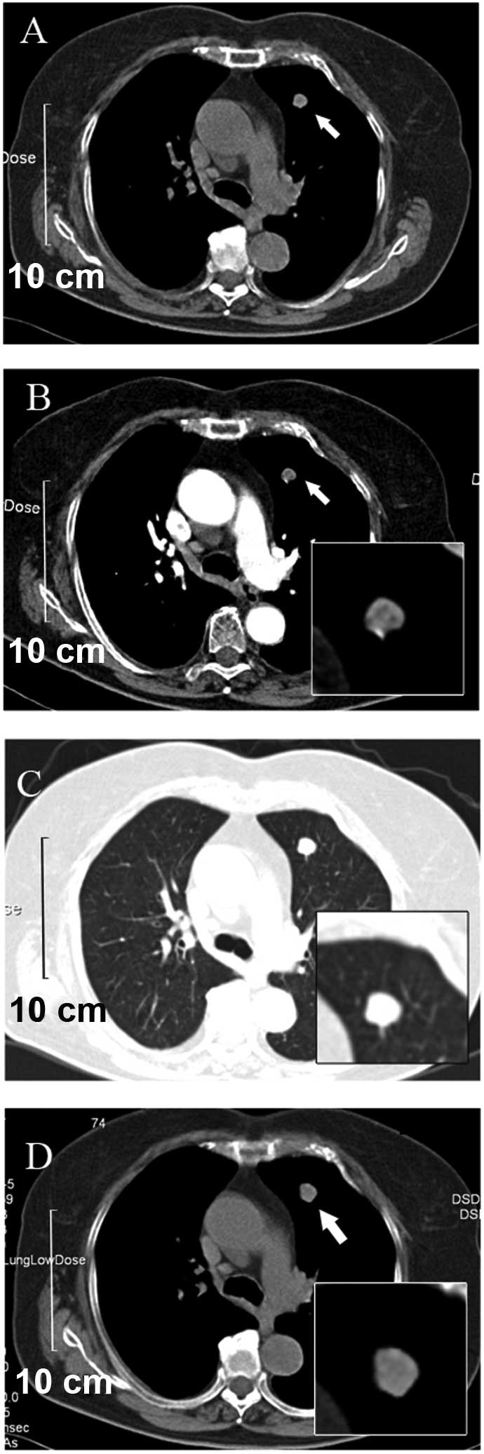
nodule that was identified on a CT scan. The scan revealed a round non-homogeneous parenchymal neoformation in the anterior segment of the left upper lobe of the lung, with a clear border (Fig. 1A). The contrast-enhanced CT that was performed four days later revealed that the neoformation contained hypodense regions with possible involvement of the vessel. In addition, non-homogeneous enhancement during the arterial phase was demonstrated. There was no indication of lobulation and spiculation. Neither the mediastinal nor hilar lymph nodes were enlarged (Fig. 1B and C).

Figure 1.

(A) CT of the chest revealed a round non-homogeneous parenchymal neoformation (arrow) that was ~1.2 cm in diameter and located in the anterius segment of the left upper lobe of the lung. The lesion was sharply demarcated. (B) The contrast-enhanced CT revealed minimal contrast-enhanced areas, and the lesion did not include any calcifications or fat. There was no sign of lobation and speculation. Neither the mediastinal nor hilar lymph nodes were observed to be enlarged in the (B) mediastinal and (C) pulmonary windows. CT, computed tomography.

Since there was no evidence of a malignant neoplasm, it was decided continue to follow-up the present patient. The following CT scan was performed two years subsequent to the first presentation (March 9, 2009). The CT scan revealed that the non-homogeneous oval mass had increased in size, to ~1.7 cm in diameter, with a regular margin and no calcification (Fig. 2). The results of physical examination and laboratory tests (serum tumor markers detection, including AFP, CEA and CA125, performed on November 6, 2010) were negative and the 
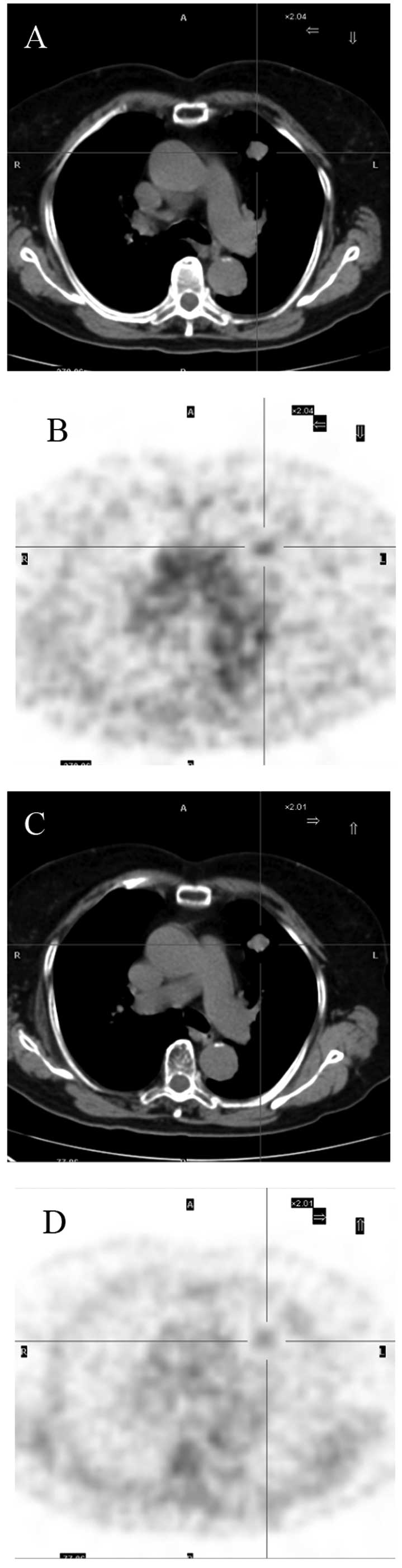
appearance of the radiology results were non-specific. Therefore, PET/CT was performed on November 8, 2010. PET/CT revealed a slightly lobulated mass in the left upper lobe of the lung that was ~1.9 cm in diameter and well defined, with involvement of the mediastinal pleura. Slight uptake of 18F-FDG was also detected. The maximum standardized uptake value (SUVmax) was found to be 2.44. Delayed scanning demonstrated that the lesion possessed diminishing metabolism, with an SUVmax of 1.64 (Fig. 3). Review of the laboratory tests, including those for serum tumor markers, remained within the normal range.

Figure 2.

The lesion was composed of fat, cartilage and smooth muscle, covered by normal bronchial epithelial cells.

Figure 3.

(A) Positron emission tomography/computed tomography revealed a slightly lobulated mass in the left upper lobe of the lung that was ~1.9 cm in diameter and well-defined, with involvement of the mediastinal pleura. (B) Slight uptake of 18F-fluorodeoxyglucose was also detected. (C and D) Delayed scanning demonstrated that the lesion possessed diminishing metabolism.

The lesion was suspected to be a pulmonary benign tumor or primary lung cancer, such as well-differentiated adenocarcinoma, but the diagnosis was unable to be confirmed pre-operatively. Since the patient requested removal of the lesion, 
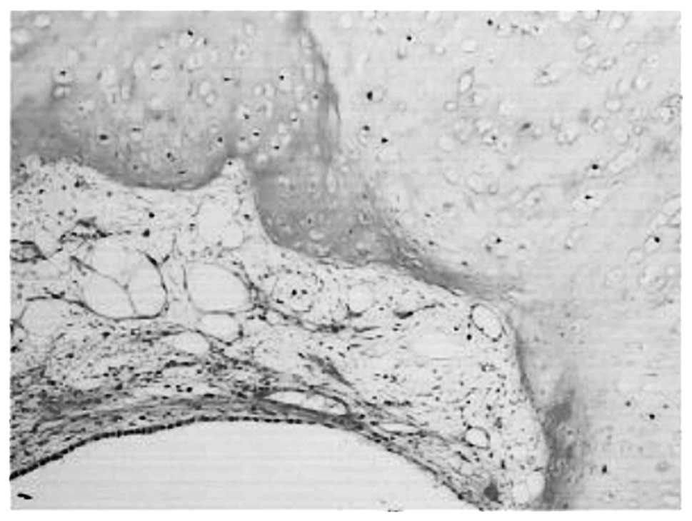
it was resected by video-assisted thoracic surgery. Gross examination of the resected specimen revealed that the tumor measured 2.5×2.0 cm, and was a white, multilobular, flexible mass without an envelope. The margin of the lesion and peripheral lung tissue was clear and there was no area demonstrating infiltrative growth. Histologically, the lesion was composed of fat, cartilage and smooth muscle, covered by normal bronchial epithelial cells (Fig. 3). On the basis of these findings, the tumor was diagnosed as a hamartoma. Six months subsequent to the surgical procedure, the patient is completely well, with no evidence of recurrence.

Discussion

Hamartomas are the third most common solitary pulmonary nodule, following granuloma and carcinoma, and usually account for 6–8% of localized parenchymal masses treated by thoracotomy (1). Individuals aged 60–79 years old develop hamartomas with the highest frequency (2). The majority of the patients with pulmonary hamartoma are free of symptoms, and the tumor is found incidentally on chest X-ray examination. On X-ray examination, a pulmonary hamartoma usually presents as a sharply demarcated coin lesion with slow growth, occasionally with calcification, but this is not diagnostic since calcifications may appear in carcinomas.

The radiological diagnosis of a pulmonary hamartoma has frequently been made using CT. In particular, CT densitometry with an advanced narrow collimated technique has been accepted as one of the best sensitive methods from eliminated partial volume averaging in detecting intranodular fat (−40 to −120 Hounsfield units), providing a highly predictive diagnosis of a pulmonary hamartoma or characteristic popcorn calcifications (1,2). However, hamartomas are extremely challenging to diagnose when the lesions possess neither fat nor calcification in 50% of pulmonary hamartomas, at most (1). This results in a diagnostic challenge, as the hamartomas cannot be differentiated from a primary or secondary lung cancer or other benign nodules. In the present study, the patient was asymptomatic and the tumor was identified incidentally on a CT scan. The mean size of hamartomas increased between 1.2 and 1.9 cm in three years, with an irregular margin and no calcification or fat identified on CT. The lesion was suspected to be a benign or primary lung cancer, such as well-differentiated adenocarcinoma, but the diagnosis was unable to be confirmed pre-operatively. 18FDG PET/CT studies may aid diagnosis when neither calcification nor fat is demonstrated on CT studies.

The use of an 18F-FDG-PET scan is based on the principle that cancer cells demonstrate an increased glucose uptake and higher rate of glycolysis compared with non-cancerous cells. In a meta-analysis reported by Gould et al, a sensitivity of 97% and a specificity of 78% were determined for the differentiation of malignant from benign pulmonary lesions using an 18F-FDG-PET scan (4). However, 18F-FDG-PET possesses a limited capacity for the detection of tumors with low glycolytic activity, including small tumors, bronchioloalveolar cell carcinoma and carcinoid tumors (5). As a result, the use of 18F-FDG-PET scans for the identification of hamartomas is limited.

To the best of our knowledge, there are only two published studies that investigated the 18F-FDG-PET scan findings in hamartomas. Teramoto et al reported that the uptake of 18F-FDG did not increase in pulmonary hamartomas (6). By contrast, in the case reported by Himpe et al, only slightly elevated uptake with an SUVmax of 3.3 and SUVmean of 1.7 was identified (7). Consistent with the findings of Himpe et al, the present patient possessed a lesion with an SUVmax of 2.44. Delayed scanning demonstrated that the lesion possessed diminishing metabolism, with an SUVmax of 1.64. The mechanism of slightly enhanced uptake of 18F-FDG in hamartomas remains unknown. Benign and slow-growing tumors usually demonstrate low glucose metabolism. Therefore, familiarity with this false positive finding would aid the interpretation of 18F-FDG-PET scan results on solitary pulmonary nodules.

The clinical features are usually benign and the prognosis subsequent to surgical excision is usually excellent. However, certain studies consider hamartomas to be a potentially low-grade malignancy, since a small number of cases of squamous cell carcinoma, adenocarcinoma or sarcoma arising from pulmonary hamartoma have been reported (8). In addition, a previous study reported that synchronous or metachronous lung cancer with hamartoma in an adjacent region (9). If a benign tumor is strongly suspected, observation of the patient is reasonable. However, if the lesion has increased in size more rapidly than usual, in addition to being large and symptomatic, patients should undergo resection, unless surgery is contraindicated due to poor pulmonary function, comorbidities, or withholding of consent. Furthermore, if malignant causes cannot be excluded and patients agree to undergo resection, the mass may be excised for diagnosis and intent to cure. In the present patient, although the nodule was strongly suspected to be benign, a malignant cause could not be completely excluded and the patient agreed to undergo resection to obtain a diagnosis. Therefore, the nodule was removed using thoracoscopic enucleation. The findings of the present case add further support to the continued use of 18F-FDG-PET scan in hamartomas

References

1 

Siegelman SS, Khouri NF, Scott WW Jr, et al: Pulmonary hamartoma: CT findings. Radiology. 160:313–317. 1986. View Article : Google Scholar : PubMed/NCBI

2 

Gjevre JA, Myers JL and Prakash UB: Pulmonary hamartomas. Mayo Clin Proc. 71:14–20. 1996. View Article : Google Scholar : PubMed/NCBI

3 

Potente G, Macori F, Caimi M, Mingazzini P and Volpino P: Noncalcified pulmonary hamartomas: computed tomography enhancement patterns with histologic correlation. J Thorac Imaging. 14:101–104. 1999. View Article : Google Scholar : PubMed/NCBI

4 

Gould MK, Maclean CC, Kuschner WG, Rydzak CE and Owens DK: Accuracy of positron emission tomography for diagnosis of pulmonary nodules and mass lesions: a meta-analysis. JAMA. 285:914–924. 2001. View Article : Google Scholar : PubMed/NCBI

5 

Chang JM, Lee HJ, Goo JM, et al: False positive and false negative FDG-PET scans in various thoracic diseases. Korean J Radiol. 7:57–69. 2006. View Article : Google Scholar : PubMed/NCBI

6 

Teramoto K and Suzumura Y: Multiple pulmonary hamartomas penetrating the visceral pleura: Report of a case. Surg Today. 37:1087–1089. 2007. View Article : Google Scholar : PubMed/NCBI

7 

Himpe U, Deroose CM, Leyn PD, Verbeken E and Vansteenkiste J: Unexpected slight fluorodeoxyglucose-uptake on positron emission tomography in a pulmonary hamartoma. J Thorac Oncol. 4:107–108. 2009. View Article : Google Scholar : PubMed/NCBI

8 

Lee BJ, Kim HR, Cheon GJ, Koh JS, Kim CH and Lee JC: Squamous cell carcinoma arising from pulmonary hamartoma. Clin Nucl Med. 36:130–131. 2011. View Article : Google Scholar : PubMed/NCBI

9 

Mahouachi R, Ben Abdelkrim I, Chtourou A, et al: Lung cancer and chondromatous hamartoma: A case report. Tunis Med. 83:789–791. 2005.(In French). PubMed/NCBI

Related Articles

  • Abstract
  • View
  • Download
  • Twitter
Copy and paste a formatted citation
Spandidos Publications style
Wang W, Song J, Shi J, Hu H, Wu Y, Yan J, Wu L and Chen Q: Slight uptake of 18F‑FDG on positron emission tomography in pulmonary hamartoma: A case report. Oncol Lett 10: 430-432, 2015.
APA
Wang, W., Song, J., Shi, J., Hu, H., Wu, Y., Yan, J. ... Chen, Q. (2015). Slight uptake of 18F‑FDG on positron emission tomography in pulmonary hamartoma: A case report. Oncology Letters, 10, 430-432. https://doi.org/10.3892/ol.2015.3245
MLA
Wang, W., Song, J., Shi, J., Hu, H., Wu, Y., Yan, J., Wu, L., Chen, Q."Slight uptake of 18F‑FDG on positron emission tomography in pulmonary hamartoma: A case report". Oncology Letters 10.1 (2015): 430-432.
Chicago
Wang, W., Song, J., Shi, J., Hu, H., Wu, Y., Yan, J., Wu, L., Chen, Q."Slight uptake of 18F‑FDG on positron emission tomography in pulmonary hamartoma: A case report". Oncology Letters 10, no. 1 (2015): 430-432. https://doi.org/10.3892/ol.2015.3245
Copy and paste a formatted citation
x
Spandidos Publications style
Wang W, Song J, Shi J, Hu H, Wu Y, Yan J, Wu L and Chen Q: Slight uptake of 18F‑FDG on positron emission tomography in pulmonary hamartoma: A case report. Oncol Lett 10: 430-432, 2015.
APA
Wang, W., Song, J., Shi, J., Hu, H., Wu, Y., Yan, J. ... Chen, Q. (2015). Slight uptake of 18F‑FDG on positron emission tomography in pulmonary hamartoma: A case report. Oncology Letters, 10, 430-432. https://doi.org/10.3892/ol.2015.3245
MLA
Wang, W., Song, J., Shi, J., Hu, H., Wu, Y., Yan, J., Wu, L., Chen, Q."Slight uptake of 18F‑FDG on positron emission tomography in pulmonary hamartoma: A case report". Oncology Letters 10.1 (2015): 430-432.
Chicago
Wang, W., Song, J., Shi, J., Hu, H., Wu, Y., Yan, J., Wu, L., Chen, Q."Slight uptake of 18F‑FDG on positron emission tomography in pulmonary hamartoma: A case report". Oncology Letters 10, no. 1 (2015): 430-432. https://doi.org/10.3892/ol.2015.3245
Follow us
  • Twitter
  • LinkedIn
  • Facebook
About
  • Spandidos Publications
  • Careers
  • Cookie Policy
  • Privacy Policy
How can we help?
  • Help
  • Live Chat
  • Contact
  • Email to our Support Team