Introduction
Papillary urothelial neoplasia is the most common
tumor of the bladder and ureter. Worldwide, urothelial bladder
cancer incidence rates are 10 per 100,000 in males and 3 per
100,000 in females, and mortality rates are 6 per 100,000 in males
and 1.3 per 100,000 in females (1).
The majority of papillary urothelial neoplasia tissues are obtained
from biopsies and transurethral resections of bladder and ureter
tumors. Maintain the integrity and morphological structure of these
tissues is challenging. However, the system proposed by the World
Health Organization in 2004 (2),
which was based on the morphological criteria of tumors, is without
a specific marker. Therefore, it can be challenging to distinguish
between the different stages and grades of papillary urothelial
neoplasia, particularly between the pathological tumor (pT)a and
pT1 neoplasia tissues. A number of markers have been extensively
investigated in order to evaluate the differentiation grades. The
results of tumor immunoreactivity analysis have suggested that
several of these markers, including cluster of differentiation
(CD)44, cytokeratin (CK)20, p53 and Ki67 (3–7), may be
useful in the diagnosis of urothelial carcinomas.
Minichromosome maintenance (MCM) proteins
participate in the initiation and elongation steps of DNA
replication (8). In total, six of the
MCM proteins (MCM2-MCM7) are highly conserved, share a 200-amino
acid nucleotide-binding region and form a range of subcomplexes
(9). For instance, the MCM4/MCM6/MCM7
trimers and MCM2-MCM7 hexamers serve as an ATPase and DNA helicase,
initiating and elongating replication forks, respectively (8). MCM complexes are recruited temporally
separated from their activation, in which the poised but inactive
MCM complex is converted into an enzymatically active helicase.
This helicase is involved in plasmid replication during late
telophase and at the beginning of the G1 phase of the
cell cycle to ensure that initiation occurs only once in every cell
division (10). The MCM complex is an
important factor involved in DNA damage-dependent regulatory
signals that control DNA replication. In addition, MCM7 interacts
with the MYCN transcription factor and participates in the
regulation of its own transcription (11,12). The
knockdown of MCM7 disrupts checkpoint signaling and leads to a
defect in an intra-S-phase checkpoint (13). Although MCM7 protein overexpression
has been identified in other malignant tumors (14,15), it
has not been previously reported in papillary urothelial
neoplasia.
The present study analyzed the expression levels of
MCM7 and MCM3 in different pathological stages and grades of
papillary urothelial neoplasia tissues. In addition, the
association of MCM7 and MCM3 with Ki67 in pTa and pT1 papillary
urothelial neoplasias was investigated.
Materials and methods
Patients
In total, 134 pT1 and pTa papillary urothelial
tumors obtained from patients admitted at the Second Hospital of
Shandong University (Jinan, China) between 2007 and 2012 were
selected for use in the present study. Of the patients, 105 were
male, the median age was 70 years and the age range was 42–87
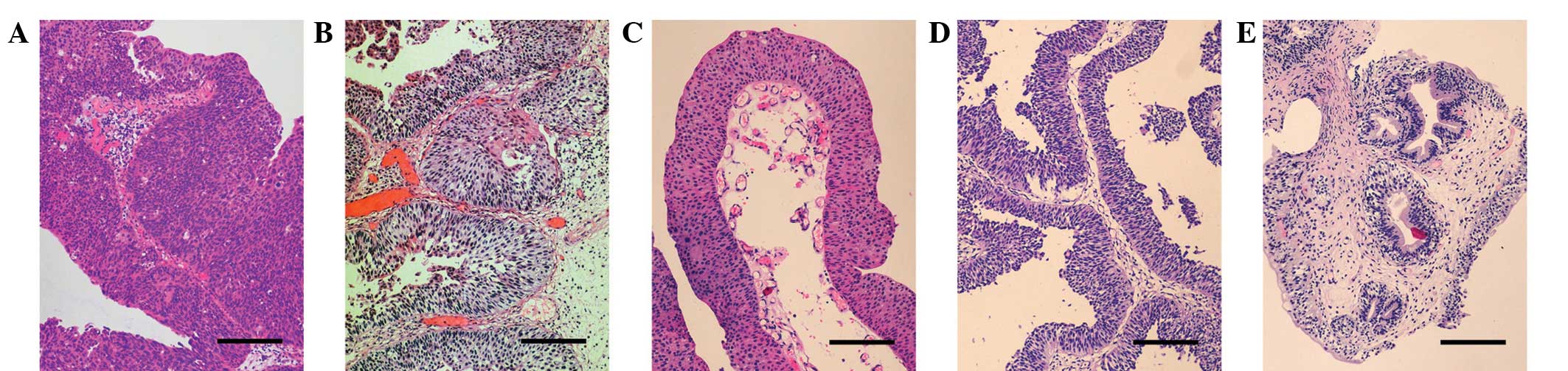
years. A total of 56 pT1 tumors (Fig.
1A) were selected, as well as 78 pTa tumors that included 46
cases of high-grade noninvasive carcinoma (Fig. 1B) and 32 cases of low-grade
noninvasive carcinoma (Fig. 1C). In
addition, 12 papillary urothelial neoplasms of low malignant
potential (PUNLMP; Fig. 1D) 9 of
which were male, with a median age of 62 years and age range of
38–82 years; and 8 inverted papillomas (Fig. 1E) 5 of which were male, with a median
age of 67 years and age range of 36–81 years, were added to the
study. The pathological samples included paraffin-embedded biopsies
and transurethral resections of bladder and ureter tumors stored at
the Second Hospital of Shandong University, which had been obtained
from previous surgical resections. Paraffin-embedded control
samples, which had been stored at the Second Hospital of Shandong
University from previous surgical resections, included malignant
mesothelioma tissues, as the positive tissue control for MCM7 and
MCM3, normal tonsil tissues, as the positive tissue control for
Ki67 and placental villi, as a negative tissue control for each
antibody. All the experimental protocols were approved by the
Medical Ethics Committee of the Second Hospital of Shandong
University and written informed consent was obtained from all
patients.
Immunohistochemical analysis
The specimens were fixed with 10% neutral
formaldehyde (Baibo Biosciences Co., Ltd., Jinan, China),
conventionally dehydrated, embedded in paraffin and then sliced in
4-µm sections using a microtome (RM2255; Leica Microsystems GmbH,
Wetzlar, Germany). The antibodies used for immunohistochemical
analysis were as follows: mouse monoclonal anti-human MCM7 (clone
47DC141; dilution, 1:100; catalog no. GTX22360; Abcam, Cambridge,
MA, USA); a rabbit polyclonal anti-human MCM3 (clone 4F7; dilution,
1:100; catalog no. 15597-1-AP; Proteintech Group, Inc., Chicago,
IL, USA); and mouse monoclonal anti-human Ki67 (clone MIB-1;
dilution, 1:50; anti-human; catalog no. F726801F; Dako, Glostrup,
Denmark) antibodies. First, the sections were deparaffinized and
rehydrated, steamed in a 0.01 mol/l citrate buffer solution (pH
6.0; ZSGB-Bio Co., Ltd., Beijing, China) for 20 min and then cooled
for 10 min. Next, the tissues were blocked by immersing the slides
in a 3% solution of hydrogen peroxide in methanol for 10 min. The
slides were then incubated with the primary antibodies in a
humidity chamber for 45 min at room temperature, followed by
incubation with the biotinylated secondary antibody (anti-mouse and
anti-rabbit; Novolink Polymer Detection System; Novocastra;
Newcastle; United Kingdom) in a humidity chamber for 40 min at
37°C. Subsequently, the slides were incubated with a streptavidin
enzyme complex (Novolink Polymer Detection System) for 25 min.
Finally, the slides were covered with 3,3′-diaminobenzidine
tetrahydrochloride solution (ZSGB-Bio Co., Ltd.) for 15 min under a
microscope (Eclipse Ci; Nikon Corporation, Tokyo, Japan), and then
counterstained with hematoxylin (Novolink Polymer Detection System)
for 1 min. The expression levels of MCM3, MCM7 and Ki67 were
assessed in tumor areas, including the basal, intermediate and
surface urothelial cells. The invasive areas were primarily
observed in the pT1 tumors. In total, five fields selected in
random were analyzed within a counting grid at a magnification of
x400. The percentage of positive cells was expressed as the mean
value of counted microvessels in the five fields.
Statistical analysis
Statistical analysis was performed using the SPSS
11.0 software package for Windows (SPSS, Inc., Chicago, IL, USA).
The Mann-Whitney U-test was used to examine the differences in the
MCM7, MCM3 and Ki67 expression levels between the pTa and pT1
papillary urothelial neoplasias. The statistical analysis of the
correlation among MCM7, MCM3 and Ki67 expression levels in each
sample was conducted with Spearman's rank correlation test. Data
are expressed as the mean ± standard deviation. A value of
P<0.05 was considered to indicate a statistically significant
difference.
Results
Immunohistochemical analysis
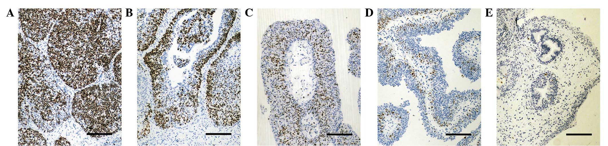
Nuclear staining patterns were demonstrated for MCM7
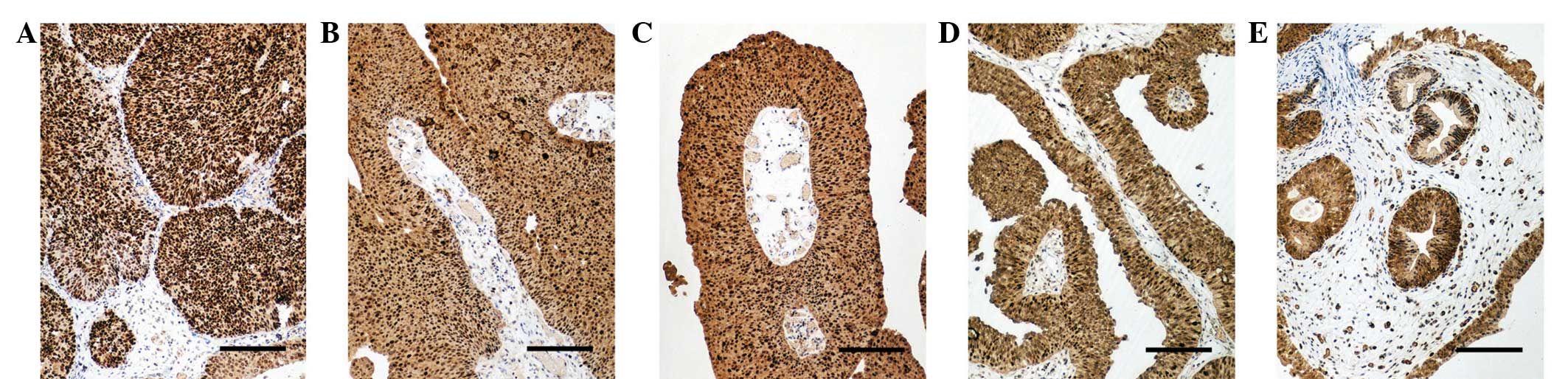
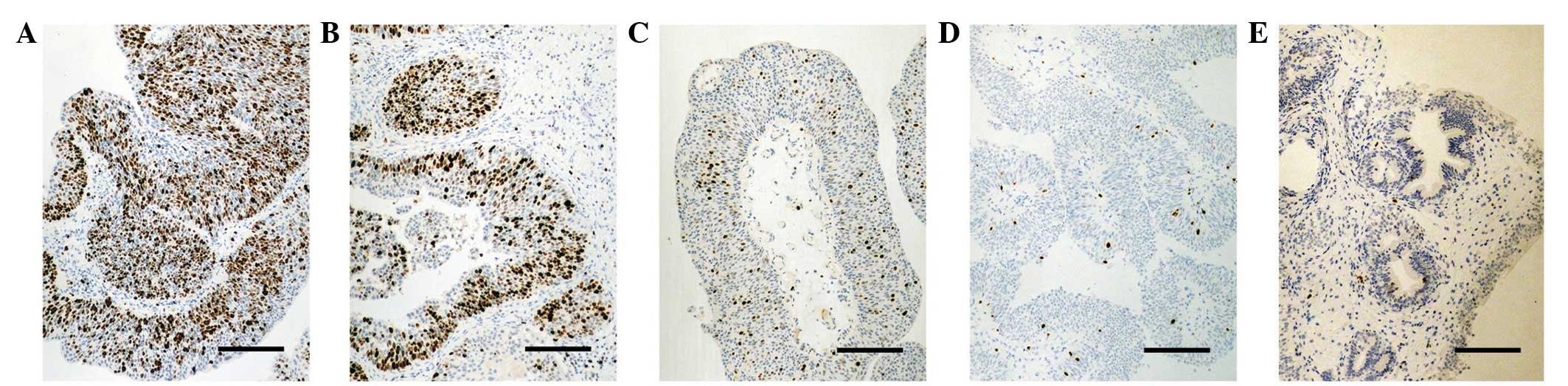
(Fig. 2), MCM3 (Fig. 3) and Ki67 (Fig. 4). The expression levels of MCM7, MCM3
and Ki67, as well as the pathological stage and grade of the 154
patients with papillary urothelial neoplasms are listed in Table I. MCM7 was highly and extensively
expressed in the epithelial layers of the pT1 tumors, highly
expressed in the basal and intermediate cells of the high-grade pTa
tumors and moderately expressed in the basal cells of the low-grade
pTa tumors and PUNLMP. MCM7 staining was limited to the basal cells
of the inverted papillomas (Fig. 2).
MCM3 was extensively expressed in the epithelial layers of pT1
tumors, pTa tumors, PUNLMP and inverted papillomas (Fig. 3). Ki67 was highly and extensively
expressed in the epithelial layers of the pT1 tumors and highly
expressed in the basal and intermediate cells of the high-grade pTa
tumors. Ki67 was expressed in a small number of cells of the
low-grade pTa tumors and PUNLMP. Ki67 staining was limited to the
basal cells of the inverted papillomas (Fig. 4).
 | Table I.Expression levels of MCM7, MCM3 and
Ki67 in urothelial neoplasia (mean ± standard deviation). |
Table I.
Expression levels of MCM7, MCM3 and
Ki67 in urothelial neoplasia (mean ± standard deviation).
| Tumor subtype | n | MCM7 | P-valuea | MCM3 | P-valuea | Ki67 | P-valuea |
|---|
| pT1 | 56 |
81.52±7.44 |
|
85.98±4.99 |
|
54.29±12.48 |
|
| pTa | 78 |
51.35±20.03 | <0.001 |
85.06±5.13 | 0.2993 |
25.96±17.76 | <0.001 |
|
High-grade | 46 |
64.80±12.69 | <0.001 |
85.22±4.83 | 0.4330 |
37.72±13.61 | <0.001 |
|
Low-grade | 32 |
32.03±10.46 | <0.001 |
84.84±5.61 | 0.3406 |
9.06±3.83 | <0.001 |
| PUNLMP | 12 |
27.50±11.38 | <0.001 |
84.58±5.42 | 0.3885 |
7.17±3.35 | <0.001 |
| Inverted
papilloma | 8 |
1.88±0.99 | <0.001 |
85.63±4.96 | 0.8489 |
1.75±0.71 | <0.001 |
| Total | 154 |
57.89±26.34 |
|
85.39±5.07 |
|
33.54±22.58 |
|
Expression of MCM7 in urothelial
neoplasia
An increase in the expression of MCM7 was observed
as the pathological stage and grade increased. MCM7 was highly and
extensively expressed in the epithelial layers of the pT1 tumors
(81.52±7.44; Fig. 2A), highly
expressed in the basal and intermediate cells of the high-grade pTa
tumors (64.80±12.69; Fig. 2B), and
moderately expressed in the basal cells of the low-grade pTa tumors
and PUNLMP (32.03±10.46 and 27.50±11.38, respectively; Fig. 2C and D). MCM7 was expressed in low
levels or individually in the basal cells of the inverted
papillomas (1.88±0.99, Fig. 2E). A
statistically significant difference in MCM7 expression was
observed between pT1 tumors, pTa tumors, PUNLMP and inverted
papillomas (Table I); however, no
statistically significant difference was detected between the
low-grade pTa tumors and PUNLMP (P=0.2294). In addition, the
expression of MCM7 in high-grade noninvasive carcinomas
(64.80±12.69) was significantly higher compared with that in
low-grade noninvasive carcinomas (32.03±10.46; P<0.001).
Expression of MCM3 in urothelial
neoplasia
MCM3 was extensively expressed in the epithelial
layers of pT1 tumors, pTa tumors, PUNLMP and inverted papillomas
(85.98±4.99, 85.06±5.13, 84.58±5.42, 85.63±4.96, respectively;
Fig. 3). However, no statistically
significant difference in the expression of MCM3 was identified
between the different pathological stages and grades (Table I).
Association of MCM7 and MCM3 with Ki67
in urothelial neoplasia
As the expression of the tumor cell proliferation
index, Ki67, increased with increasing tumor grade, a significant
difference was observed in the type of urothelial neoplasia,
progressed from inverted papilloma to pT1 tumor (P<0.001
Fig. 4). Furthermore, there was a
significant positive correlation between MCM7 and Ki67 expression
levels in the urothelial neoplasia tissues
(rs=0.9106; P<0.001); however, no significant
correlation was detected between MCM3 and MCM7 or Ki67
(rs=0.0734, P=0.3657;
rs=0.0638, P=0.4318; Table II).
| Table II.Correlation between MCM7, MCM3 and
Ki67 in urothelial neoplasia. |
Table II.
Correlation between MCM7, MCM3 and
Ki67 in urothelial neoplasia.
|
| MCM7 | MCM3 | Ki67 |
|---|
|
|
|
|
|
|---|
|
| rs | P-value | rs | P-value | rs | P-value |
|---|
| MCM7 | – | – | 0.0734 | 0.3657 | 0.9106 | <0.001 |
| MCM3 | 0.0734 | 0.3657 | – | – | 0.0638 | 0.4318 |
Discussion
Previous studies have used molecular genetics to
compare noninvasive (pTa) and superficially-invasive (pT1)
carcinomas (2–4,6). In pTa
tumors, FGFR3 gene mutations are extremely common (>70%),
whereas p53 gene mutations occur in <5% of cases. By
contrast, pT1 tumors frequently exhibit p53 mutations, while
FGFR3 gene mutations are reported in ~30% of cases (16,17). This
indicates that different mechanisms are involved in the development
of pTa and pT1 tumors. The latter are associated with a higher risk
of recurrence and poorer survival rates. Therefore, a correct
pathological diagnosis is important for clinical treatment and
prognosis (18–20).
The present study identified a statistically
significant and progressive increase in the expression of MCM7 with
increasing grade and stage of papillary urothelial neoplasms. MCM7
was expressed at a low level or individually in the basal cells of
normal urothelium and inverted papilloma, but overexpressed in
urothelial carcinomas, particularly in pT1 tumors. The results in
the present study suggested that MCM7 can promote the proliferation
and even invasion of tumors. In addition, the overexpression of
MCM7 with higher Ki67 expression in pT1 tumors indicated that the
ratio of G1 phase cells increased as the pathological
stage and grade increased. The overexpression of MCM7 is therefore
considered to induce aberrant DNA replication and in this process,
the tumor invasion should be promoted. A significant difference in
MCM7 expression was also observed between pT1 and pTa tumors
(P<0.001). Tumor invasion is known to be important for patient
prognosis. Therefore, MCM7 in particular may be a reliable marker
for the differential diagnosis of pTa and pT1 papillary urothelial
neoplasms.
In addition, the current study revealed that MCM7
expression was significantly positively correlated with Ki67
expression in urothelial neoplasia tissues
(rs=0.9106, P<0.001). In previous studies,
Ki67 was identified to be a potential prognostic marker that
improved the risk stratification and a reliable indicator of
biological aggressiveness in urothelial neoplasia (5,7).
Therefore, MCM7 may have the potential to predict the prognosis and
behavior of pTa and pT1 papillary urothelial neoplasms.
In conclusion, MCM7 was identified to be
significantly associated with tumor grade and Ki67 expression.
Therefore, MCM7 may be a useful marker for predicting the prognosis
and behavior of pTa and pT1 papillary urothelial neoplasms. Further
investigation of MCM7 expression may aid in clarifying the
molecular pathways involved in papillary urinary neoplasia.
References
|
1
|
Ferlay J, Randi G, Bosetti C, Levi F,
Negri E, Boyle P and Vecchia CL: Declining mortality from bladder
cancer in Europe. BJU Int. 101:11–9. 2008.PubMed/NCBI
|
|
2
|
Eble JN, Sauter G, Epstein JI and
Sesterhenn IA: Pathology and genetics of tumours of the urinary
system and male genital organsWorld Health Organization
Classification of Tumors. IARC Press; Lyon: pp. 90–126. 2004
|
|
3
|
Gonzalez-Campora R, Davalos-Casanova G,
Beato-Moreno A, Luque RJ, Alvarez-Kindelan J, Requena MJ, Montironi
R and Lopez-Beltrán A: Apoptotic and proliferation indexes in
primary superficial bladder tumors. Cancer Lett. 242:266–272. 2006.
View Article : Google Scholar : PubMed/NCBI
|
|
4
|
Desai S, Lim SD, Jimenez RE, Chun T, Keane
TE, McKenney JK, Zavala-Pompa A, Cohen C, Young RH and Amin MB:
Relationship of cytokeratin 20 and CD44 protein expression with
WHO/ISUP grade in pTa and pT1 papillary urothelial neoplasia. Mod
Pathol. 13:1315–1323. 2000. View Article : Google Scholar : PubMed/NCBI
|
|
5
|
Bertz S, Otto W, Denzinger S, Wieland WF,
Burger M, Stöhr R, Link S, Hofstädter F and Hartmann A: Combination
of CK20 and Ki-67 immunostaining analysis predicts recurrence,
progression and cancer-Specific survival in pT1 urothelial bladder
cancer. Eur Urol. 65:218–226. 2014. View Article : Google Scholar : PubMed/NCBI
|
|
6
|
Southgate J, Harnden P and Trejdosiewicz
LK: Cytokeratin expression patterns in normal and malignant
urothelium: a review of the biological and diagnostic implications.
Histol Histopathol. 14:657–664. 1999.PubMed/NCBI
|
|
7
|
Van Oers JM, Wild PJ, Burger M, Denzinger
S, Stoehr R, Rosskopf E, Hofstaedter F, Steyerberg EW,
Klinkhammer-Schalke M, Zwarthoff EC, et al: FGFR3 mutations and a
normal CK20 staining pattern define low-grade noninvasive
urothelial bladder tumours. Eur Urol. 52:760–768. 2007. View Article : Google Scholar : PubMed/NCBI
|
|
8
|
Lei M and Tye BK: Initiating DNA
synthesis: from recruiting to activating the MCM complex. J Cell
Sci. 114:1447–1454. 2001.PubMed/NCBI
|
|
9
|
Koonin EV: A common set of conserved
motifs in a vast variety of putative nucleic acid-dependent ATPases
including MCM proteins involved in the initiation of eukaryotic DNA
replication. Nucleic Acids Res. 21:2541–2547. 1993. View Article : Google Scholar : PubMed/NCBI
|
|
10
|
Dimitrova DS, Prokhorova TA, Blow JJ,
Todorov IT and Gilbert DM: Mammalian nuclei become licensed for DNA
replication during late telophase. J Cell Sci. 115:51–59.
2002.PubMed/NCBI
|
|
11
|
Shohet JM, Hicks MJ, Plon SE, Burlingame
SM, Stuart S, Chen SY, Brenner MK and Nuchtern JG: Minichromosome
maintenance protein MCM7 Is a direct target of the MYCN
transcription factor in neuroblastoma. Cancer Res. 62:1123–1128.
2002.PubMed/NCBI
|
|
12
|
Fitch MJ, Donato JJ and Tye BK: Mcm7, a
subunit of the presumptive MCM helicase, modulates its own
expression in conjunction with Mcm1. J Biol Chem. 278:25408–25416.
2003. View Article : Google Scholar : PubMed/NCBI
|
|
13
|
Cortez D, Glick G and Elledge SJ:
Minichromosome maintenance proteins are direct targets of the ATM
and ATR checkpoint kinases. Proc Natl Acad Sci USA.
101:10078–10083. 2004. View Article : Google Scholar : PubMed/NCBI
|
|
14
|
Kimura F, Okayasu I, Kakinuma H, Satoh Y,
Kuwao S, Saegusa M and Watanabe J: Differential diagnosis of
reactive mesothelial cells and malignant mesothelioma cells using
the cell proliferation markers minichromosome maintenance protein
7, geminin, topoisomerase II alpha and Ki-67. Acta Cytol.
57:384–390. 2013. View Article : Google Scholar : PubMed/NCBI
|
|
15
|
Ren B, Yu G, Tseng GC, Cieply K, Gavel T,
Nelson J, Michalopoulos G, Yu YP and Luo JH: MCM7 amplification and
overexpression are associated with prostate cancer progression.
Oncogene. 25:1090–1098. 2006. View Article : Google Scholar : PubMed/NCBI
|
|
16
|
Bakkar AA, Wallerand H, Radvanyi F, et al:
FGFR3 and TP53 gene mutations define two distinct pathways in
urothelial cell carcinoma of the bladder. Cancer Res. 63:8108–8112.
2003.PubMed/NCBI
|
|
17
|
van Rhijn BW, Vis AN, van der Kwast TH, et
al: Molecular grading of urothelial cell carcinoma with fibroblast
growth factor receptor 3 and MIB-1 is superior to pathologic grade
for the prediction of clinical outcome. J Clin Oncol. 21:1912–1921.
2003. View Article : Google Scholar : PubMed/NCBI
|
|
18
|
Oh IJ, Kim HE, Song SY, et al: Diagnostic
value of serum glutathione peroxidase 3 levels in patients with
lung cancer. Thoracic Cancer. 5:425–430. 2014. View Article : Google Scholar
|
|
19
|
Liu YT, Shi YK, Hao XZ, et al: Analysis of
clinicopathological features of the echinoderm
microtubule-associated protein-like-4-anaplastic lymphoma kinase
fusion gene in Chinese patients with advanced non-small-cell lung
cancer. Thoracic Cancer. 5:255–260. 2014. View Article : Google Scholar
|
|
20
|
Üstündag S and Zencirci AD: Factors
affecting the quality of life of cancer patients undergoing
chemotherapy: A questionnaire study. Asia-Pacific J Oncol Nurs.
2:17–25. 2015. View Article : Google Scholar
|