Spandidos Publications Logo
  • About
    • About Spandidos
    • Aims and Scopes
    • Abstracting and Indexing
    • Editorial Policies
    • Reprints and Permissions
    • Job Opportunities
    • Terms and Conditions
    • Contact
  • Journals
    • All Journals
    • Oncology Letters
      • Oncology Letters
      • Information for Authors
      • Editorial Policies
      • Editorial Board
      • Aims and Scope
      • Abstracting and Indexing
      • Bibliographic Information
      • Archive
    • International Journal of Oncology
      • International Journal of Oncology
      • Information for Authors
      • Editorial Policies
      • Editorial Board
      • Aims and Scope
      • Abstracting and Indexing
      • Bibliographic Information
      • Archive
    • Molecular and Clinical Oncology
      • Molecular and Clinical Oncology
      • Information for Authors
      • Editorial Policies
      • Editorial Board
      • Aims and Scope
      • Abstracting and Indexing
      • Bibliographic Information
      • Archive
    • Experimental and Therapeutic Medicine
      • Experimental and Therapeutic Medicine
      • Information for Authors
      • Editorial Policies
      • Editorial Board
      • Aims and Scope
      • Abstracting and Indexing
      • Bibliographic Information
      • Archive
    • International Journal of Molecular Medicine
      • International Journal of Molecular Medicine
      • Information for Authors
      • Editorial Policies
      • Editorial Board
      • Aims and Scope
      • Abstracting and Indexing
      • Bibliographic Information
      • Archive
    • Biomedical Reports
      • Biomedical Reports
      • Information for Authors
      • Editorial Policies
      • Editorial Board
      • Aims and Scope
      • Abstracting and Indexing
      • Bibliographic Information
      • Archive
    • Oncology Reports
      • Oncology Reports
      • Information for Authors
      • Editorial Policies
      • Editorial Board
      • Aims and Scope
      • Abstracting and Indexing
      • Bibliographic Information
      • Archive
    • Molecular Medicine Reports
      • Molecular Medicine Reports
      • Information for Authors
      • Editorial Policies
      • Editorial Board
      • Aims and Scope
      • Abstracting and Indexing
      • Bibliographic Information
      • Archive
    • World Academy of Sciences Journal
      • World Academy of Sciences Journal
      • Information for Authors
      • Editorial Policies
      • Editorial Board
      • Aims and Scope
      • Abstracting and Indexing
      • Bibliographic Information
      • Archive
    • International Journal of Functional Nutrition
      • International Journal of Functional Nutrition
      • Information for Authors
      • Editorial Policies
      • Editorial Board
      • Aims and Scope
      • Abstracting and Indexing
      • Bibliographic Information
      • Archive
    • International Journal of Epigenetics
      • International Journal of Epigenetics
      • Information for Authors
      • Editorial Policies
      • Editorial Board
      • Aims and Scope
      • Abstracting and Indexing
      • Bibliographic Information
      • Archive
    • Medicine International
      • Medicine International
      • Information for Authors
      • Editorial Policies
      • Editorial Board
      • Aims and Scope
      • Abstracting and Indexing
      • Bibliographic Information
      • Archive
  • Articles
  • Information
    • Information for Authors
    • Information for Reviewers
    • Information for Librarians
    • Information for Advertisers
    • Conferences
  • Language Editing
Spandidos Publications Logo
  • About
    • About Spandidos
    • Aims and Scopes
    • Abstracting and Indexing
    • Editorial Policies
    • Reprints and Permissions
    • Job Opportunities
    • Terms and Conditions
    • Contact
  • Journals
    • All Journals
    • Biomedical Reports
      • Information for Authors
      • Editorial Policies
      • Editorial Board
      • Aims and Scope
      • Abstracting and Indexing
      • Bibliographic Information
      • Archive
    • Experimental and Therapeutic Medicine
      • Information for Authors
      • Editorial Policies
      • Editorial Board
      • Aims and Scope
      • Abstracting and Indexing
      • Bibliographic Information
      • Archive
    • International Journal of Epigenetics
      • Information for Authors
      • Editorial Policies
      • Editorial Board
      • Aims and Scope
      • Abstracting and Indexing
      • Bibliographic Information
      • Archive
    • International Journal of Functional Nutrition
      • Information for Authors
      • Editorial Policies
      • Editorial Board
      • Aims and Scope
      • Abstracting and Indexing
      • Bibliographic Information
      • Archive
    • International Journal of Molecular Medicine
      • Information for Authors
      • Editorial Policies
      • Editorial Board
      • Aims and Scope
      • Abstracting and Indexing
      • Bibliographic Information
      • Archive
    • International Journal of Oncology
      • Information for Authors
      • Editorial Policies
      • Editorial Board
      • Aims and Scope
      • Abstracting and Indexing
      • Bibliographic Information
      • Archive
    • Medicine International
      • Information for Authors
      • Editorial Policies
      • Editorial Board
      • Aims and Scope
      • Abstracting and Indexing
      • Bibliographic Information
      • Archive
    • Molecular and Clinical Oncology
      • Information for Authors
      • Editorial Policies
      • Editorial Board
      • Aims and Scope
      • Abstracting and Indexing
      • Bibliographic Information
      • Archive
    • Molecular Medicine Reports
      • Information for Authors
      • Editorial Policies
      • Editorial Board
      • Aims and Scope
      • Abstracting and Indexing
      • Bibliographic Information
      • Archive
    • Oncology Letters
      • Information for Authors
      • Editorial Policies
      • Editorial Board
      • Aims and Scope
      • Abstracting and Indexing
      • Bibliographic Information
      • Archive
    • Oncology Reports
      • Information for Authors
      • Editorial Policies
      • Editorial Board
      • Aims and Scope
      • Abstracting and Indexing
      • Bibliographic Information
      • Archive
    • World Academy of Sciences Journal
      • Information for Authors
      • Editorial Policies
      • Editorial Board
      • Aims and Scope
      • Abstracting and Indexing
      • Bibliographic Information
      • Archive
  • Articles
  • Information
    • For Authors
    • For Reviewers
    • For Librarians
    • For Advertisers
    • Conferences
  • Language Editing
Login Register Submit
  • This site uses cookies
  • You can change your cookie settings at any time by following the instructions in our Cookie Policy. To find out more, you may read our Privacy Policy.

    I agree
Search articles by DOI, keyword, author or affiliation
Search
Advanced Search
presentation
Oncology Letters
Join Editorial Board Propose a Special Issue
Print ISSN: 1792-1074 Online ISSN: 1792-1082
Journal Cover
September-2015 Volume 10 Issue 3

Full Size Image

Sign up for eToc alerts
Recommend to Library

Journals

International Journal of Molecular Medicine

International Journal of Molecular Medicine

International Journal of Molecular Medicine is an international journal devoted to molecular mechanisms of human disease.

International Journal of Oncology

International Journal of Oncology

International Journal of Oncology is an international journal devoted to oncology research and cancer treatment.

Molecular Medicine Reports

Molecular Medicine Reports

Covers molecular medicine topics such as pharmacology, pathology, genetics, neuroscience, infectious diseases, molecular cardiology, and molecular surgery.

Oncology Reports

Oncology Reports

Oncology Reports is an international journal devoted to fundamental and applied research in Oncology.

Experimental and Therapeutic Medicine

Experimental and Therapeutic Medicine

Experimental and Therapeutic Medicine is an international journal devoted to laboratory and clinical medicine.

Oncology Letters

Oncology Letters

Oncology Letters is an international journal devoted to Experimental and Clinical Oncology.

Biomedical Reports

Biomedical Reports

Explores a wide range of biological and medical fields, including pharmacology, genetics, microbiology, neuroscience, and molecular cardiology.

Molecular and Clinical Oncology

Molecular and Clinical Oncology

International journal addressing all aspects of oncology research, from tumorigenesis and oncogenes to chemotherapy and metastasis.

World Academy of Sciences Journal

World Academy of Sciences Journal

Multidisciplinary open-access journal spanning biochemistry, genetics, neuroscience, environmental health, and synthetic biology.

International Journal of Functional Nutrition

International Journal of Functional Nutrition

Open-access journal combining biochemistry, pharmacology, immunology, and genetics to advance health through functional nutrition.

International Journal of Epigenetics

International Journal of Epigenetics

Publishes open-access research on using epigenetics to advance understanding and treatment of human disease.

Medicine International

Medicine International

An International Open Access Journal Devoted to General Medicine.

Journal Cover
September-2015 Volume 10 Issue 3

Full Size Image

Sign up for eToc alerts
Recommend to Library

  • Article
  • Citations
    • Cite This Article
    • Download Citation
    • Create Citation Alert
    • Remove Citation Alert
    • Cited By
  • Similar Articles
    • Related Articles (in Spandidos Publications)
    • Similar Articles (Google Scholar)
    • Similar Articles (PubMed)
  • Download PDF
  • Download XML
  • View XML
Case Report

Atypical teratoid/rhabdoid tumor with hereditary multiple exostoses in an 18-year-old male: A case report

  • Authors:
    • Qian Wu
    • Bo Xiao
    • Li Li
    • Li Feng
  • View Affiliations / Copyright

    Affiliations: Department of Neurology, Xiangya Hospital, Central South University, Changsha, Hunan 410008, P.R. China
  • Pages: 1561-1564
    |
    Published online on: June 17, 2015
       https://doi.org/10.3892/ol.2015.3389
  • Expand metrics +
Metrics: Total Views: 0 (Spandidos Publications: | PMC Statistics: )
Metrics: Total PDF Downloads: 0 (Spandidos Publications: | PMC Statistics: )
Cited By (CrossRef): 0 citations Loading Articles...

This article is mentioned in:


Abstract

Atypical teratoid/rhabdoid tumor (AT/RT) is an aggressive embryonic brain tumor that is predominantly found in young children. The present study reports the unusual case of an adult male AT/RT patient with a history of another type of tumor, hereditary multiple exostoses (EXT or HME), who survived for 18 years. The patient's first and chief complaint was a 1‑month history of progressive projectile vomiting. The patient was admitted twice for treatment, and on the second admission, a craniotomy was performed to remove a intracranial mass. However, no radiotherapy or chemotherapy treatment was administered. Pathology revealed monotonous medium‑ to large‑sized neoplastic cells. The patient succumbed to a pulmonary infection and respiratory failure 14 days after the initial treatment. The prognosis for AT/RT is extremely poor; furthermore, the patient presented with another tumor. There may have been an association between the two tumors that worsened the clinical phenotype and prognosis of this patient. Additionally, symptomatic treatment for this condition is insufficient; early surgery and radiotherapy may be more useful for preventing the aggressive progression of these tumors.

Introduction

Intracranial atypical teratoid/rhabdoid tumor (AT/RT) is a rare malignant embryonic neoplasm that usually occurs in children aged <3 years and accounts for a total incidence of 1–2% of all brain tumors in children and >10% of central nervous system tumors in infants (1). The clinical course is dismal, with a median survival time from diagnosis to mortality that spans only a few months (2). The tumor is typically associated with a chromosome 22q11.2 mutation or deletion, leading to the loss of nuclear expression of integrase interactor 1; in adults, the clinical presentation varies with tumor location (1). Hereditary multiple exostoses (EXT) is the most common benign bone tumor and is an autosomal dominant disorder, which is characterized by the formation of cartilage-capped bone projections (exostoses) localized mainly in the juxta-epiphyseal region of the long bones. EXT is associated with two loci, 8q24.1 (EXT1) and 11p11-p13 (EXT2) (3,4). The condition is characterized by multiple osteochondromas and is usually painless. The present study reports an adult case of intracranial AT/RT with a history of EXT and investigates the possible association between the two tumors. Written informed consent was obtained from the patient in accordance with the Declaration of Helsinki. The Ethics Committee of the Xiangya Hospital of Central South University (Changsha, Hunan, China) approved all experiments described in the study.

Case report

On October 11 2012, an 18-year-old male was first admitted to the Xiangya Hospital of Central South University following one month of progressive projectile vomiting. At this time, the patient did not complain of a headache or stomach 
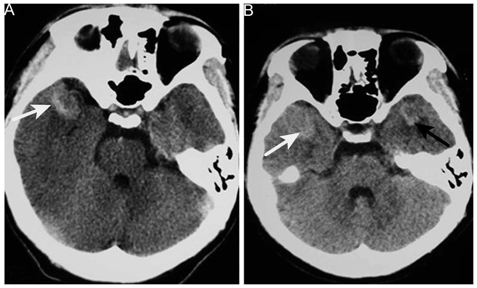
discomfort. Brain computed tomography (CT) scans that had been obtained a few days after the symptoms arose revealed a hyperintense lesion on the right side of the temporal lobe; which was suspected to be an intracranial hemorrhage at the time. However, a month of corresponding therapy did not relieve the vomiting. The patient's past medical history included EXT detected at 10 years old, and the current examination noted two protuberant bony deformities over the right scapula and distal humerus. Upon admission, the neurological examination results were normal. Hematological and biochemical examination, and cerebrospinal fluid routine testing were normal; the opening pressure was 100 mm H2O (normal range, 80–180 mm H2O). Repeat CT of the brain revealed no changes in the CT value of the lesion 
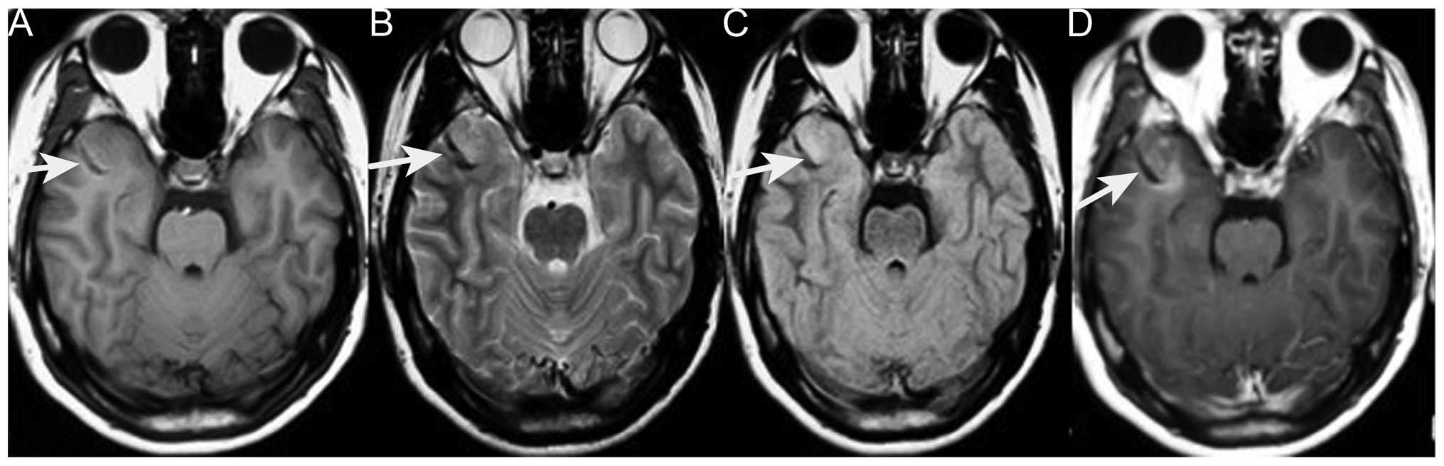
(Fig. 1). Brain magnetic resonance imaging (MRI) revealed an abnormally low signal intensity on T1- and T2-weighted images and a high signal intensity on gadolinium-enhancing T1 in the right lateral fissure and adjacent temporal region (Fig. 2). Based on these results, the patient was diagnosed with an intracranial tumor. Surgery was suggested for removing the mass, which the patient refused, only accepting carbamazepine (0.1 g, 3 times/day, October 17–23, 2012) and ondansetron (8 mg, 2 times/day, October 12–16, 2012; 8 mg, 4 times/day, October 17–20, 2012) therapy. Upon leaving the hospital, the vomiting had been alleviated.

Figure 1.

Brain computed tomography. (A) High-density tumor in the right anterior temporal region (white arrow). (B) Image obtained 6 months later showing another tumor in the opposite temporal region (black arrow).

Figure 2.

Images obtained during the first admission. (A) Axial T1-weighted MRI of a low-signal mass in the right temporal region (white arrow). (B) T2-weighted MRI of a low-intensity signal mass (white arrow). (C) Fluid-attenuated inversion recovery MRI of a low-intensity signal mass (white arrow). (D) Axial T1-weighted contrast-enhanced MRI of the mass with mild enhancement (white arrow). MRI, magnetic resonance imaging.

However, 6 months later, the patient was admitted again due to worsening vomiting and new symptoms of bilateral blurred vision, and severe neck and waist pain. A neurological examination revealed sixth cranial nerve palsy and bilateral papilledema. The lumbar puncture had an opening pressure of >400 mm H2O. Cerebrospinal fluid testing revealed nothing abnormal and no cancer cells. Brain CT revealed a new hyperintense lesion in the left temporal region (Fig. 
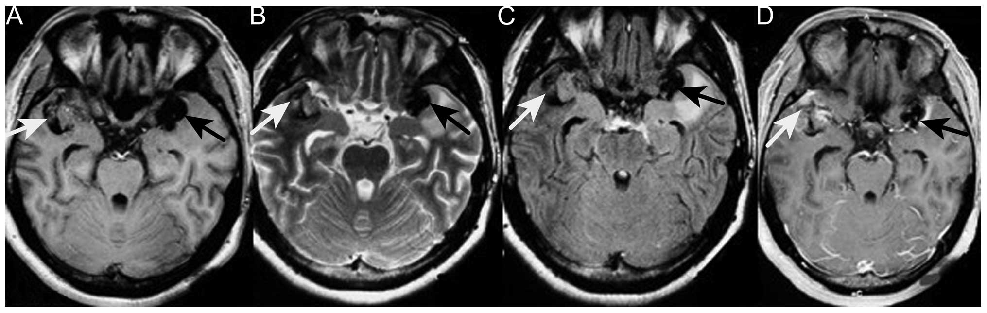
1). Repeat MRI revealed two enhancing masses, with a low intensity signal on T1-weighted imaging and a heterogeneous signal on T2-weighted imaging, which had progressed more quickly compared with the lesion detected 6 months earlier. Moderate peripheral edema was also observed (Fig. 3). Digital subtraction angiography disclosed diffuse poor filling of all venous sinuses during the venous phase. Surgery was performed, but the patient's condition precluded the use of radiotherapy or chemotherapy. CT angiography revealed that the tumors were 2.9×1.9 cm (left temporal region) and 2.2×1.9 cm (right temporal region); surgical microscopy revealed surrounding strong angiogenic 
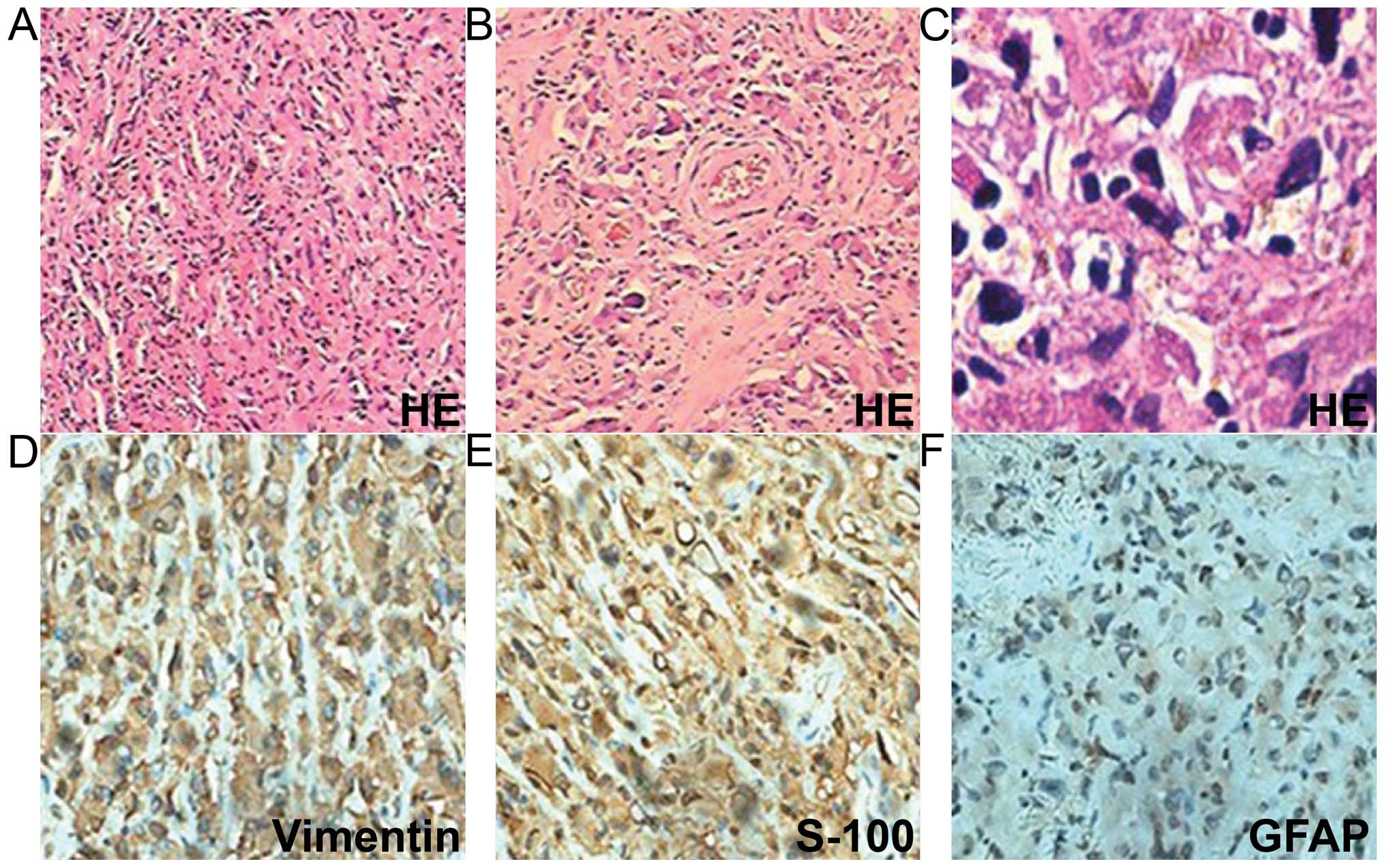
adhesion. The tumor pathology revealed sheets of monotonous medium- to large-sized neoplastic cells, with abundant eosinophilic cytoplasm and clear eccentric nucleus with prominent nucleoli. The tumors were positive for vimentin, glial fibrillary acidic protein and S-100 (Fig. 4). The Ki-67 labeling index was >2%, and the tumors were negative for cluster of differentiation (CD)138, CD38, pan-cytokeratin, desmin, muscle cell actin (HHF35), melanoma marker (HMB45), leukocyte common antigen, melan-A and myogenin. The diagnosis of intracranial AT/RT was verified, however, the patient succumbed to pulmonary infection at 14 days post-surgery.

Figure 3.

Images obtained during the second admission. A new tumor was present in the left temporal region (black arrow); the tumor on the right was larger than observed 6 months earlier (white arrow). The two tumors were symmetrical around the lateral fissure. (A) T1-weighted MRI of the low-intenisty signal tumors. (B) T2-weighted MRI of the heterogeneous tumors. (C) Fluid-attenuated inversion recovery MRI of the tumor. (D) T1-weighted contrast MRI of moderate heterogeneous enhancement. The tumors, the one on the left in particular, show moderate edema and marked calcification. MRI, magnetic resonance imaging.

Figure 4.

Tumor pathology. (A) Sheets of tumor cells (H&E staining; magnification, x40). (B and C) Rhabdoid cells with eccentric prominent nuclei and abundant eosinophilic cytoplasm (H&E staining; magnification, x200 and x400, respectively). (D) Tumor cells demonstrating diffuse cytoplasmic immunoreactivity for vimentin (magnification, x400). (E) Tumor cells demonstrating cytoplasmic immunoreactivity for S-100 (magnification, x400). (F) Tumor cells demonstrating cytoplasmic immunoreactivity for GFAP (magnification, x400). H&E, hematoxylin and eosin; GFAP, glial fibrillary acidic protein.

Discussion

The case reported in the present study is of a rare adult supratentorial AT/RT, associated with the chief complaint of vomiting and nausea. The patient's symptoms and physical examination results cannot be fully attributed to the mass itself and are more associated with cranial hypertension. To the best of our knowledge, this is the first description of an AT/RT tumor of the brain and comorbidity with EXT. Upon first admission, our primary consideration was intracranial chondrosarcoma. CT and MRI for intracranial chondrosarcoma also often disclose a mass lesion that includes areas of calcification and heterogeneous contrast enhancement. Malignant transformation of exostoses into chondrosarcoma occurs in 5% of cases (5). Brain metastasis is exceptionally rare, with few documented cases; lung and breast cancer are the primary sources of brain metastasis, together accounting for nearly two-thirds of total cases (6). Suspicion of malignant transformation is indicated by growth of the tumor following puberty, the presence of pain, a >1-cm thick cartilaginous cap in adults, or hints from radiography, bone scintigraphy (7), and enhanced CT or MRI. In the present case, the lesion did not increase in size and bone scintigraphy did not reveal focally increased radiotracer accumulation. There was no enhancement of the scapula tumor and no cardiopulmonary neoplasm on enhanced chest CT. With the exception of metastatic chondrosarcoma, intracranial primary chondrosarcoma mostly originates from skull base synchondroses and tends to grow extradurally. These neoplasms include classic, myxoid and mesenchymal chondrosarcoma (8). The peak incidence of classic chondrosarcoma is in patients in their sixth decade, where the tumor typically arises in the skull base. Mesenchymal chondrosarcoma tends to occur in the second and third decades of life, and is typically supratentorial, but most often located in the frontoparietal region and attached to the meninges, particularly the falx cerebri. The tumor has a biphasic pattern of undifferentiated mesenchymal cells and well-differentiated cartilage, and the typical transition between the two states is clear. Myxoid chondrosarcoma is the rarest form, and reported locations include the petrous bone, posterior fossa and falx cerebri. Histologically, the tumor is characterized by prominent mucinous supporting stroma. Intracranial primary chondrosarcoma has a tendency to locate in the skull base or the falx cerebri. However, the tumor in the present case was near neither the skull nor the falx cerebri. The differential diagnosis included oligodendroglioma, germinoma and other tumors containing calcification.

The present case was an uncommon example of an AT/RT tumor with typical histological features, but presenting in the supratentorial compartment of an adult. The most notable aspect was that the patient had a history of EXT. From the literature, we infer that the two tumors may be genetically correlated. Several studies reported that AT/RT was associated with insulin-like growth factor II (IGF2) and IGF receptor type 1, and that autocrine/paracrine stimulation of cell growth by IGF2, which is on chromosome 11p15 and near EXT2, may be a mechanism involved in AT/RT tumorigenesis (9–12). Therefore, AT/RT can theoretically occur together with skeletal dysplasia. However, given the scarcity of cases and limitations of the experimental infrastructure, conclusions cannot be derived at present. Instead, the current case report is presented to improve the approach to diagnosing AT/RT and aid more patients.

References

1 

Shonka NA, Armstrong TS, Prabhu SS, Childress A, Choi S, Langford LA and Gilbert MR: Atypical teratoid/rhabdoid tumors in adults: a case report and treatment-focused review. J Clin Med Res. 3:85–92. 2011.PubMed/NCBI

2 

Lafay-Cousin L, Hawkins C, Carret AS, Johnston D, Zelcer S, Wilson B, Jabado N, Scheinemann K, Eisenstat D, Fryer C, et al: Central nervous system atypical teratoid rhabdoid tumours: The Canadian Paediatric Brain Tumour Consortium experience. Eur J Cancer. 48:353–359. 2012. View Article : Google Scholar : PubMed/NCBI

3 

Solomon L: Hereditary multiple exostosis. J Bone Joint Surg. 45:292–304. 1963.

4 

Li Y1, Wang D, Wang W, Wang J, Li H, Wang J, Wang X and Fu Q: Identification of four novel EXT1 and EXT2 mutations in five Chinese pedigrees with hereditary multiple exostoses. Genet Test Mol Biomarkers. 13:825–830. 2009. View Article : Google Scholar : PubMed/NCBI

5 

Altay M, Bayrakci K, Yildiz Y, Erekul S and Saglik Y: Secondary chondrosarcoma in cartilage bone tumors: Report of 32 patients. J Orthop Sci. 12:415–423. 2007. View Article : Google Scholar : PubMed/NCBI

6 

Valiente M, Obenauf AC, Jin X, Chen Q, Zhang XH, Lee DJ, Chaft JE, Kris MG, Huse JT, Brogi E and Massagué J: Serpins promote cancer cell survival and vascular co-option in brain metastasis. Cell. 156:1002–1016. 2014. View Article : Google Scholar : PubMed/NCBI

7 

Sánchez-Rodríguez V, Medina-Romero F, Gómez Rodríguez-Bethencourt MÁ, González Díaz MA, González Soto MJ and Alarcó Hernández R: Value of the bone scintigraphy in multiple osteochrondromatosis with sarcomatous degeneration. Rev Esp Med Nucl Imagen Mol. 31:270–274. 2012.PubMed/NCBI

8 

Im SH, Kim DG, Park IA and Chi JG: Primary intracranial myxoid chondrosarcoma: Report of a case and review of the literature. J Korean Med Sci. 18:301–307. 2003. View Article : Google Scholar : PubMed/NCBI

9 

Ogino S, Cohen ML and Abdul-Karim FW: Atypical teratoid/rhabdoid tumor of the CNS: Cytopathology and immunohistochemistry of insulin-like growth factor-II, insulin-like growth factor receptor type 1, cathepsin D, and Ki-67. Mod Pathol. 12:379–385. 1999.PubMed/NCBI

10 

Tessema M, Länger F, Bock O, Seltsam A, Metzig K, Hasemeier B, Kreipe H and Lehmann U: Down-regulation of the IGF-2/H19 locus during normal and malignant hematopoiesis is independent of the imprinting pattern. Int J Oncol. 26:499–507. 2005.PubMed/NCBI

11 

Lopez-Gines C, Cerda-Nicolas M, Kepes J, Donat J, Gil-Benso R and Llombart-Bosch A: Complex rearrangement of chromosomes 6 and 11 as the sole anomaly in atypical teratoid/rhabdoid tumors of the central nervous system. Cancer Genet Cytogenet. 122:149–152. 2000. View Article : Google Scholar : PubMed/NCBI

12 

Narendran A, Coppes L, Jayanthan A, Coppes M, Teja B, Bernoux D, George D and Strother D: Establishment of atypical-teratoid/rhabdoid tumor (AT/RT) cell cultures from disseminated CSF cells: A model to elucidate biology and potential targeted therapeutics. J Neurooncol. 90:171–180. 2008. View Article : Google Scholar : PubMed/NCBI

Related Articles

  • Abstract
  • View
  • Download
  • Twitter
Copy and paste a formatted citation
Spandidos Publications style
Wu Q, Xiao B, Li L and Feng L: Atypical teratoid/rhabdoid tumor with hereditary multiple exostoses in an 18-year-old male: A case report. Oncol Lett 10: 1561-1564, 2015.
APA
Wu, Q., Xiao, B., Li, L., & Feng, L. (2015). Atypical teratoid/rhabdoid tumor with hereditary multiple exostoses in an 18-year-old male: A case report. Oncology Letters, 10, 1561-1564. https://doi.org/10.3892/ol.2015.3389
MLA
Wu, Q., Xiao, B., Li, L., Feng, L."Atypical teratoid/rhabdoid tumor with hereditary multiple exostoses in an 18-year-old male: A case report". Oncology Letters 10.3 (2015): 1561-1564.
Chicago
Wu, Q., Xiao, B., Li, L., Feng, L."Atypical teratoid/rhabdoid tumor with hereditary multiple exostoses in an 18-year-old male: A case report". Oncology Letters 10, no. 3 (2015): 1561-1564. https://doi.org/10.3892/ol.2015.3389
Copy and paste a formatted citation
x
Spandidos Publications style
Wu Q, Xiao B, Li L and Feng L: Atypical teratoid/rhabdoid tumor with hereditary multiple exostoses in an 18-year-old male: A case report. Oncol Lett 10: 1561-1564, 2015.
APA
Wu, Q., Xiao, B., Li, L., & Feng, L. (2015). Atypical teratoid/rhabdoid tumor with hereditary multiple exostoses in an 18-year-old male: A case report. Oncology Letters, 10, 1561-1564. https://doi.org/10.3892/ol.2015.3389
MLA
Wu, Q., Xiao, B., Li, L., Feng, L."Atypical teratoid/rhabdoid tumor with hereditary multiple exostoses in an 18-year-old male: A case report". Oncology Letters 10.3 (2015): 1561-1564.
Chicago
Wu, Q., Xiao, B., Li, L., Feng, L."Atypical teratoid/rhabdoid tumor with hereditary multiple exostoses in an 18-year-old male: A case report". Oncology Letters 10, no. 3 (2015): 1561-1564. https://doi.org/10.3892/ol.2015.3389
Follow us
  • Twitter
  • LinkedIn
  • Facebook
About
  • Spandidos Publications
  • Careers
  • Cookie Policy
  • Privacy Policy
How can we help?
  • Help
  • Live Chat
  • Contact
  • Email to our Support Team