Introduction
Head and neck squamous cell carcinoma (HNSCC) is the
eighth leading cause of cancer-related mortality worldwide
(1). Laryngeal squamous cell
carcinoma (LSCC), the second most common malignant neoplasm of the
upper respiratory tract, is a subtype of HNSCC (2). Approximately 10,000 new cases of LSCC
are diagnosed each year in the United States (3). In China, the incidence of LSCC is
increasing, particularly in the country's Northeast region
(4). Significant predisposing factors
to the development and progression of LSCC, include alcohol abuse
and tobacco (5). Early stage LSCC may
be effectively treated with surgery or radiotherapy (6). When diagnosed at an advanced stage, this
disease usually requires a combination of treatment modalities.
However, although such combined therapy has improved local control
and overall quality of life, the local recurrence rate varies from
10–50%, depending on tumor stage and the overall survival rate has
not improved significantly over two decades (7,8).
Therefore, it is necessary to identify novel biomarkers for use in
the diagnosis of LSCC. In addition, the study of the molecular
mechanisms underlying the development of LSCC may improve treatment
and increase survival for patients with this disease.
Forkhead box Q1 (FOXQ1, also termed HFH1) is a
member of the forkhead transcription factor family (9), which is involved in a variety of
biological processes, including epithelial differentiation
(10), cell cycle progression
(11), embryonic stem cell
differentiation (12), metabolism
(13,14) and carcinogenesis (15–17). As
one of the first forkhead genes to be investigated, FOXQ1 has been
demonstrated to be involved in metabolism, aging (18) and carcinogenesis (19). Overexpression of the FOXQ1 protein is
associated with epithelial-mesenchymal transition (EMT) and a poor
prognosis in certain types of cancer, such as non-small cell lung
cancer (20) and breast cancer
(21). However, little is known
regarding FOXQ1 expression in LSCC and its involvement in the
pathogenesis of this disease. The present study aimed to
investigate the effect of FOXQ1 expression on the development and
progression of LSCC, by measuring its expression in LSCC tissue
samples, and investigating its effect on cell proliferation, cell
cycle progression and cell migration.
Materials and methods
Patients and tissue samples
Thirty pairs of LSCC and corresponding adjacent
normal tissues, used for reverse transcription-quantitative
polymerase chain reaction (RT-qPCR) and western blotting, were
collected from the Ear, Nose and Throat department of The 463
Hospital of PLA (Shenyang, China) between January 2009 and December
2014, following receipt of written informed consent. Tissue samples
were obtained from 24 males and 6 females (mean age, 64.72 years;
range, 45–83 years) and included 6 cases of stage I LSCC, 6 cases
of stage II LSCC, 8 cases of stage III LSCC and 10 cases of stage
IV LSCC. Tumors were staged according to the International Union
Against Cancer TNM classification for malignant tumors (22). All tissue samples, including cancer
tissues and matched adjacent normal tissues (typically removed from
areas 4–15 mm from the tumors), were obtained during surgery. All
specimens were frozen and stored at −80°C prior to use. Approval
for this study was obtained from the Ethics Committee of China
Medical University (Shenyang, China).
Cell culture
The Hep2 human laryngeal carcinoma cell line, was
obtained from the Shanghai Institute for Biochemistry, Chinese
Academy of Sciences (Shanghai, China). Cells were cultured in
RPMI-1640 (Gibco Life Technologies, Carlsbad, CA, USA),
supplemented with 10% fetal bovine serum, 100 µg/ml penicillin and
100 µg/ml streptomycin (all obtained from GE Healthcare Life
Sciences, Logan, UT, USA) in humidified 5% CO2 at 37°C.
Trypsin solution (0.25%; GE Healthcare Life Sciences) was used to
detach cells from the culture flask.
Transient transfection with
FOXQ1-specific small interfering RNA (siRNA)
Three siRNAs targeting human FOXQ1 and a negative
control siRNA (FOXQ1-NC), were designed and obtained from
GenePharma Co., Ltd. (Shanghai, China). The siRNAs and FOXQ1-NC
siRNA (FOXQ1-siRNA1, 5′-CGCGGACTTTGCACTTTGA-3′; FOXQ1-siRNA2,
5′-AGGGAACCTTTCCACACTA-3′; FOXQ1-siRNA3, 5′-CCATCAAACGTGCCTTAAA-3′;
and FOXQ1-NC siRNA, 5′-TTCTCCGAACGTGTCACGT-3′) were used to inhibit
the expression of FOXQ1. Preliminary experiments indicated that
FOXQ1-siRNA1 most effectively down-regulated FOXQ1 expression. This
sequence was therefore selected for subsequent experiments. Hep2
cell were seeded in 6-well plates at a density of
0.5×10⁶ cells/well. FOXQ1-siRNA, FOXQ1-NC and mock group
(blank control ± transfection reagent) were transfected into Hep2
cells using lipofectamine 2000 transfection reagent (Invitrogen
Life Technologies, Carlsbad, CA, USA), according to the
manufacturer's instructions. Following transfection for 72 h, cells
were collected for subsequent experiments.
RNA isolation and RT-qPCR
Total RNA was extracted using TRIzol™ reagent
(Invitrogen Life Technologies) for analysis of FOXQ1 and GAPDH mRNA
expression, according to the manufacturer's instructions. RNA was
reverse transcribed, using the Reverse Transcription PCR kit with
Oligo-dT primers and RT-qPCR was conducted, using SYBR-Premix Ex
Taq™ (Takara Bio Inc., Shiga, Japan), according to the
manufacturer's instructions. For detection of FOXQ1-mRNA
expression, qPCR was performed under the following conditions:
Denaturation at 95°C for 30 sec, followed by 40 cycles of
amplification (annealing at 95°C for 5 sec and elongation at 60°C
for 30 sec). GAPDH was used to normalize FOXQ1-mRNA expression
levels using the 2−ΔΔCt method. The following primers
were used: Forward, 5′-ATTTCTTGCTATTGACCGATGC-3′ and reverse,
5′-CCCAAGGAGACCACAGTTAGAG-3′ for FOXQ1 and forward,
5′-GGAAGATGGTGATGGGATT-3′ and reverse, 5′-GGATTTGGTCGTATTGGG-3′ for
GAPDH. All primers were purchased from Takara Bio, Inc.
Western blotting
Western blot analysis was performed according to
standard procedures. In brief, protein was isolated from tissue
samples or cells. Protein concentration was determined using a
bicinchoninic acid Protein Assay kit (Pierce Biotechnology, Inc.,
Rockford, IL, USA). Proteins were fractionated using SDS-PAGE
(Invitrogen Life Technologies) and transferred to PVDF membranes
(Beyotime Institute of Biotechnology, Haimen, China). After
blocking with 5% milk in Tris-buffered saline with Tween-20 (TBST;
Invitrogen Life Technologies), membranes were incubated with a
polyclonal rabbit anti-human FOXQ1 antibody (cat. no. sc-134549;
Santa Cruz Biotechnology, Inc., Dallas, TX, USA) at a 1:1,000
dilution over 3 h. The membranes were then washed thrice with TBST,
and incubated with horseradish peroxidase-conjugated polyclonal
goat anti-rabbit (cat. no. KC-MM-095) or goat anti-mouse (cat. no.
KC-MM-035) secondary antibodies (KangCheng, Shanghai, China) at a
1:2,000 dilution for 2 h at room temperature. The membranes were
also stripped and blotted with a monoclonal mouse anti-human
β-actin antibody (cat. no. A5316; Sigma-Aldrich, St. Louis, MO,
USA) at a 1:1,000 dilution, as a loading control. Blots were
developed with enhanced chemiluminescence and chemiluminescence
detection film (Beyotime Institute of Biotechnology).
Cell proliferation assay
Hep2 cells were transfected with mock, FOXQ1-NC and
FOXQ1-siRNA, and cells were seeded in 96-well plates at 4,000 cells
per well. The proliferating cells were measured using a Cell
Counting Kit-8 (CCK-8) assay (Beyotime Institute of Biotechnology),
at 2, 4, 6 and 8 days following transfection. Cells were incubated
at 37°C for 2 h following the addition of 10 µl CCK-8 to each well
and the absorbance at 450 nm was detected using a microplate reader
(MK3; Thermo Fisher Scientific, Inc., Waltham, MA, USA).
Cell cycle and apoptosis assay
For analysis of cell cycle progression and
apoptosis, mock and transfected cells were fixed in 70% cold
ethanol for 30 min. After washing with cold phosphate-buffered
saline (PBS) 3 times, the samples were centrifuged at 500 × g for 5
min. The pellets were then suspended and stained with 10 mg/l
propidium iodide and 100 mg/l RNase for 20 min. The distribution of
cells in each phase of the cell cycle and the proportion of
apoptotic cells were analyzed using FACScan cytometry (Becton
Dickinson, San Jose, CA, USA).
Matrigel invasion assay
Following transfection for 24 h, 2×10⁵
Hep2 cells were suspended in culture medium with 1% FBS and plated
in the upper chamber of the Transwell plate with matrigel-coated
membrane (Becton Dickinson). Cells were incubated for 36 h,
following which, cells that had not invaded through the filter were
removed. Cells on the lower surface of the membrane were fixed with
4% paraformaldehyde for 15 min, then washed with PBS and stained
using hematoxylin and eosin, according to the manufacturer's
instructions. The number of cells on the membrane were counted
under a microscope (CX31; Olympus Corporation, Tokyo, Japan). The
number of migrated cells was expressed as the mean value of five
randomly-selected fields. Each experiment was repeated three
times.
Statistical analysis
All values in the present study are reported as the
mean ± standard deviation of three independent experiments. The
paired samples t-test was used to compare the expression of FOXQ1
mRNA and protein between LSCC and adjacent tissues, while one-way
analysis of variance and Student's t-test were used to
compare values between the experimental and control groups, using
SPSS 13.0 (SPSS, Inc., Chicago, IL, USA). P<0.05 was considered
to indicate a statistically significant difference.
Results
FOXQ1 expression in LSCC tissues and
adjacent normal tissues
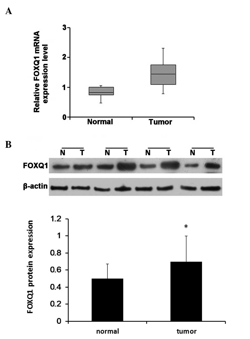
Total RNA was extracted from 30 pairs of LSCC
tissues and adjacent normal tissues and subjected to RT-qPCR in
order to measure the expression of FOXQ1 mRNA. Following
normalization to GAPDH, the mean expression of FOXQ1 mRNA in LSCC
tissues was significantly higher than that in adjacent normal
tissues (1.54±0.66 vs. 0.75±0.28; P<0.05; Fig. 1A). FOXQ1 protein expression was also
measured by western blotting in the same samples in which FOXQ1
mRNA expression was measured. The results demonstrated that FOXQ1
protein expression was increased in 19 of 30 LSCC tissues (~63%),
compared with matched adjacent normal tissues. FOXQ1 protein
expression was higher in LSCC tissues than that in adjacent normal
tissues (Fig. 1B; P<0.05). These
findings were in accordance with the FOXQ1 mRNA expression data. By
contrast, analysis of the association of FOXQ1 expression with
characteristics, such as patient age, gender and tumor stage,
revealed no significant associations between these variable (data
not shown).
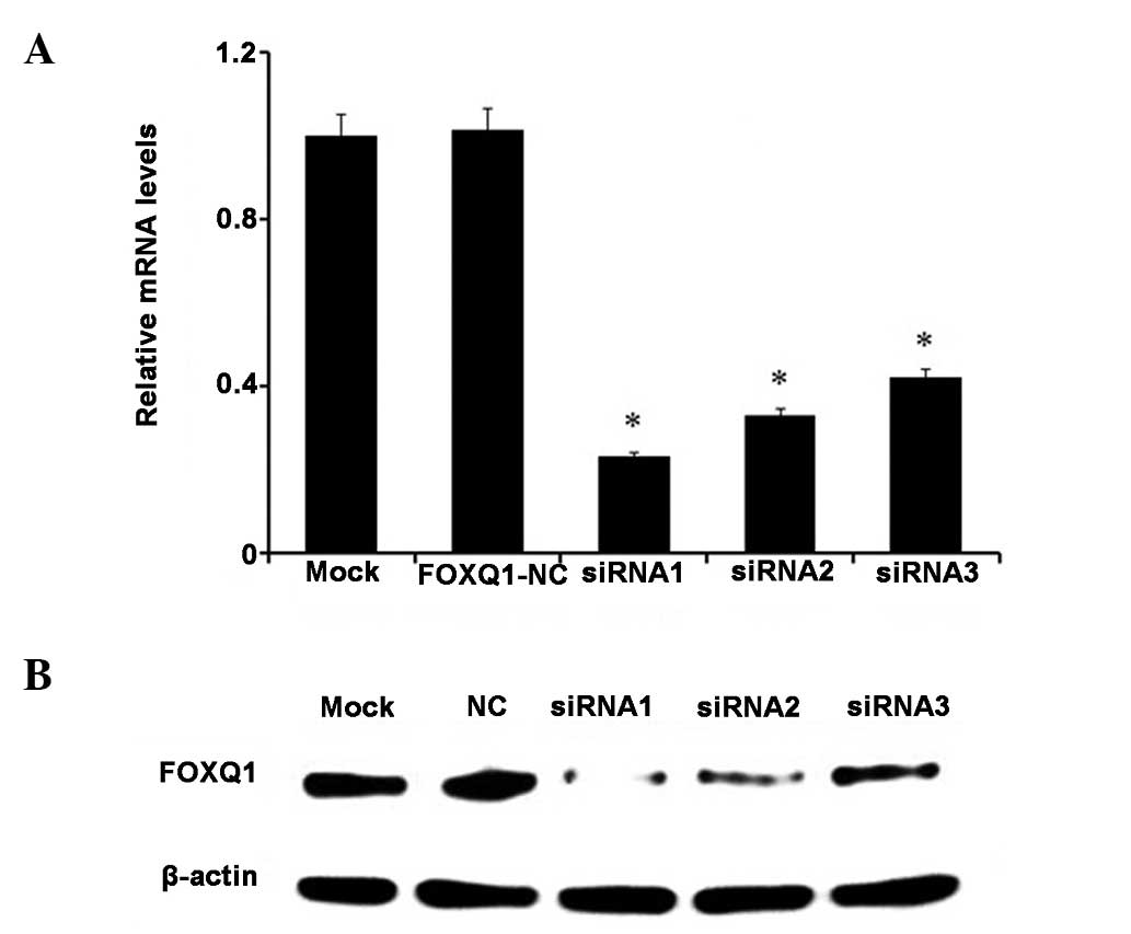
Inhibition of FOXQ1 following siRNA
transfection in Hep2 cells
Following transfection of Hep2 cell with FOXQ1 siRNA
for 72 h, the expression of FOXQ1 mRNA and protein was
detected by RT-qPCR and western blotting. Cells were also
transfected with FOXQ1-NC as a negative control. The results are
shown in (Fig. 2). Following
transfection with FOXQ1 siRNA, Hep2 cells exhibited
significant downregulation of FOXQ1 expression at the mRNA
and protein levels (Fig. 2A and B;
P<0.05).
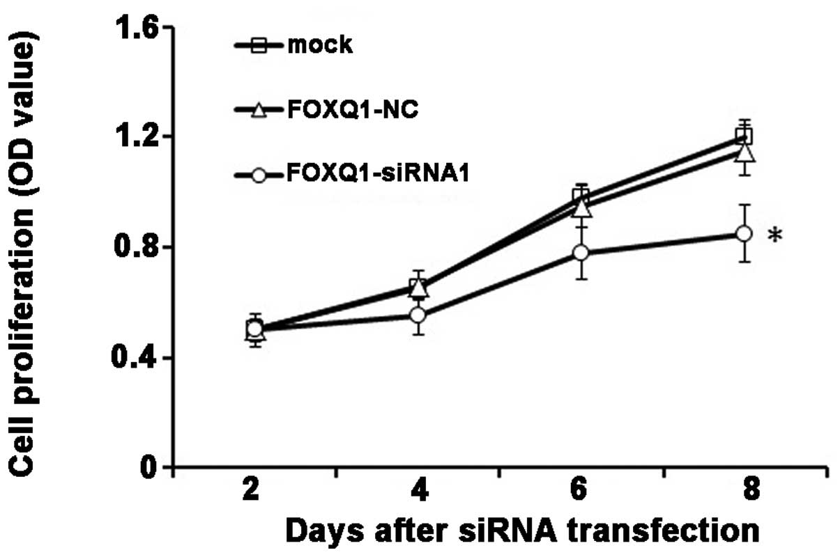
Downregulation of FOXQ1 expression
reduces proliferation of Hep2 cells
Cell proliferation was determined using a CCK-8
assay. The results demonstrated that downregulation of FOXQ1 in
Hep2 cells resulted in a significant reduction in cellular
proliferation at 4, 6 and 8 d after transfection (P<0.05). This
indicates that suppression of FOXQ1 correlates with decreased
proliferation of Hep2 cells (Fig.
3).
Inhibition of FOXQ1 induces G0/G1
arrest, while it has no effect on apoptosis in Hep2 cells
Flow cytometric analysis of the cell cycle
demonstrated that inhibition of FOXQ1 in Hep2 cells reduced the
proportion of cells in the S and G2/M phases, and more cells were
arrested in the G0/G1 phase compared with cells in the control
group (Table I). Furthermore,
apoptosis of FOXQ1-NC- and FOXQ1-siRNA-transfected cells was
examined using flow cytometry. As shown in Table I, after 4 days, 2.42% and 2.84% of
control cells and FOXQ1-NC cells were apoptotic, respectively,
while 2.95% of FOXQ1-siRNA cells were apoptotic. No significant
difference in the level of apoptosis in Hep2 cells was detected
among these groups.
 | Table I.Flow cytometry analysis of cell cycle
progression and apoptosis. |
Table I.
Flow cytometry analysis of cell cycle
progression and apoptosis.
| Group | G0/G1 phase (%) | S phase (%) | G2/M phase (%) | Apoptosis (%) |
|---|
| Mock |
47.64±3.61 |
27.55±1.54 |
22.39±3.44 |
2.42±0.78 |
| FOXQ1-NC |
46.65±0.89 |
28.64±1.85 |
21.87±1.23 |
2.84±2.64 |
| FOXQ1-siRNA |
58.03±3.45a |
23.35±0.65 |
15.67±2.15 |
2.95±1.65 |
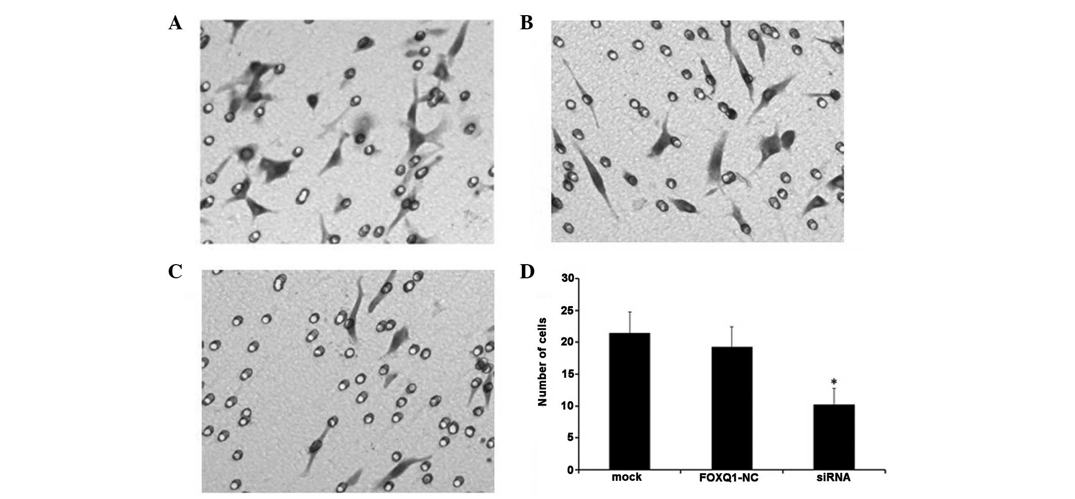
Effect of FOXQ1 silencing on cell
invasion in Hep2 cell lines
The results of the matrigel invasion assay
demonstrated that the number of migrating cells was significantly
decreased in the FOXQ1-siRNA transfection group, compared with that
in the control group. The numbers of invading cells in the mock and
FOXQ1-NC groups were 21.46±3.35 and 19.29±3.16, respectively, which
were significantly higher than the number in the FOXQ1-siRNA group
(10.24±2.52; P<0.01; Fig. 4).
Discussion
FOXQ1 belongs to the forkhead transcription factor
family. Previous studies have demonstrated that FOXQ1 is a
downstream target of homeobox C13. Each of these may affect
medullary differentiation through a common regulatory pathway
(23,24). A recent study reported that FOXQ1
promotes glioma cell proliferation and migration by suppressing the
promoter activity of neurexin-3-α (NRXN3) (25). Overexpression of FOXQ1 may enhance
tumor growth and tumorigenicity of colorectal cancer (19). Furthermore, overexpression of FOXQ1 is
associated with a poor prognosis in non-small cell lung cancer
(20) and with EMT regulation, via
inhibition of E-cadherin transcription (26). To date, little is known regarding the
mechanism underlying the effect of FOXQ1 on the development of
human laryngeal cancer.
In the present study, FOXQ1 expression was
upregulated at the mRNA and protein level in LSCC tissues, compared
with adjacent normal tissues. However, no significant association
was detected between FOXQ1 expression level, and gender, age or
tumor stage in patients with LSCC. In order to examine whether
FOXQ1 is involved in the development and progression of LSCC, RNA
interference was used to reduce the expression of FOXQ1 in cultured
Hep2 cells. Significant inhibition expression of FOXQ1 was observed
with RT-qPCR and western blotting. In vitro suppression of
Hep2 cell proliferation was analyzed, and the results demonstrated
that, compared with FOXQ1-NC and mock cell groups, the
proliferation of Hep2 cells was significantly inhibited following
transfection with FOXQ1-siRNA.
In order to measure the effect of FOXQ1 on cell
cycle progression, FACS analyses was performed, following
transfection of FOXQ1-siRNA. The results indicated that
siRNA-mediated knockdown of FOXQ1, led to cell cycle arrest in the
G0/G1 phase. Gao et al (27)
obtained similar results and suggested that expression of FOXQ1 may
affect levels of cell cycle regulators; depletion of FOXQ1 reduced
the expression of cyclin E and CDK4 and increased that of the
cyclin dependent kinase inhibitors (CKDIs), p27Kip1 and
p21Cip1, which together prevented cell cycle
progression.
Kaneda et al (19) and Qin et al (28) demonstrated that apoptosis was
inhibited in H1299 cells overexpressing FOXQ1 and in the 7721
hepatocellular carcinoma cell line, respectively. However, in the
present study, no significant difference in the level of apoptosis
was detected, following suppression of FOXQ1, among the Hep2 cell
groups. These findings are in accordance with those of Gao et
al (27), which were conducted in
the SKOV3 ovarian cancer cell line. It is therefore hypothesized
that the effect of FOXQ1 on apoptosis may vary among different
types of carcinoma. The mechanism underlying the influence of FOXQ1
on apoptosis requires further investigation.
The primary cause of death in almost all forms of
cancer, including breast (29) and
colorectal cancer (30), is cancer
cell metastasis to distant organs. The initial step in metastasis
is the invasion of surrounding tissues by cancer cells, and tissue
invasion and metastasis are hallmarks of malignant tumors.
Suppression of the pathways involved in invasion and metastasis in
cancer cells may be a treatment option for patients with cancer.
The results of the transwell assay in the present study, suggested
that deletion of FOXQ1 in Hep2 cells transfected with siRNA may
significantly reduce cell invasiveness, which further indicates
that FOXQ1 is associated with the aggressiveness of LSCC cells. Sun
et al (25) showed that FOXQ1
expression directly affected glioma cell migration in an
NRXN3-dependent manner in vitro and in vivo. Zhu
et al (31) demonstrated that
suppression of FOXQ1 expression reversed the process of EMT, in
association with the upregulation of E-cadherin, and that it also
caused T24 bladder cancer cells to acquire an epithelial
cobblestone phenotype, resulting in significantly reduced
invasiveness. These results suggest that FOXQ1 is involved in tumor
invasion and metastasis.
The present study demonstrated that the mRNA and
protein expression of FOXQ1 was increased in LSCC tissues, compared
with normal adjacent tissues. The results also showed that
inhibition of FOXQ1 by transfection of siRNA into Hep2 cells
significantly reduced cell growth and migration, and arrested Hep2
cells in the G0/G1 phase, in contrast to the control groups. These
results indicate that FOXQ1 exhibits an oncogenic role in LSCC,
which is in accordance with the results of previous studies,
conducted in different types of tumors.
In conclusion, the current study has demonstrated
that FOXQ1 is overexpressed in LSCC tissues, and that it may affect
Hep2 cell growth, cell cycle progression and cell migration. These
results suggest that FOXQ1 is a potential therapeutic target in
laryngeal cancer. However, the siRNA was only transiently
transfected, no long-term effects on cells were examined, and in
vitro effects may differ from in vivo effects. Therefore
further in vivo evaluation is required.
Acknowledgements
This study was supported by a grant from the
Medicine Summit Project of Liaoning Province (grant no.
4010218).
References
|
1
|
Ragin CC, Modugno F and Gollin SM: The
epidemiology and risk factors of head and neck cancer: A focus on
human papillomavirus. J Dent Res. 86:104–114. 2007. View Article : Google Scholar : PubMed/NCBI
|
|
2
|
Jemal A, Siegel R, Ward E, Murray T, Xu J
and Thun MJ: Cancer statistics, 2007. CA Cancer J Clin. 57:43–66.
2007. View Article : Google Scholar : PubMed/NCBI
|
|
3
|
Knab BR, Salama JK, Solanki A, Stenson KM,
Cohen EE, Witt ME, Haraf DJ and Vokes EE: Functional organ
preservation with definitive chemoradiotherapy for T4 laryngeal
squamous cell carcinoma. Ann Oncol. 19:1650–1654. 2008. View Article : Google Scholar : PubMed/NCBI
|
|
4
|
Du LB, Mao WM, Chen WQ, Zhang SW, Yu CD,
Zheng RS, Xia QM and Wang XH: Incidence and mortality of larynx
cancer in China during 2003–2007. Zhonghua Liu Xing Bing Xue Za
Zhi. 33:395–398, (In Chinese). PubMed/NCBI
|
|
5
|
Cripps C, Winquist E, Devries MC,
StysNorman D and Gilbert R: Head and Neck Cancer Disease Site
Group: Epidermal growth factor receptor targeted therapy in stages
III and IV head and neck cancer. Curr Oncol. 17:37–48. 2010.
View Article : Google Scholar : PubMed/NCBI
|
|
6
|
Ji W, Guan C and Pan Z: Analysis of
curative effects on laryngeal carcinoma patients in the northeast
region of China. Acta Otolaryngol. 128:574–577. 2008. View Article : Google Scholar : PubMed/NCBI
|
|
7
|
Ozdek A, Sarac S, Akyol MU, Sungur A and
Yilmaz T: c-myc and bcl-2 Expression in supraglottic squamous cell
carcinoma of the larynx. Otolaryngol Head Neck Surg. 131:77–83.
2004. View Article : Google Scholar : PubMed/NCBI
|
|
8
|
Jain P, Kumar P, Pai VR and Parikh PM:
Neoadjuvant chemotherapy or chemoradiotherapy in head and neck
cancer. Indian J Cancer. 45:83–89. 2008. View Article : Google Scholar : PubMed/NCBI
|
|
9
|
Bieller A, Pasche B, Frank S, Gläser B,
Kunz J, Witt K and Zoll B: Isolation and characterization of the
human forkhead gene FOXQ1. DNA Cell Biol. 20:555–561. 2001.
View Article : Google Scholar : PubMed/NCBI
|
|
10
|
Feuerborn A, Srivastava PK, Küffer S,
Grandy WA, Sijmonsma TP, Gretz N, Brors B and Gröne HJ: The
Forkhead factor FoxQ1 influences epithelial differentiation. J Cell
Physiol. 226:710–719. 2011. View Article : Google Scholar : PubMed/NCBI
|
|
11
|
Korver W, Roose J and Clevers H: The
winged-helix transcription factor Trident is expressed in cycling
cells. Nucleic Acids Res. 25:1715–1719. 1997. View Article : Google Scholar : PubMed/NCBI
|
|
12
|
Ogaki S, Harada S, Shiraki N, Kume K and
Kume S: An expression profile analysis of ES cell-derived
definitive endodermal cells and Pdx1-expressing cells. BMC Dev
Biol. 11:132011. View Article : Google Scholar : PubMed/NCBI
|
|
13
|
Carlsson P and Mahlapuu M: Forkhead
transcription factors: Key players in development and metabolism.
Dev Biol. 250:1–23. 2002. View Article : Google Scholar : PubMed/NCBI
|
|
14
|
Candelario J, Chen LY, Marjoram P, Reddy S
and Comai L: A filtering strategy identifies FOXQ1 as a potential
effector of lamin A dysfunction. Aging (Albany NY). 4:567–577.
2012.PubMed/NCBI
|
|
15
|
Sizemore ST and Keri RA: The forkhead box
transcription factor FOXC1 promotes breast cancer invasion by
inducing matrix metalloprotease 7 (MMP7) expression. J Biol Chem.
287:24631–24640. 2012. View Article : Google Scholar : PubMed/NCBI
|
|
16
|
Wendling DS, Lück C, von Schweinitz D and
Kappler R: Characteristic overexpression of the forkhead box
transcription factor Foxf1 in Patched-associated tumors. Int J Mol
Med. 22:787–792. 2008.PubMed/NCBI
|
|
17
|
Xia L, Huang W, Tian D, Zhu H, Qi X, Chen
Z, Zhang Y, Hu H, Fan D, Nie Y, et al: Overexpression of forkhead
box C1 promotes tumor metastasis and indicates poor prognosis in
hepatocellular carcinoma. Hepatology. 57:610–624. 2013. View Article : Google Scholar : PubMed/NCBI
|
|
18
|
Jonsson H and Peng SL: Forkhead
transcription factors in immunology. Cell Mol Life Sci. 62:397–409.
2005. View Article : Google Scholar : PubMed/NCBI
|
|
19
|
Kaneda H, Arao T, Tanaka K, Tamura D,
Aomatsu K, Kudo K, Sakai K, De Velasco MA, Matsumoto K, Fujita Y,
et al: FOXQ1 is overexpressed in colorectal cancer and enhances
tumorigenicity and tumor growth. Cancer Res. 70:2053–2063. 2010.
View Article : Google Scholar : PubMed/NCBI
|
|
20
|
Feng J, Zhang X, Zhu H, Wang X, Ni S and
Huang J: FoxQ1 overexpression influences poor prognosis in
non-small cell lung cancer, associates with the phenomenon of EMT.
PLoS One. 7:e399372012. View Article : Google Scholar : PubMed/NCBI
|
|
21
|
Zhang H, Meng F, Liu G, Zhang B, Zhu J, Wu
F, Ethier SP, Miller F and Wu G: Forkhead transcription factor
foxq1 promotes epithelial-mesenchymal transition and breast cancer
metastasis. Cancer Res. 71:1292–1301. 2011. View Article : Google Scholar : PubMed/NCBI
|
|
22
|
Sobin LH and Wittekind C: Head and neck
tumors, larynxTNM Classification of Malignant Tumors. 6th.
Wiley-Liss; New York, NY: pp. 52–56. 2002
|
|
23
|
Potter CS, Peterson RL, Barth JL, Pruett
ND, Jacobs DF, Kern MJ, Argraves WS, Sundberg JP and Awgulewitsch
A: Evidence that the satin hair mutant gene Foxq1 is among multiple
and functionally diverse regulatory targets for Hoxc13 during hair
follicle differentiation. J Biol Chem. 281:29245–29255. 2006.
View Article : Google Scholar : PubMed/NCBI
|
|
24
|
Hong HK, Noveroske JK, Headon DJ, Liu T,
Sy MS, Justice MJ and Chakravarti A: The winged helix/forkhead
transcription factor Foxq1 regulates differentiation of hair in
satin mice. Genesis. 29:163–171. 2001. View Article : Google Scholar : PubMed/NCBI
|
|
25
|
Sun HT, Cheng SX, Tu Y, Li XH and Zhang S:
FoxQ1 promotes glioma cells proliferation and migration by
regulating NRXN3 expression. PLoS One. 8:e556932013. View Article : Google Scholar : PubMed/NCBI
|
|
26
|
Qiao Y, Jiang X, Lee ST, Karuturi RK, Hooi
SC and Yu Q: FOXQ1 regulates epithelial-mesenchymal transition in
human cancers. Cancer Res. 71:3076–3086. 2011. View Article : Google Scholar : PubMed/NCBI
|
|
27
|
Gao M, Shih IeM and Wang TL: The role of
forkhead box Q1 transcription factor in ovarian epithelial
carcinomas. Int J Mol Sci. 13:13881–13893. 2012. View Article : Google Scholar : PubMed/NCBI
|
|
28
|
Qin J, Xu Y, Li X, Wu Y, Zhou J, Wang G
and Chen L: Effects of lentiviral-mediated Foxp1 and Foxq1 RNAi on
the hepatocarcinoma cell. Exp Mol Pathol. 96:1–8. 2014. View Article : Google Scholar : PubMed/NCBI
|
|
29
|
Piccolo S, Enzo E and Montagner M: p63,
Sharp1, and HIFs: Master regulators of metastasis in
triple-negative breast cancer. Cancer Res. 73:4978–4981. 2013.
View Article : Google Scholar : PubMed/NCBI
|
|
30
|
Agarwal E, Brattain MG and Chowdhury S:
Cell survival and metastasis regulation by Akt signaling in
colorectal cancer. Cell Signal. 25:1711–1719. 2013. View Article : Google Scholar : PubMed/NCBI
|
|
31
|
Zhu Z, Pang Z, Xing Y, Wan F, Lan D and
Wang H: Short hairpin RNA targeting FOXQ1 inhibits invasion and
metastasis via the reversal of epithelial-mesenchymal transition in
bladder cancer. Int J Oncol. 42:1271–1278. 2013.PubMed/NCBI
|