Introduction
Gastric cancer is the second highest cause of
cancer-associated mortality worldwide. Despite the advances in
diagnosis and treatment, the prognosis of patients with gastric
cancer remains poor. The median survival time of patients with
advanced gastric cancer is ≤10 months, and only 10–20% of patients
survive >5 years.
Kangai 1 (KAI1), also termed cluster of
differentiation (CD)82, is a tumor metastasis suppressor that was
first identified as a metastasis suppressor for prostate cancer.
KAI1 is located on human chromosome 11p11.2 (1) and is ~80 kb in length, containing 10
exons and 9 introns. This gene encodes a 267-amino acid protein
that is a member of the transmembrane 4 superfamily (TM4SF). KAI1
has been found to be downregulated in numerous types of human
cancers, including prostate, breast and ovarian cancers. A previous
study has identified that p53 plays a role in the positive
regulation of the expression of the KAI1 gene and may activate KAI1
through the consensus binding sequence in the promoter (2). In addition, the major cause of the poor
prognosis of patients with gastric cancer is the reduction in the
expression of these two proteins (3).
It has also been reported that the expression of KAI is
downregulated in advanced cancer, and more so in metastatic cancer
(4–6).
Consequently, KAI1 expression plays an important role in cancer
progression and may also be a potential target for the inhibition
of cancer metastasis. In order to detect the role of KAI1 in the
progression and prognosis of gastric cancer, immunohistochemistry
and in situ hybridization were used in the present study to
evaluate KAI1 expression in various stages of gastric cancer.
At present, no specific studies have been conducted
to investigate the effects and mechanisms of the KAI1 gene on the
migration and invasion of gastric carcinoma cells. To detect these
aspects, the pEGFP-N1-KAI1 plasmid was transfected into the gastric
carcinoma SGC7901 cells through liposomes in the present study.
Materials and methods
Patients
Tissue specimens obtained from 128 patients with
gastric adenocarcinoma that underwent resection at the Shandong
Cancer Hospital (Jinan, Shandong, China) between January 2007 and
April 2009 were used in the present study. The patients consisted
of 81 males and 47 females, aged between 30 and 74 years (median,
48 years). The inclusion criteria for the present study were as
follows: Complete surgical R0 resection of the primary tumor;
pathologically confirmed diagnosis of gastric adenocarcinoma; no
chemotherapy or radiotherapy administered; and the absence of
secondary malignancies. All patient records contained complete
clinical, pathological and follow-up data. Normal gastric mucosa
tissue (≤5 cm) adjacent to the tumor was excised and confirmed to
be tumor-free by pathological analysis.
Tumor histology was determined according to the
criteria provided by the World Health Organization (7). The pathological tumor-node-metastasis
(TNM) stage was assessed according to the Unified International
Gastric Cancer Staging Classification System, as incorporated in
the UICC TNM classification manual (8). The clinical outcome of the patients was
followed up from the date of surgery to either the date of
mortality or April 20, 2014, resulting in a follow-up period of
1–60 months (mean, 40 months). The present study was conducted in
accordance with the Declaration of Helsinki (9), and the Ethics Committee of the
Affiliated Hospital of Shandong Academy of Medical Sciences (Jinan,
Shandong, China) approved the present experimental protocols.
Written informed consent was obtained from all patients.
Immunohistochemistry
The tissue sections were conventionally dewaxed,
hydrated and subjected to antigen repair with EDTA. The monoclonal
mouse anti-human KAI1/CD82 antibody (cat no. 564341; BD
Biosciences, San Jose, CA, USA) was diluted at 1:200. The
immunohistochemical staining was performed using the of
streptavidin-peroxidase two-stage method, according to the
instructions of the kits (Fuzhou Maixin Biotech Co., Ltd., Fuzhou,
Fujian, China). Negative controls were stained following the same
procedure, with the exception that the primary antibody was
replaced with PBS. The KAI1-positive tissue provided by Fuzhou
Maixin Biotech Co., Ltd. was used as a positive control.
The staining intensity and percentage of cells
stained for KAI1 expression were evaluated in a blind manner by
three pathologists simultaneously, and a consensus was reached for
each score. Cells positive for the expression of KAI1 were
considered to be cells with brown plasma membranes and cytoplasm.
The presence of KAI1 expression was assessed through the ratio of
stained to non-stained cells. At least nine visual fields were
observed for each section under a high power lens (H600L; Nikon,
Tokyo, Japan). The staining intensity was judged based on the ratio
of KAI1-positive to total cell numbers observed in the visual
field. Sections with ≤10% KAI1-positive cells were considered to
not express KAI1 and sections with >10% KAI1-positive cells were
considered to express KAI1.
In situ hybridization
The mRNA sequence of the KAI1 gene was retrieved
from the National Center for Biotechnology Information database
(U.S. National Library of Medicine, Bethesda, MD, USA). The
oligonucleotide probe sequences were designed using Primer3
software (Whitehead Institute for Biomedical Research, Cambridge,
MA, USA) as follows: 5′-CAGCCTTTCTGTGAGGAAGG-3′ (800–819 bp);
5′-GATGGTCCTGTCCATCTGCT-3′ (983–1,002 bp); and
5′-GCAGTCACTATGCTCAT-3′ (438–454 bp) (Sangon Biotech Co., Ltd.,
Shanghai, China). The primers were marked by digoxin. The tissue
sections were conventionally dewaxed and underwent gradient
alcoholic dehydration. The slides were incubated in 3%
H2O2 at room temperature for 10 min, and were
then digested using Proteinase K, diluted in 3% saline sodium
citrate, at 37°C for 20 min. The in situ hybridization was
performed according to the instructions for the kits (Roche, Basel,
Switzerland). Blank controls were operated following the same
procedure, but without the probe. The expression of KAI1 was
indicated by in situ hybridization as clear yellow brown
granular material, which was located on the cell membrane. The KAI1
staining intensity was scored as follows: 0, absent; 1, weak; 2,
moderate; and 3, strong. The percentage of KAI1-positive cells was
scored into four categories, as follows: 1, 0–10%; 2, 11–30%; 3,
31–60%; and 4, 61–100%. The sum of these two scores was classified
as follows: 1–3, absent; 4–5, positive; and 6–7, strongly
positive.
Eukaryotic expression plasmid vector,
cell culture, plasmid transfection and reverse
transcription-semi-quantitative polymerase chain reaction
(RT-sqPCR) assay
pEGFP-N1 is a eukaryotic expression plasmid without
the objective gene, and this plasmid possesses a selectable marker
gene for G418 resistance. The eukaryotic expression plasmid vector
was supplied by Proteintech Group Inc. (Chicago, IL, USA).
Cell culture
The SGC7901 cell line was obtained from the Cell
Bank of the Chinese Academy of Sciences (Shanghai, China). The
cells were cultured in Dulbecco's modified Eagle's medium (DMEM;
Sigma-Aldrich, St. Louis, MO, USA) containing 10% fetal bovine
serum (FBS; HyClone, Logan, UT, USA) at 37°C in an incubator with a
humidified 5% CO2 atmosphere.
Plasmid transfection
Once the cells had reached 70–90% confluency, the
pEGFP-N1-KAI1 plasmid was transfected into the SGC7901 cells using
Lipofectamine 2000 (Invitrogen, Carlsbad, CA, USA), in accordance
with the manufacturer's instructions. The vector control plate was
transfected with the pEGFP-N1 plasmid, and cells without
transfection acted as a blank control.
RT-sqPCR assay
The effect of KAI1 gene transfection was measured
using an RT-sqPCR assay. Total RNA with isolated using TRIzol
(Invitrogen, Carlsbad, CA, USA) using an RNeasy Mini kit (cat no.
74104; Qiagen, Dusseldorf, Germany) and cDNA was generated by
reverse transcription, according to the instructions of the Reverse
Transcription System kit (Promega, Madison, WI, USA). The primers
used for KAI1 PCR were as follows: Forward,
5′-CCCCAAGTACTGAGGCAGC-3′, and reverse, 5′-AACCACAGAACAGCCAGGG-3′.
This generated a 217-bp product (1040–1256 bp) (Fig. 2A). The PCR mixture contained 5 µl 2X
HiFi PCR Master Mix, 0.5 µl forward primer (2 µM), 0.5 µl reverse
primer (2 µM), 3.5 µl RNase-free double-distilled H2O,
and 0.5 µl cDNA template. The PCR conditions were as follows: 95°C
for 3 min; 30 cycles at 94°C for 20 sec, 55°C for 20 sec and 72°C
for 20 sec; and 72°C for 5 min.
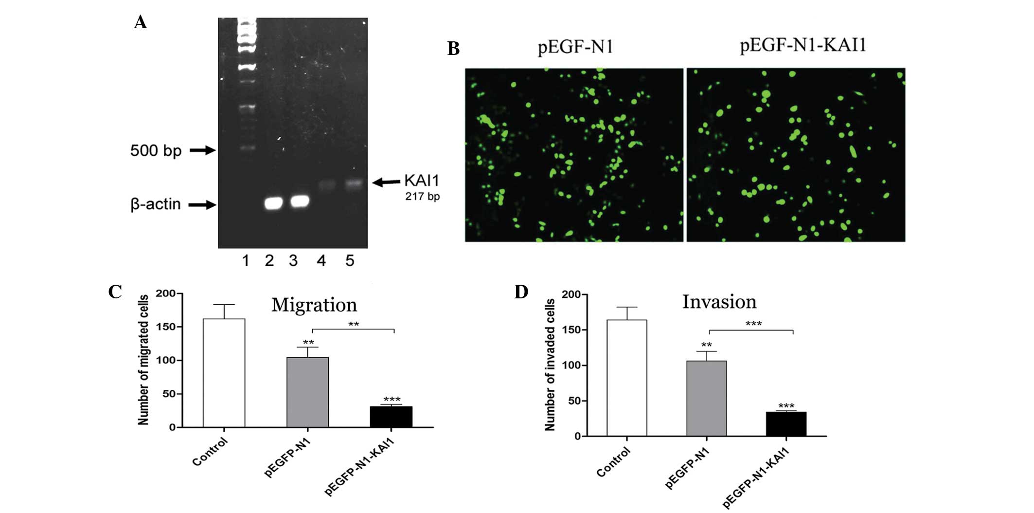 | Figure 2.Findings of the RT-sqPCR,
transfection, migration and invasion assays. (A) The pEGFP-N1-KAI1
plasmid was transfected into human gastric carcinoma SGC7901 cells
by liposome. KAI1 was clearly overexpressed in pEGFP-N1-KAI1
transfected cells (lane 5) compared to the control cells (lane 4).
Lanes 1, 100 bp+1kb ladder; lane 2, β-actin expression in
pEGFP-N1-transfected SGC7901 cells; lane 3, β-actin expression in
pEGFP-N1-KAI1-transfected SGC7901 cells; lane 4, KAI1 expression in
pEGFP-N1-transfected SGC7901 cells; and lane 5, KAI1 expression in
pEGFP-N1-KAI1-transfected SGC7901 cells. KAI1 was clearly more
highly expressed in lane 5 compared with lane 4. (B) Fluorescent
expression of pEGFP-N1-KAI1 and pEGFP-N1 in transfected SGC7901
cells, revealing that transfection with the KAI1 gene inhibited the
migration and invasion activity of SGC7901 cells. (C) The invasion
activity of pEGFP-N1-KAI1 cells was significantly downregulated.
(D) The migratory activity of pEGFP-N1-KAI1 cells was significantly
decreased. RT-sqPCR, reverse transcription-semi-quantitative
polymerase chain reaction; KAI1, Kangai 1. |
Cell migration and invasion
assays
A Transwell chamber assay was used to perform the
cell migration analysis. The cells were fasted for 24 h with
serum-free medium DMEM containing 0.1% bovine serum albumin (BSA),
and then trypsinized and resuspended with medium to a density of
1×10⁶/ml. The three groups of cells were seeded in the
upper chamber of the Transwell insert, and 600 µl DMEM containing
10% FBS was added to the lower chamber. The cells were cultured at
37°C in a humidified 5% CO2 incubator for 24 h, the
inserts were washed with PBS. A cotton swab was used to remove
adherent cells on the inner side of the upper chamber membrane. The
upper chamber was then dried naturally and stained with 0.5%
hematoxylin. Six visual fields of each insert were randomly counted
under an upright light microscope (BX51; Olympus, Tokyo, Japan),
and the average cell number was calculated.
Cell invasion assay
The Transwell chamber invasion assay was performed
to investigate the role of KAI1 in gastric cancer cells. Laminins
(20 µg/ml; EMD Millipore, Billerica, MA, USA) were diluted in
serum- and BSA-free DMEM and were then added to a 24-well plate at
0.95 ml/well (10 µg/cm2). This process was repeated 3
times. The plate was then incubated at 4°C overnight. The
subsequent experimental steps were in accordance with the cell
migration assay.
RT-sqPCR
Total RNA was obtained using TRIzol extraction
(Invitrogen, Carlsbad, CA, USA) and reverse transcribed into
complementary DNA using the kit (Takara RT-PCR), according to the
manufacturer's instructions. RT-sqPCR was performed by using the
Roche Cobas 4800 System (Roche). The sequences of the
HIF-1αx, MMP-2x, MMP-9x,
bFGFx and uPA primers are described in Table I.
 | Table I.Polymerase chain reaction primers for
HIF-1α, MMP-2, MMP-9, bFGF, uPA and β-actin. |
Table I.
Polymerase chain reaction primers for
HIF-1α, MMP-2, MMP-9, bFGF, uPA and β-actin.
| Gene | Direction | Primer sequence
(5′-3′) |
|---|
| HIF-1α | F |
AGCCAGACGATCATGCAGCTACTA |
|
| R |
TGTGGTAATCCACTTTCATCCATTG |
| MMP-2 | F |
TGTCGCCCCCAAAACGGACA |
|
| R |
ATGCTCCCAGCGGCCAAAGT |
| MMP-9 | F |
TGCTGGGCTGCTGCTTTGCT |
|
| R |
CGGGCAAAGGCGTCGTCAAT |
| bFGF | F |
GAACGGGGGCTTCTTCCT |
|
| R |
CCCAGTTCGTTTCAGTGCC |
| uPA | F |
TGAGCGACTCCAAAGGCAGCA |
|
| R |
TGAAGCAGTGTGTGGCGCTGA |
| β-actin | F |
GGCATCGTGATGGACTCCG |
|
| R |
GCTGGAAGGTGGACAGCGA |
Statistical analysis
The PEMS 3.1 software (Jingyuan Guangzhou
Pharmaceutical Research Ltd., Guangzhou, China) was used for the
statistical analysis. All data were expressed as the mean ±
standard deviation from at least three independent experiments. The
KAI1 staining level in tissues from various stages of gastric
cancer was compared using the χ2 test. The Kaplan-Meier
survival curve and log-rank test were used to analyze the
association between KAI1 expression and patient survival. The
correlation analysis was performed using Spearman's rank
correlation coefficient test, and the inspection level was α=0.05.
P<0.05 was considered to indicate a statistically significant
difference.
Results
KAI1 protein expression in gastric
cancer tissue and the correlation with clinical pathology
The KAI1 protein was found to be expressed in normal
gastric mucosa tissues, with an expression rate of 22% in gastric
cancer tissue, and the difference between the expression rate of
KAI1 in cancer and normal tissues was statistically significant
(χ2=24.382; P=0.000). The expression of KAI1 was
associated with the differentiation degree of gastric cancer tumors
(Table II). The deeper the degree of
gastric cancer invasion, the lower the rate of KAI1 expression
(Table III). Correlation analysis
was used to further detect the association between the various
clinical stages and the positive expression of the KAI1 protein.
The present results indicated a negative correlation between the
clinical stages and the positive expression of KAI1 (r=-0.9890;
P=0.0110; Table III).
| Table II.Association between KAI1 expression
and the differentiation of gastric cancer tumors. |
Table II.
Association between KAI1 expression
and the differentiation of gastric cancer tumors.
| Differentiation | Total, n | KAI1-positive, n | Occupancy, % | χ2
value | P-value |
|---|
| Superior | 44 | 18 | 40.9 | 5.5110 | 0.0189 |
| Inferior | 84 | 10 | 11.9 |
|
|
| Table III.Association between KAI1 expression
and other clinicopathological features of gastric cancer. |
Table III.
Association between KAI1 expression
and other clinicopathological features of gastric cancer.
| Clinicopathological
feature | Total, n | KAI1-positive, n | Positive rates of
KAI1, % | χ2
value | P-value |
|---|
| Depth of
invasion |
|
| Mucosa
and submucosa | 16 | 12 | 75.0 | 16.9004 | 0.0007 |
| Muscular
layer | 48 | 10 | 20.8 |
|
|
|
Serosa | 44 | 6 | 13.6 |
|
|
| Out of
serosa | 20 | 0 | 0.0 |
|
|
| Lymphatic
metastasis |
|
| Yes | 98 | 12 | 12.0 |
9.0682 | 0.0026 |
| No | 30 | 16 | 53.0 |
|
|
| Distant
metastasis |
|
|
Yes | 12 | 0 |
0.0 |
0.7104 | 0.3993 |
| No | 116 | 28 | 24.0 |
|
|
| TNM stage |
|
| I | 32 | 14 | 43.8 |
4.3881 | 0.0362 |
| II | 40 | 10 | 25.0 |
|
|
|
III | 44 |
4a |
9.1 |
|
|
| IV | 12 | 0 |
0.0 |
|
|
KAI1 mRNA expression in gastric cancer
tissue and the correlation with clinical pathology
KAI1 mRNA expression was present in all normal
gastric mucosa samples and in 40 out of 128 gastric carcinoma
tissue samples, which was a statistically significant difference
(P=0.0001). There was no significant difference (P>0.05) between
the presence of KAI1 mRNA expression and the tumor location, or
between the age and gender of the patients. The rate of KAI1 mRNA
expression in the superior differentiation group, which consisted
of well- and moderately-differentiated adenocarcinoma, was
increased compared with the expression in the inferior
differentiation group, which consisted of poorly-differentiated and
mucinous adenocarcinoma and signet-ring cell carcinoma (P<0.05).
Correlation analysis was used to analyze the association between
the depth of invasion and KAI1 mRNA expression, and there was a
significant negative correlation between these variables
(r=-0.9558; P=0.044 2). Similarly, there was a significant negative
correlation between the TNM stage and KAI1 mRNA expression
(r=-0.9891; P=0.0109). The rate of KAI1 mRNA expression in gastric
cancer patients with lymph node metastasis was markedly decreased
compared with the rate in gastric cancer patients without lymph
node metastasis, and the difference was statistically significant
(P<0.05; Table IV).
| Table IV.KAI1 mRNA expression in gastric
cancer tissue and its correlation with clinical pathology. |
Table IV.
KAI1 mRNA expression in gastric
cancer tissue and its correlation with clinical pathology.
|
|
| KAI1 mRNA
expression |
|
|
|---|
|
|
|
|
|
|
|---|
| Clinicopathological
factor | Total, n | Present, n | Rate, % | χ2
value | P-value |
|---|
| Histological
classification |
|
|
Superior differentiation | 44 | 24 | 54.5 | 7.5416 | 0.0060 |
|
Inferior differentiation | 84 | 16 | 19.0 | |
|
|
| Depth of
invasion |
|
|
T1 | 16 | 12 | 75.0 |
|
|
|
T2 | 48 | 18 | 37.5 | 2.0497 | 0.1522 |
|
T3 | 44 | 8 | 18.2 | 5.8058 |
0.0131a |
|
T4 | 20 | 2 | 10.0 | 5.4029 |
0.0201a |
| Lymphatic
metastasis |
|
|
Yes | 98 | 16 | 16.3 | 18.8097 | 0.0000 |
| No | 30 | 24 | 80.0 | |
|
|
| TNM staging |
|
| I | 32 | 20 | 62.5 |
|
|
| II | 40 | 14 | 35.0 | 1.7067 |
0.1914b |
|
III | 44 | 6 | 13.6 | 7.7757 |
0.0053b |
| IV | 12 | 0 |
0.0 | 4.5852 |
0.0322b |
Correlation between KAI1 expression
and prognosis in patients with gastric cancer
The result of Kaplan-Meier analysis indicated that
the survival time of the group that expressed KAI1 was
significantly longer compared with the group that did not express
KAI1. The log-rank test indicated that the difference between the
two groups was statistically significant (χ2=11.523;
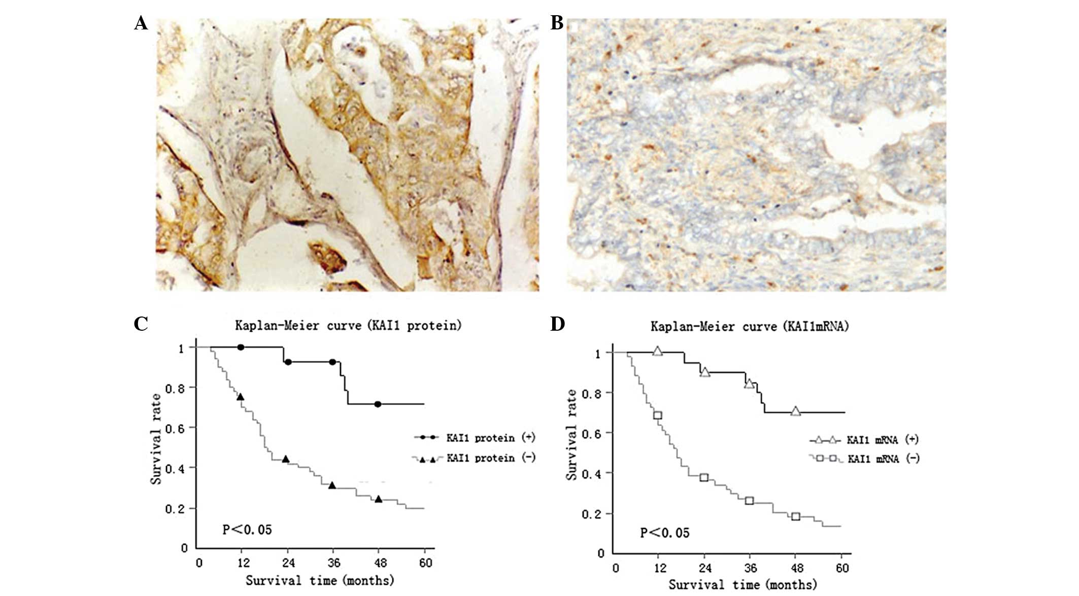
v=1; P<0.05; Fig. 1C).
In addition, the difference between the five-year
survival rates of the groups expressing and not expressing the KAI1
protein was statistically significant (P<0.05; Table V). Thus, patients expressing the KAI1
protein demonstrated an improved prognosis.
| Table V.Association between KAI1 expression
and the survival time of patients. |
Table V.
Association between KAI1 expression
and the survival time of patients.
|
|
| Expression of
KAI1 |
|
|
|---|
|
|
|
|
|
|
|---|
| Survival time,
years | Total, n | Present, n | Occupancy, % | Absent, n | Occupancy,% | χ2
value | P-value |
|---|
| >5 | 40 | 20 | 71 | 20 | 20 | 42.4261 | 0.000 |
| <5 | 88 | 8 | 29 | 80 | 80 |
|
|
KAI1 mRNA expression in gastric cancer
tissue and its correlation with the prognosis of gastric cancer
patients
The statistical results of Kaplan-Meier indicated
that the survival time of the group expressing KAI1 mRNA was longer
compared with the group without KAI1 mRNA expression, and the
difference was statistically significant (P<0.05; Fig. 1D). However, the difference between the
five-year survival rate of the group expressing KAI1 mRNA and group
without KAI1 expression was statistically significant (P<0.05;
Table VI). Therefore, patients
demonstrating KAI1 mRNA expression had an improved prognosis, and
this result was consistent with the aforementioned conclusion.
| Table VI.The association between KAI1 mRNA
expression and the survival time of patients. |
Table VI.
The association between KAI1 mRNA
expression and the survival time of patients.
|
|
| Expression of KAI1
mRNA |
|
|
|---|
|
|
|
|
|
|
|---|
| Survival time,
years | Total, n | Present, n | Occupancy, % | Absent, n | Occupancy, % | χ2
value | P-value |
|---|
| >5 | 40 | 28 | 70 | 12 | 13.6 | 19.1733 | 0.0007 |
| <5 | 88 | 12 | 30 | 76 | 86.4 |
|
|
Transfection of SGC7901 cells
To examine the effect of the expression of KAI1 on
the SGC7901 cells, gastric cancer SGC7901 cells were transiently
transfected with the pEGFP-N1-KAI1 and pEGFP-N1 plasmids.
Electrophoresis of the RT-sqPCR products revealed that KAI1 was
evidently overexpressed in the cells transfected with pEGFP-N1-KAI1
compared with the cells transfected with pEGFP-N1 (Fig. 2A). Fluorescent expression was observed
in the cells transfected with pEGFP-N1-KAI1 and those transfected
with pEGFP-N1 (Fig. 2B).
Cell migration and invasion
The migratory and invasive ability of SGC-7901 cells
was detected using a Transwell assay. The number of cells that had
traversed the membrane was counted subsequent to transfection for
48 h. Compared with the cells transfected with the pEGFP-N1
plasmid, the migratory and invasive activity of the cells
transfected with the pEGFP-N1-KAI1 plasmid was significantly
decreased (Fig. 2C and D).
Expression of metastasis-associated
genes in gastric cancer cells
RT-sqPCR was used to measure the effects of KAI1
transfection on the expression of metastasis-associated genes in
SGC7901 cells. Table VII reports
that the expression of HIF-1α was slightly increased following
transfection with the KAI1 expression plasmid. The expression of
MMP-9 was slightly reduced, but this effect was not statistically
significant (P>0.05). The expression of MMP-2 was significantly
increased in gastric carcinoma SGC7901 cells by KAI1 expression,
whereas the expression of uPA was significantly downregulated. The
expression of bFGF was also significantly decreased
(P<0.05).
| Table VII.Effect of KAl1 gene transfection on
the expression of genes associated with gastric cancer
metastasis. |
Table VII.
Effect of KAl1 gene transfection on
the expression of genes associated with gastric cancer
metastasis.
|
| KAI1
expression |
|
|---|
|
|
|
|
|---|
| Gene | Present | Absent | Fold change in
expression |
|---|
| HIF-1α | 1.7260 | 1.6700 | 1.034 |
| MMP-2 | 7.9120 | 0.2715 | 28.76 |
| MMP-9 | 1.2210 | 1.2740 | 0.958 |
| bFGF | 0.4565 | 0.5805 | 0.786 |
| uPA | 0.1621 | 7.5990 | 0.021 |
Discussion
KAI1 was first identified as a metastasis suppressor
in prostate cancer (1). Subsequent
studies demonstrated that this gene suppresses invasion and
metastasis in various cancers during disease progression (4,10). The
present study used immunohistochemistry and in situ
hybridization (MK2158; Wuhan Boster Biological Technology Co.,
Ltd., Wuhan, China) to examine 16 cases of well-differentiated
adenocarcinoma, 28 cases of moderately-differentiated
adenocarcinoma, 66 cases of poorly-differentiated adenocarcinoma,
10 cases of mucinous adenocarcinoma and 8 cases of signet-ring cell
carcinoma. In addition, the effects of KAI1 expression on the
migration and invasion of gastric cancer cells were investigated,
as migration and invasion are the critical steps in cancer
progression. The results of the present study demonstrated that
KAI1 staining occurs mainly in the cytoplasm (Fig. 1A), which is consistent with the
findings of previous studies (4,10,11).
Statistical analysis of the positive rate of KAI1
revealed a significant reduction in cytoplasmic KAI1 expression in
internal gastric cancer compared with superficial gastric cancer,
indicating that reduced KAI1 expression may be critical for the
progression of gastric cancer. The present data revealed that
decreased KAI1 expression was significantly associated with the
presence of lymphatic metastasis, TNM stage, patient survival time,
depth of invasion and the degree of gastric cancer differentiation.
The results also indicated that reduced KAI1 expression was not
involved in distant metastasis, but significantly stimulated
gastric cancer cell migration (Fig.
2C) and invasion (Fig. 2D), the
two important events in the process of tumor progression into
metastasis (12). This indicates that
KAI1 is a potent gastric cancer metastasis suppressor. In addition,
analysis of the survival curves in the present study revealed that
the expression of KAI1 was positively associated with the survival
of patients, which is consistent with the conclusions from studies
performed on malignant melanoma (11), non-small-cell lung cancer (13) and prostate cancer (14).
The KAI1 protein plays an important role in
signaling pathways, as it mediates the signal transduction between
cells and the surrounding environment, which affects the movement
and differentiation of cells and ultimately inhibits the invasion
and metastasis of tumor cells. KAI1 may regulate the infiltration
and metastasis of tumors through the following four mechanisms.
First, KAI1 binds with integrin to form a complex and affects cell
adhesion by regulating the functions of integrin. Second, the
protein promotes adhesion between tumor cells by acting on the
signal pathway of Src kinase (15).
Third, KAI1/CD82-associated surface protein is involved in the KAI1
signaling pathway (16). Fourth, the
inhibition of KAI1 is mediated by numerous other signaling pathways
and factors, including FAK, Rac and GTPase (17,18).
In addition, KAI1 is a highly glycosylated protein,
and a deficiency in the glycosylation process leads to a lack of
KAI1 function, without affecting the expression of the protein
(13). The signaling pathway mediated
by INF may activate nuclear factor-κB and increase the expression
of KAI1 (19).
In the present study, the expression of HIF-1α,
MMP-2, MMP-9, bFGF and uPA was detected in order to further reveal
the underlying mechanism of the function of the KAI1 gene in
gastric cancer. The current study identified that the inhibitory
effect of KAI1 expression on the cell migration and invasion of
gastric cancer was not mediated by HIF-1α, MMP-2 and MMP-9, but may
be associated with the reduction in uPA and bFGF expression.
bFGF may promote angiogenesis and degrade the
extracellular matrix by upregulating the expression of MMPs
(20). bFGF may also directly induce
the tumor cells to secrete a variety of protein decomposing enzymes
and collagenases, in order to promote tumor metastasis and
invasion. Notably, a previous study revealed that uPAR ligation
enhanced the migration of human lung fibroblasts by recruiting α5β1
integrin and lipid rafts through caveolin-Fyn-Shc signaling
(21). This mechanism may be
applicable to the present study. It was therefore hypothesized that
in the current study, uPAR ligation increased the migration of
gastric cancer cells, possibly through the recruitment of α5β1
integrin complexes, alongside the activation of the lipid
raft-localized caveolin-Fyn-Shc pathway. This, in turn, led to a
hypermigratory phenotype. As a result, the reduced expression of
uPA may limit the activation of this pathway and therefore,
decrease the migratory ability of cancer cells. However, the
underlying specific mechanism requires additional
investigation.
The present study demonstrated that KAI1 may be used
as a prognostic marker in human gastric cancer, which is not
consistent with the findings of a previous study by Knoener et
al (22). This is possibly due to
the various characteristics of the respective studies, including
the ethnicity, experimental specimens, surgical technology and
reagents. And the main difference is specimen. As we all know,
specimens kept for too long might lead to an antigen loss.
Therefore, we could collect more recent cases to detect whether
KAI1 is a prognostic marker or not.
Furthermore, the experimental data revealed that the
expression of MMP-2 significantly increased following transfection
with the KAI1 gene, which was not consistent with the
anti-metastatic role. This discrepancy may be due to the initiation
of malignant tumor development being an extremely complicated
process that involves multiple anomalies of signaling pathways,
including the induction of apoptosis, regulation of the cell cycle
and inhibition of tumor cell adhesion. Proto-oncogenes,
anti-oncogenes and multiple other factors participate in this
process. The association between these factors is complex, and the
effect may be negative, positive or modulatory, and in various
forms. In addition, the association may change with the progression
of tumors. Anti-oncogenes may not inhibit all the cocarcinogens.
Therefore, it is not notable that the expression of MMP-2
significantly increased subsequent to transfection with the KAI1
gene. This result also indicated that the inhibitory effect of KAI1
may not be mediated by MMP-2.
The present results demonstrated clearly that the
invasive ability of gastric cancer SGC7901 cells was significantly
decreased subsequent to transfection with the KAI1 gene. In
addition, the KAI1 gene may suppress the migration and invasion of
SGC7901 cells by reducing the expression of uPA and bFGF.
Therefore, the present data indicated that the expression level of
KAI1 protein may affect the migration and invasion ability of
gastric cancer cells. Upregulation of KAI1 gene expression in
gastric cancer cells may represent an important direction to
inhibit the invasion and metastasis of gastric cancer, which
requires additional investigation.
In summary, the present study confirmed that KAI1
plays an important role in the inhibition of metastasis and
invasion of gastric carcinoma, and downregulated KAI1 expression
was significantly associated with advanced gastric cancer and a
poor five-year survival rate in patients. The present study
provides a theoretical and experimental basis for the investigation
and clinical application of KAI1. It was concluded that KAI1 may be
used as a potential therapeutic target and is a promising
prognostic marker for gastric cancer.
Acknowledgements
This study was supported by the Natural Science
Foundation of Shandong Province (grant no., Y2007C025).
References
|
1
|
Dong JT, Lamb PW, RinkerSchaeffer CW,
Vukanovic J, Ichikawa T, Isaacs JT and Barrett JC: KAI1, a
metastasis suppressor gene for prostate cancer on human chromosome
11p11.2. Science. 268:884–886. 1995. View Article : Google Scholar : PubMed/NCBI
|
|
2
|
Mashimo T, Watabe M, Hirota S, Hosobe S,
Miura K, Tegtmeyer PJ, RinkerShaeffer CW and Watabe K: The
expression of the KAI1 gene, a tumor metastasis suppressor, is
directly activated by p53. Proc Natl Acad Sci USA. 95:11307–11311.
1998. View Article : Google Scholar : PubMed/NCBI
|
|
3
|
Guo C, Liu QG, Zhang L, Song T and Yang X:
Expression and clinical significance of p53, JunB and KAI1/CD82 in
human hepatocellular carcinoma. Hepatobiliary Pancreat Dis Int.
8:389–396. 2009.PubMed/NCBI
|
|
4
|
Liu FS, Dong JT, Chen JT, Hsieh YT, Ho E,
Hung MJ, Lu CH and Chiou LC: KAI1 metastasis suppressor protein is
down-regulated during the progression of human endometrial cancer.
Clin Cancer Res. 9:1393–1398. 2003.PubMed/NCBI
|
|
5
|
Lee HS, Lee HK, Kim HS, Yang HK and Kim
WH: Tumour suppressor gene expression correlates with gastric
cancer prognosis. J Pathol. 200:39–46. 2003. View Article : Google Scholar : PubMed/NCBI
|
|
6
|
Lee JH, Seo YW, Park SR, Kim YJ and Kim
KK: Expression of a splice variant of KAI1, a tumor metastasis
suppressor gene, influences tumor invasion and progression. Cancer
Res. 63:7247–7255. 2003.PubMed/NCBI
|
|
7
|
Hamilton SR, Bosman FT, Boffetta P, et al:
Tumor pathological type: Carcinoma of the colon and rectumWHO
Classification of Tumours of the Digestive System. Bosman FT,
Cameiro F, Hruban RH and Theise ND: IARC Press; Lyon: pp. 134–135.
2010
|
|
8
|
Leslie HS, Mary KG and Christian W: TNM
Classification of Malignant Tumours. 7th. John Wiley & Sons;
New York, NY: pp. 78–80. 2009
|
|
9
|
World Medical Association, . World Medical
Association Declaration of Helsinki: Ethical principles for medical
research involving human subjects. JAMA. 310:2191–2194. 2013.
View Article : Google Scholar : PubMed/NCBI
|
|
10
|
Liu X, Guo XZ, Li HY, Chen J, Ren LN and
Wu CY: KAI1 inhibits lymphangiogenesis and lymphatic metastasis of
pancreatic cancer in vivo. Hepatobiliary Pancreat Dis Int.
13:87–92. 2014. View Article : Google Scholar : PubMed/NCBI
|
|
11
|
Tang Y, Cheng Y, Martinka M, Ong CJ and Li
G: Prognostic significance of KAI1/CD82 in human melanoma and its
role in cell migration and invasion through the regulation of ING4.
Carcinogenesis. 35:86–95. 2014. View Article : Google Scholar : PubMed/NCBI
|
|
12
|
Gupta GP and Massagué J: Cancer
metastasis: Building a framework. Cell. 127:679–695. 2006.
View Article : Google Scholar : PubMed/NCBI
|
|
13
|
Adachi M, Taki T, Ieki Y, Huang CL,
Higashiyama M and Miyake M: Correlation of KAI1/CD82 gene
expression with good prognosis in patients with non-small cell lung
cancer. Cancer Res. 56:1751–1755. 1996.PubMed/NCBI
|
|
14
|
Liu W, IiizumiGairani M, Okuda H,
Kobayashi A, Watabe M, Pai SK, Pandey PR, Xing F, Fukuda K and
Modur V: KAI1 gene is engaged in NDRG1 gene-mediated metastasis
suppression through the ATF3-NFkappaB complex in human prostate
cancer. J Biol Chem. 286:18949–18959. 2011. View Article : Google Scholar : PubMed/NCBI
|
|
15
|
Jee B, Jin K, Hahu JH, Song HG and Lee H:
Metastasis-suppressor KAI1/CD82 induces homotypic aggregation of
human prostate cancer cells through Src-dependent pathway. Exp Mol
Med. 35:30–37. 2003. View Article : Google Scholar : PubMed/NCBI
|
|
16
|
Zhang XA, Lane WS, Charrin S, Rubinstein E
and Liu L: EWI2/PGRL associates with the metastasis suppressor
KAI1/CD82 and inhibits the migration of prostate cancer cells.
Cancer Res. 63:2665–2674. 2003.PubMed/NCBI
|
|
17
|
Zhang XA, He B, Zhou B and Liu L:
Requirement of the p130CAS-Crk coupling for metastasis suppressor
KAI1/CD82 mediated inhibition of cell migration. J Biol Chem.
278:27319–27328. 2003. View Article : Google Scholar : PubMed/NCBI
|
|
18
|
Kauffman EC, Robinson VL, Stadler WM,
Sokoloff MH and Rinker-Schaeffer CW: Metastasis suppression the
evolving role of metastasis suppresson genes for regulating cancer
cell growth at the secondary site. J Urol. 169:1122–1133. 2003.
View Article : Google Scholar : PubMed/NCBI
|
|
19
|
Shinohara T, Mike T, Nshimura N, Nokihara
H, Hamada H, Mukaida N and Sone S: Nuclear factor-kappaB-dependent
expression of metastasis suppressor KAI1/CD82 gene in lung cancer
cell lines expressing mutant p53. Cancer Res. 61:673–678.
2001.PubMed/NCBI
|
|
20
|
Feng JG and Nan XR: The distribution and
expression of bFGF in oral squamous cell carcinoma. Chinese
Remedies & Clinics. 8:3882008.
|
|
21
|
Grove LM, Southern BD, Jin TH, White KE,
Paruchuri S, Harel E, Wei Y, Rahaman SO, Gladson CL, Ding Q, et al:
Urokinase-type plasminogen activator receptor (uPAR) ligation
induces a raft-localized integrin signaling switch that mediates
the hypermotile phenotype of fibrotic fibroblasts. J Biol Chem.
289:12791–12804. 2014. View Article : Google Scholar : PubMed/NCBI
|
|
22
|
Knoener M, Krech T, Puls F, Lehmann U,
Kreipe H and Christgen M: Limited value of KAI1/CD82 protein
expression as a prognostic marker in human gastric cancer. Dis
Markers. 32:337–342. 2012. View Article : Google Scholar : PubMed/NCBI
|