Mazabraud's syndrome (MS) is a rare benign syndrome
that is reported to be associated with single or multiple
intramuscular myxomas and fibrous dysplasia occurring in a single
or multiple bones. Intramuscular myxoma in itself is a relatively
uncommon benign mesenchymal tumor (1), while fibrous dysplasias are more
frequent benign lesions (2). To the
best of our knowledge, 92 cases of MS have been reported in the
medical literature thus far. However, only 9 cases have reported
the combination of a solitary myxoma and monostotic fibrous
dysplasia (3–10). MS involving the mandible and the
adjacent soft tissue of the bone lesions was reported by Logel
(11). However, MS of the masseter
and the mandible are rarely reported compared with other regions.
The aim of the current study was to present a novel case of
solitary myxoma and monostotic fibrous dysplasia involving the jaw
and the masseter. Additionally, an overview of the 92 reported
cases of MS is also presented.
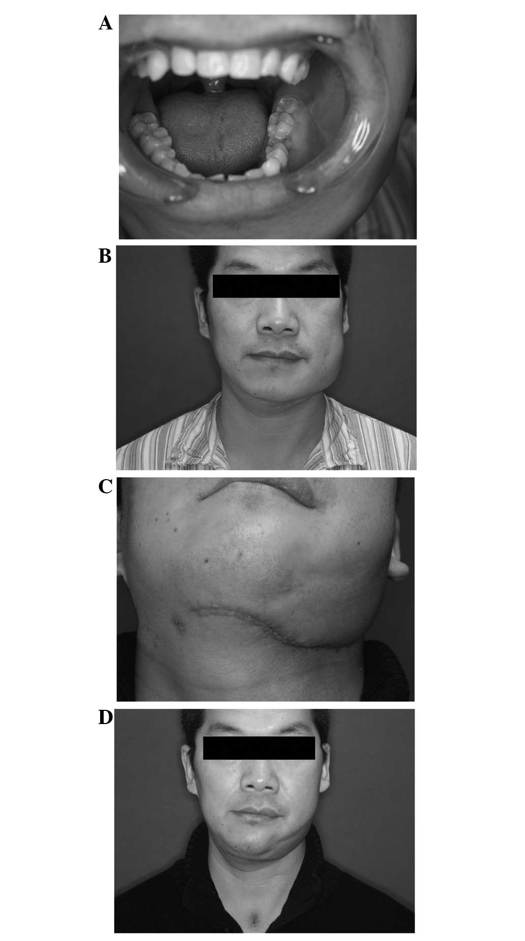
On February 8th, 2014, a 38-year-old male patient
presented to the Shanghai Ninth People's Hospital (Shanghai, China)
with a history of a slowly growing painless swelling in the left
mandibular area that had been present for 23 years. A definitive
diagnosis had not been reached upon examination at a local hospital
(Zhengzhou, China) in 2011. The patient had undergone a
pathological biopsy prior to the current examination and was
diagnosed with ossifying fibroma in another hospital (Zhongshan
Hospital, Zhengzhou, China). The current physical examination
identified two masses, one on the left mandible and one on the left
musculus buccinator. The left mandibular lump measured ~5 cm in
diameter, was hard in nature and protruded from the surface with
ill-defined margins. There was no visible disease activity and no
palpable tenderness. The soft-tissue swelling located in the
musculus buccinator near the left mandibular measured ~3 cm in
diameter; the lesion exhibited no visible ulceration and no
palpable tenderness (Fig. 1).
Conventional panoramic radiography demonstrated a hazy shadow of
low but uneven density, often with ill-defined margins in the
mandible. The patient did not present with any bony deformities or
café-au-lait spots. Computed tomography (CT) and three-dimensional
CT reconstruction in the oral and maxillofacial regions showed that
the mass exhibited a localized ‘ground-glass’ pattern on the left
mandibular body and mandibular ramus, and an uneven high-density
shadow was identified (Fig. 2). The
buccal bone cortex of the mass was discontinuous. Adjacent soft
tissue was markedly thickened, with uniform density.
A pre-operative diagnosis of single intramuscular
myxoma and a benign bone lesion was determined and
histopathologically confirmed following resection of the lesions.
The left mandible lesions and outermost region of the bony plates
of the ramus were completely resected, while conserving the
condyle. Next, a free fibular osteocutaneous flap with skin island
reconstruction was performed. During the surgery, a soft-tissue
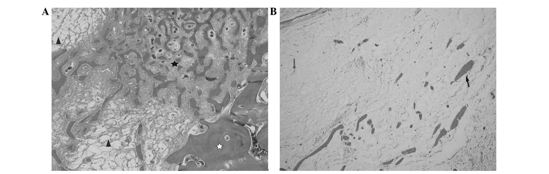
mass of 3.5×2.5×1.0 cm was resected. Histological examination of
the left mandibular lesions showed immature woven bone trabeculae
devoid of osteoblastic lining laid in proliferative fibrous
connective tissue, and the lesions were characterized by some
mucous degeneration and were hypercellular enough to be
characterized as fibrous dysplasia. The histopathological
examination of the buccal mass revealed a tumor extending into the
striated muscle, formed from hypocellular tissue with bland
stellate cells and spindle-shaped cells within a polysaccharide
abundant myxoid matrix. A histopathological diagnosis of
intramuscular myxoma was formed. Immunohistochemical analysis
showed positive staining for vimentin and cluster of
differentiation (CD)99, and negative staining for desmin, smooth
muscle actin, B-cell lymphoma-2, S-100 protein and CD34. The
intramuscular myxoma with adjacent fibrous dysplasia was diagnosed
as MS (Fig. 3). Follow-up
examinations were planned every 3–6 months for 3 years. Three and
six months after surgery, MRI follow-up examinations revealed that
the patient exhibited no clear evidence of recurrence of myxoma and
fibrous dysplasia. However, the patient has subsequently been lost
to follow-up.
This study was approved by the Shanghai Ninth
People's Hospital Institutional Review Board and the patient
provided written informed consent.
Six of these cases were excluded from the study
because we were not get detailed information (3,6,9,15–17), therefore, including the present case,
a total of 87 cases were reviewed. Table
I reveals that the majority of affected patients were female
(61 cases). While only 26 cases were male, indicating an ~2.3-fold
greater incidence in females than in males. The most common onset
of fibrous dysplasia is polyostotic, present in 70 cases (80.46%).
In 17 cases, fibrous dysplasia was monostotic (19.54%). However,
these myxomas were solitary in 40 cases (45.98%). Monostotic
fibrous dysplasia and solitary myxoma, when associated, is a rare
condition. Only a few cases were mentioned in the literature.
Another common finding of MS that was recorded in the literature
was its localization in the lower extremities, which occurred in
82.71% of cases (67 in the lower extremities, 35 in the upper
extremities, 19 in all four extremities and in 11 in the head and
neck; in 6 cases, detailed information was not recorded). These
tumors were bilateral in 30 cases and unilateral in 51 cases. This
finding is in agreement with other studies, which confirm that the
majority patients with MS have multiple intramuscular myxomas and
polyostotic fibrous dysplasia that tend to be located in the lower
extremities, with a particular predilection for the lower right
limb (18). The age of onset of MS in
the 87 patients studied ranged between 17 and 82 years, with an
average age of 46.25 years. MS was most commonly diagnosed in the
middle-aged population.
The case reported in the present study differs from
the majority of the previously reported cases due to its rare
localization in the head and neck, specifically the mandible, in
addition to the unusual association of monostotic fibrous dysplasia
and solitary myxoma. As the mandible is an important aesthetic and
functional organ in the oral and maxillofacial region, an accurate
pre-operative estimation of mandibular invasion remains
challenging.
Another unusual aspect of the present study was the
complex appearance of histopathological findings of the mucous
degeneration of fibrous dysplasia. It is rare to find the mucous
transformation of fibrous dysplasia that was observed in this case
in patients with MS. Although uncommon, malignant transformation of
fibrous dysplasia can also occur in patients with MS (76).
Intramuscular myxomas are rare benign tumors of the
musculoskeletal system that were first recognized as a definite
clinicopathologic entity by Enzinger in 1965. The study presented a
brief compilation of 34 examples of intramuscular myxoma and
demonstrated the benign clinical course of the tumor, as relatively
few of the cases exhibited a tendency to metastasize (77). While intramuscular myxoma is rare,
fibrous dysplasia is not. Fibrous dysplasia is a congenital disease
characterized by a condition affecting one, several or numerous
bones, leading to osteolytic lesions, deformities and fractures
(78). Intramuscular myxoma is
usually a solitary lesion, and exhibits an association with
multiple fibrous dysplasia (19).
Occasionally, MS is associated with McCune-Albright syndrome (MAS)
(20). MAS is a rare syndrome with
the three combinated characteristics of polyostotic fibrous
dysplasia, endocrine dysfunction and café-au-lait spots (79).
The clinical presentation of MS is similar to that
of fibrous dysplasia and intramuscular myxoma. Myxoma may be
diagnosed in adolescents or young adults with symptoms, but fibrous
dysplasia is incidentally found on imaging studies. In a series of
66 patients, Zoccali et al (14) reported that the myxoma develops 12
years prior to the fibrous dysplasia diagnosis. Intramuscular
myxoma is a rare, benign tumor of the musculoskeletal system, with
a low incidence. The tumor may occur in isolation or in association
with fibrous dysplasia or McCune-Albright syndrome (21). Pain and facial palsy are uncommon. The
tumor can occur at any location and tends to involve the large
muscles of the thighs, followed by the buttocks, arms, and chest
wall and shoulders. The majority of intramuscular myxomas are two
or more, painless, palpable masses that are firm, slightly movable
and discrete. Moreover, the myxomas are most typically located in
the are of the bone lesions (21).
Fibrous dysplasia of the bone is a disease that is characterized by
bone deformities, pain and pathological fractures. Patients can,
however, be asymptomatic. Fibrous dysplasia can involve one bone
(monostatic) or several bones (polyostotic) (80). There are few conditions which present
as a solitary intramuscular mass in association with monostotic
fibrous dysplasia. MS occurs at a younger age, may be associated
with additional symptoms (café-au-lait spots and endocrine
dysfunction) (81) and is often
operable. The patient may also experience a long survival time with
the tumor.
Imaging is useful in the verification of the
lesions, particularly for the diagnosis of fibrous dysplasia.
Ultrasound examination is safe for the patient and is necessary for
the diagnosis of intramuscular myxoma. The typical ultrasound
features of MS are considered to be well-defined, hypoechoic masses
that are formed from numerous small, fluid-filled spaces that join
to form a microcystic pattern (22).
CT scans, particularly coronal view scans, are useful in
determining the size and quantity of fibrous dysplasia lesions, as
well as verifying any bony involvement.
The optimal treatment for MS is surgical excision.
Although MS is a benign tumor, it has a tendency to recur, with an
accompanying risk of malignant transformation if incompletely
excised and commonly requiring ‘second-look’ surgery (82). The rate of recurrence depends on the
surgical approach. The surgical treatment of fibrous dysplasia is
mostly indicated where there are progressive deformities, or a risk
of fracture or pathological fracture. When the lesions occur in the
jaw, the main aim of surgery is tumor resection, with good safety
margins, and the reconstruction of the resultant defect, as this is
important for the aesthetics and function of the oral and
maxillofacial region (83). In the
present case, free fibular osteocutaneous flap reconstruction was
performed. For myxoma, a total excision is necessary and curative
(84). When excluding differentials,
such as primary malignancy or metastatic tumors, a full
histopathological investigation should be performed for the
lesions.
The precise etiology of fibrous dysplasia in
association with myxoma remains unclear. The G
protein/cAMP/adenylate cyclase signaling pathway that is central to
the tissues involved in MAS led to the conclusion that regulatory
Gsα protein (encoded by the GNAS gene) mutations were the
underlying molecular etiology of MS (84). This theory is gradually becoming
accepted.
In conclusion, clinicians should be aware of
Mazabraud's syndrome in order to successfully manage patients with
fibrous dysplasia in association with soft tissue myxomas and
prevent misdiagnosis of this benign clinical entity as a malignant
condition. In addition, differential diagnosis must be continued
during clinical treatment when evaluating intramuscular lesions in
the context of associated fibrous dysplasia.
|
1
|
Yaligod V and Ajoy SM: Intramuscular
myxoma - a rare tumor. J Orthop Case Rep. 3:38–41. 2013.
|
|
2
|
Chapurlat RD and Meunier PJ: Fibrous
dysplasia of bone. Best Pract Res Clin Rheumatol. 14:385–398. 2000.
View Article : Google Scholar
|
|
3
|
Segev Z and Reiner S: Intramuscular myxoma
associated with fibrous dysplasia of adjacent bone. Harefuah.
108:493–494. 1985.(In Hebrew). PubMed/NCBI
|
|
4
|
Arishima Y, Setoguchi T, Abematsu M,
Tominaga H, Fukunaga E and Komiya S: Mazabraud's syndrome with
solitary myxoma and monostotic fibrous dysplasia. J Orthop Sci.
15:144–147. 2010. View Article : Google Scholar : PubMed/NCBI
|
|
5
|
Braunwarth K: Simultaneous occurrence of
fibrous dysplasia with extraosseous fibromyxomas. Fortschr Geb
Rontgenstr. 78:589–594. 1953.(In German). View Article : Google Scholar : PubMed/NCBI
|
|
6
|
Berkhoff WB, ter Bruggen Hugenholtz FI and
Ingenhoes R: A patient with multiple intramuscular myxomas as an
extra-osseous manifestation of fibrous dysplasia. Ned Tijdschr
Geneeskd. 125:1460–1462. 1981.(In Dutch). PubMed/NCBI
|
|
7
|
Limouzy F, Durroux R, Chiron P, Tricoire
JL, Puget J and Utheza G: Fibrous dysplasia associated with
intramuscular myxoma. Rev Chir Orthop Reparatrice Appar Mot.
82:336–339. 1996.(In French). PubMed/NCBI
|
|
8
|
Struk DW, Munk PL and Lee MJ:
Musculoskeletal case 8. Mazabraud's syndrome - intramuscular myxoma
associated with fibrous dysplasia. Can J Surg. 43:15, 62–63.
2000.
|
|
9
|
Miyake M, Tateishi U, Maeda T, Arai Y,
Seki K, Hasegawa T and Sugimura K: F-18 fluorodeoxyglucose positron
emission tomography finding of Mazabraud syndrome. Clin Nucl Med.
31:627–629. 2006. View Article : Google Scholar : PubMed/NCBI
|
|
10
|
Zhao H, Zhang H and Jiang Z:
Clinicopathological characteristics of Mazabraud syndrome. Lin
Chuang Yu Bing Li Xue Za Zhi. 24:47–49. 2008.(In Chinese).
|
|
11
|
Logel RJ: Recurrent intramuscular myxoma
associated with Albright's syndrome. J Bone Joint Surg Am.
58:565–568. 1976.PubMed/NCBI
|
|
12
|
Henschen F: Fall von ostitis Fibrosa mit
multiplen Tumoren in der umgebenden Muskulatur. Verh Dtsch Ges
Pathol. 21:93–97. 1926.(In German).
|
|
13
|
Mazabraud A, Semat P and Roze R: Apropos
of the association of fibromyxomas of the soft tissues with fibrous
dysplasia of the bones. Presse Med. 75:2223–2228. 1967.(In French).
PubMed/NCBI
|
|
14
|
Zoccali C, Teori G, Prencipe U and Erba F:
Mazabraud's syndrome: A new case and review of the literature. Int
Orthop. 33:605–610. 2009. View Article : Google Scholar : PubMed/NCBI
|
|
15
|
Beele X, Delanote G, Buyse G and Claikens
B: Mazabraud syndrome. JBR-BTR. 91:84–85. 2008.PubMed/NCBI
|
|
16
|
Tang J, Zhao HY, Zheng L, Zhang HZ and
Jiang ZM: Abnormal expression of c-myc, p53, p16 protein and GNAS1
gene mutation in fibrous dysplasia. Zhonghua Bing Li Xue Za Zhi.
38:292–297. 2009.(In Chinese). PubMed/NCBI
|
|
17
|
Yang XD, Xu G, Lü B, Li K and Xu Y:
Mazabraud syndrome: Report of a case. Zhonghua Bing Li Xue Za Zhi.
40:274–276. 2011.(In Chinese). PubMed/NCBI
|
|
18
|
Munksgaard PS, Salkus G, Iyer VV and
Fisker RV: Mazabraud's syndrome: Case report and literature review.
Acta Radiol Short Rep. 2:20479816134925322013.PubMed/NCBI
|
|
19
|
Kabukcuoglu F, Kabukcuoglu Y, Yilmaz B,
Erdem Y and Evren I: Mazabraud's syndrome: Intramuscular myxoma
associated with fibrous dysplasia. Pathol Oncol Res. 10:121–123.
2004. View Article : Google Scholar : PubMed/NCBI
|
|
20
|
Kitagawa Y, Ishihara Y, Hayashi M, Kim Y,
Fujii N and Ito H: Mazabraud syndrome associated with
McCune-Albright syndrome. J Orthop Sci. 16:129–132. 2011.
View Article : Google Scholar : PubMed/NCBI
|
|
21
|
Lopez-Ben R, Pitt MJ, Jaffe KA and Siegal
GP: Osteosarcoma in a patient with McCune-Albright syndrome and
Mazabraud's syndrome. Skeletal Radiol. 28:522–526. 1999. View Article : Google Scholar : PubMed/NCBI
|
|
22
|
Szendrói M, Rahóty P, Antal I and Kiss J:
Fibrous dysplasia associated with intramuscular myxoma (Mazabraud's
syndrome): A long-term follow-up of three cases. J Cancer Res Clin
Oncol. 124:401–406. 1998. View Article : Google Scholar : PubMed/NCBI
|
|
23
|
Krogius A: A case of osteitis fibrosa with
multiple fibromyxomatoesen muscle tumors. Acta Chir Scan.
64:465–471. 1928.(In German).
|
|
24
|
Uehlinger E: Osteofibrosis deformans
juvenilis. (Polyostotische fibroise dysplasie Jaffé-Lichtenstein.).
Virchows Arch Path Anat. 306:255–299. 1940.(In German). View Article : Google Scholar
|
|
25
|
Mazabraud A and Girard J: A peculiar case
of fibrous dysplasia with osseous and tendinous localizations. Rev
Rhum Mal Osteoartic. 24:652–659. 1957.(In French). PubMed/NCBI
|
|
26
|
Heinemann G and Worth D: Zur Osteofibrosis
deformans juvenilis. Bruns Beitr Klin Chir. 197:327–336. 1958.(In
German). PubMed/NCBI
|
|
27
|
Laporte F, Leger H and Malchair G:
Albright's syndrome. J Chir (Paris). 82:457–475. 1961.(In French).
PubMed/NCBI
|
|
28
|
Lick RF and Viehweger G: A contribution to
the diagnosis of fibrous dysplasia of the skeletal system
(Jaffe-Lichtenstein-Uehlinger). A roentgenological and pathologic
anatomical study. Fortschr Geb Rontgenstr Nuklearmed. 97:33–38.
1962.(In German). View Article : Google Scholar : PubMed/NCBI
|
|
29
|
Roze R, Mazabraud A and Semat P: Fibrous
dysplasia of bone and myxomas of the soft tissues. Localized
sarcomatous degeneration. J Radiol Electrol Med Nucl. 48:527–536.
1967.(In French). PubMed/NCBI
|
|
30
|
Semat P, Roze R and Mazabraud A: Fibrous
dysplasia of bone associated with soft tissue myxma. J Radiol
Electrol Med Nucl. 50:287–292. 1969.(In French). PubMed/NCBI
|
|
31
|
Wirth WA, Leavitt D and Enzinger FM:
Multiple intramuscular myxomas. Another extraskeletal manifestation
of fibrous dysplasia. Cancer. 27:1167–1173. 1971. View Article : Google Scholar : PubMed/NCBI
|
|
32
|
Lejeune E, Bouvier M, Vauzelle JL, Queneau
P, Thomas JD, Chaudy J, Leung TK and Deplante JP: Fibrous dysplasia
and soft tissue myxomas. Rev Rhum Mal Osteoartic. 39:281–288.
1972.(In French). PubMed/NCBI
|
|
33
|
Ireland DC, Soule EH and Ivins JC: Myxoma
of somatic soft tissues. A report of 58 patients, 3 with multiple
tumors and fibrous dysplasia of bone. Mayo Clin Proc. 48:401–410.
1973.PubMed/NCBI
|
|
34
|
Sedmak DD, Hart WR, Belhobek GH and Marks
KE: Massive intramuscular myxoma associated with fibrous dysplasia
of bone. Cleve Clin Q. 50:469–472. 1983. View Article : Google Scholar : PubMed/NCBI
|
|
35
|
Lever EG and Pettingale KW: Albright's
syndrome associated with a soft-tissue myxoma and hypophosphataemic
osteomalacia. Report of a case and review of the literature. J Bone
Joint Surg Br. 65:621–626. 1983.PubMed/NCBI
|
|
36
|
Witkin GB, Guilford WB and Siegal GP:
Osteogenic sarcoma and soft tissue myxoma in a patient with fibrous
dysplasia and hemoglobins JBaltimore and S. Clin Orthop Relat Res.
204:245–252. 1986.PubMed/NCBI
|
|
37
|
Blasier RD, Ryan JR and Schaldenbrand MF:
Multiple myxomata of soft tissue associated with polyostotic
fibrous dysplasia. A case report. Clin Orthop Relat Res.
206:211–214. 1986.PubMed/NCBI
|
|
38
|
Biagini R, Ruggieri P, Boriani S and Picci
P: The Mazabraud syndrome: Case report and review of the
literature. Ital J Orthop Traumatol. 13:105–111. 1987.PubMed/NCBI
|
|
39
|
Glass-Royal MC, Nelson MC, Albert F, Lack
EE and Bogumill GP: Case report 557: Solitary intramuscular myxoma
in a patient with polyostotic fibrous dysplasia. Skeletal Radiol.
18:392–398. 1989. View Article : Google Scholar : PubMed/NCBI
|
|
40
|
Sundaram M, McDonald DJ and Merenda G:
Intramuscular myxoma: A rare but important association with fibrous
dysplasia of bone. AJR Am J Roentgenol. 153:107–108. 1989.
View Article : Google Scholar : PubMed/NCBI
|
|
41
|
Gianoutsos MP, Thompson JF and Marsden FW:
Mazabraud's syndrome: Intramuscular myxoma associated with fibrous
dysplasia of bone. Aust N Z J Surg. 60:825–828. 1990. View Article : Google Scholar : PubMed/NCBI
|
|
42
|
Prayson MA and Leeson MC: Soft-tissue
myxomas and fibrous dysplasia of bone. A case report and review of
the literature. Clin Orthop Relat Res. 291:222–228. 1993.PubMed/NCBI
|
|
43
|
Gober GA and Nicholas RW: Case report 800:
Skeletal fibrous dysplasia associated with intramuscular myxoma
(Mazabraud's syndrome). Skeletal Radiol. 22:452–455. 1993.
View Article : Google Scholar : PubMed/NCBI
|
|
44
|
Aoki T, Kouho H, Hisaoka M, Hashimoto H,
Nakata H and Sakai A: Intramuscular myxoma with fibrous dysplasia:
A report of two cases with a review of the literature. Pathol Int.
45:165–171. 1995. View Article : Google Scholar : PubMed/NCBI
|
|
45
|
Fujii K, Inoue M, Araki Y and Ishida O:
Multiple intramuscular myxomas associated with polyostotic fibrous
dysplasia. Eur J Radiol. 22:152–154. 1996. View Article : Google Scholar : PubMed/NCBI
|
|
46
|
Court-Payen M, Jensen L Ingemann,
Bjerregaard B, Lausten G Schwarz and Skjoldbye B: Intramuscular
myxoma and fibrous dysplasia of bone - Mazabraud's syndrome. A case
report. Acta Radiol. 38:368–371. 1997. View Article : Google Scholar : PubMed/NCBI
|
|
47
|
Cabral CE, Guedes P, Fonseca T, Rezende
JF, Júnior LC Cruz and Smith J: Polyostotic fibrous dysplasia
associated with intramuscular myxomas: Mazabraud's syndrome.
Skeletal Radiol. 27:278–282. 1998. View Article : Google Scholar : PubMed/NCBI
|
|
48
|
Thomachot B, Daumen-Legre V, Pham T,
Acquaviva PC and Lafforgue P: Fibrous dysplasia with intramuscular
myxoma (Mazabraud's syndrome). Report of a case and review of the
literature. Rev Rhum Engl Ed. 66:180–183. 1999.PubMed/NCBI
|
|
49
|
Walker RE, Schwartz RK and Gale DR:
Musculoskeletal case of the day. Mazabraud's syndrome
(intramuscular myxomas associated with fibrous dysplasia of bone).
AJR Am J Roentgenol. 173(797): 800–802. 1999.
|
|
50
|
Kransdorf MJ and Murphey MD: Diagnosis
please. Case 12: Mazabraud syndrome. Radiology. 212:129–132. 1999.
View Article : Google Scholar : PubMed/NCBI
|
|
51
|
Okamoto S, Hisaoka M, Ushijima M, Nakahara
S, Toyoshima S and Hashimoto H: Activating Gs(alpha) mutation in
intramuscular myxomas with and without fibrous dysplasia of bone.
Virchows Arch. 437:133–137. 2000. View Article : Google Scholar : PubMed/NCBI
|
|
52
|
Faivre L, Nivelon-Chevallier A, Kottler
ML, Robinet C, Van Kien P Khau, Lorcerie B, Munnich A, Maroteaux P,
Cormier-Daire V and Le Merrer M: Mazabraud syndrome in two
patients: Clinical overlap with McCune-Albright syndrome. Am J Med
Genet. 99:132–136. 2001. View Article : Google Scholar : PubMed/NCBI
|
|
53
|
Delabrousse E, Couvreur M, Bartholomot B,
Lucas X and Kastler B: Mazabraud syndrome: A case diagnosed with
MRI. J Radiol. 82:165–167. 2001.(In French). PubMed/NCBI
|
|
54
|
Pollandt K, Lohmann CH, Werner M and
Delling G: Clinical pathological aspects of Mazabraud's syndrome.
Pathologe. 23:357–360. 2002.(In German). View Article : Google Scholar : PubMed/NCBI
|
|
55
|
Iwasko N, Steinbach LS, Disler D, Pathria
M, Hottya GA, Kattapuram S, Varma DG and Kumar R: Imaging findings
in Mazabraud's syndrome: Seven new cases. Skeletal Radiol.
31:81–87. 2002. View Article : Google Scholar : PubMed/NCBI
|
|
56
|
Tsitouridis I, Sayegh FE, Natsis K,
Goutsaridou F, Melidis D and Emmanouilidou M: Mazabraud's syndrome:
MRI evaluation. Eur J Radiol Extra. 45:83–87. 2003. View Article : Google Scholar
|
|
57
|
Fang AS, Riley G, Huang SJ and O'Donnell
RJA: A 35-year-old man with bony deformities and a soft tissue
mass. Clin Orthop Relat Res. 417:313–320. 2003.PubMed/NCBI
|
|
58
|
Jhala DN, Eltoum I, Carroll AJ, Lopez-Ben
R, Lopez-Terrada D, Rao PH, Pettenati MJ and Siegal GP:
Osteosarcoma in a patient with McCune-Albright syndrome and
Mazabraud's syndrome: A case report emphasizing the cytological and
cytogenetic findings. Hum Pathol. 34:1354–1357. 2003. View Article : Google Scholar : PubMed/NCBI
|
|
59
|
Nguyen BD and Ram PC: Mazabraud's syndrome
with sarcomatous transformation: Scintigraphic and radiologic
imaging. Clin Nucl Med. 30:829–830. 2005. View Article : Google Scholar : PubMed/NCBI
|
|
60
|
Martin S, Rapariz JM, Osés MJ and Martinez
C: A possible cause of multiple intramuscular masses: Mazabraud's
syndrome (2007: 11b). Eur Radiol. 18:417–421. 2008. View Article : Google Scholar : PubMed/NCBI
|
|
61
|
McLaughlin A, Stalley P, Magee M, Soper J
and Van der Wall H: Correlative imaging in an atypical case of
Mazabraud syndrome. AJR Am J Roentgenol. 189:W353–W356. 2007.
View Article : Google Scholar : PubMed/NCBI
|
|
62
|
Singnurkar A, Phancao JP, Chatha DS and
Stern J: The appearance of Mazabraud's syndrome on 18F-FDG PET/CT.
Skeletal Radiol. 36:1085–1089. 2007. View Article : Google Scholar : PubMed/NCBI
|
|
63
|
Calisir C, Inan U, Yavas US, Isiksoy S and
Kaya T: Mazabraud's syndrome coexisting with a uterine tumor
resembling an ovarian sex cord tumor (UTROSCT): A case report.
Korean J Radiol. 8:438–442. 2007. View Article : Google Scholar : PubMed/NCBI
|
|
64
|
Macfarlane P, Lew W and Neuhaus S: An
aggressive case of Mazabraud's syndrome. Eur J Surg Oncol.
33:1087–1089. 2007. View Article : Google Scholar : PubMed/NCBI
|
|
65
|
Schepers S, Brys P and Samson I: Case
6532. Mazabraud's Syndrome. http://www.eurorad.org/eurorad/case.php?id=6532Accessed.
May 8–2008
|
|
66
|
Santos CT, Choo CT and Loh AH: Orbital
fibrous dysplasia with soft tissue hamartoma - a variant of
Mazabraud's syndrome. Orbit. 27:207–209. 2008. View Article : Google Scholar : PubMed/NCBI
|
|
67
|
Tagliafico A, Succio G, Martinoli C and
Serafini G: Clinical overlap between Mazabraud and McCune-Albright
syndromes. J Ultrasound Med. 28:397–399. 2009.PubMed/NCBI
|
|
68
|
Case DB, Chapman CN Jr, Freeman JK and
Polga JP: Best cases from the AFIP: Atypical presentation of
polyostotic fibrous dysplasia with myxoma (Mazabraud syndrome).
Radiographics. 30:827–832. 2010. View Article : Google Scholar : PubMed/NCBI
|
|
69
|
IJpma FF, Moll FC, Mostert AK, Flach HZ
and de Vries JE: Mazabraud syndrome - benign intramuscular myxoma
with fibrous skeletal dysplasia. Ned Tijdschr Geneeskd.
155:A25132011.(In Dutch). PubMed/NCBI
|
|
70
|
van der Wal WA, Unal H, de Rooy JW, Flucke
U and Veth RP: Fibrous dysplasia of bone associated with
soft-tissue myxomas as well as an intra-osseous myxoma in a woman
with Mazabraud's syndrome: A case report. J Med Case Reports.
5:2392011. View Article : Google Scholar
|
|
71
|
Gaumétou E, Tomeno B and Anract P:
Mazabraud's syndrome. A case with multiple myxomas. Orthop
Traumatol Surg Res. 98:455–460. 2012. View Article : Google Scholar : PubMed/NCBI
|
|
72
|
Schimmöller L, Lehwald N, Antoch G and
Kröpil P: Polyostotic fibro-osseus lesions associated with
intramuscular soft tissue neoplasms. Radiologe. 52:934–936.
2012.(In German). View Article : Google Scholar : PubMed/NCBI
|
|
73
|
John AM, Behera KK, Mathai T, Parmar H and
Paul TV: Mazabraud syndrome. Indian J Endocrinol Metab. 17:740–742.
2013. View Article : Google Scholar : PubMed/NCBI
|
|
74
|
Tsourdi E, Hamann C and Hofbauer LC:
Skeletal and soft tissue involvement in Mazabraud syndrome. J Clin
Endocrinol Metab. 98:E1381–E1382. 2013. View Article : Google Scholar : PubMed/NCBI
|
|
75
|
Endo M, Kawai A, Kobayashi E, et al:
Solitary intramuscular myxoma with monostotic fibrous dysplasia as
a rare variant of Mazabraud's syndrome. Skeletal Radiol.
36:523–529. 2007.Crawford EA, Brooks JS and Ogilvie CM:
Osteosarcoma of the proximal part of the radius in Mazabraud
syndrome. A case report. J Bone Joint Surg Am 91: 955-960, 2009.
View Article : Google Scholar : PubMed/NCBI
|
|
76
|
Enzinger FM: Intramuscular myxoma: A
review and follow-up study of 34 cases. Am J Clin Pathol.
43:104–113. 1965.PubMed/NCBI
|
|
77
|
Lichtenstein L and Jaffé HL: Fibrous
dysplasia of bone. Arch Pathol (Chic). 33:777–816. 1942.
|
|
78
|
Alves C and Silva SF: Partial benefit of
anastrozole in the long-term treatment of precocious puberty in
McCune-Albright syndrome. J Pediatr Endocrinol Metab. 25:323–325.
2012. View Article : Google Scholar : PubMed/NCBI
|
|
79
|
Adetayo OA, Salcedo SE, Borad V, Richards
SS, Workman AD and Ray AO: Fibrous dysplasia: An overview of
disease process, indications for surgical management, and a case
report. Eplasty. 15:e62015.PubMed/NCBI
|
|
80
|
Fornage BD and Romsdahl MM: Intramuscular
myxoma: Sonographic appearance and sonographically guided needle
biopsy. J Ultrasound Med. 13:91–94. 1994.PubMed/NCBI
|
|
81
|
He Y, Zhang ZY, Zhu HG, Sader R, He J and
Kovacs AF: Free fibula osteocutaneous flap for primary
reconstruction of T3-T4 gingival carcinoma. J Craniofac Surg.
21:301–305. 2010. View Article : Google Scholar : PubMed/NCBI
|
|
82
|
Dreizin D, Glen C and Jose J: Mazabraud
syndrome. Am J Orthop (Belle Mead NJ). 41:332–335. 2012.PubMed/NCBI
|
|
83
|
Coloma J, García E and Rodríguez A:
Intramuscular myxoma. Report of three cases. Acta Ortop Mex.
28:244–247. 2014.PubMed/NCBI
|
|
84
|
Walther I, Walther BM, Chen Y and Petersen
I: Analysis of GNAS1 mutations in myxoid soft tissue and bone
tumors. Pathol Res Pract. 210:1–4. 2014. View Article : Google Scholar : PubMed/NCBI
|