Spandidos Publications Logo
  • About
    • About Spandidos
    • Aims and Scopes
    • Abstracting and Indexing
    • Editorial Policies
    • Reprints and Permissions
    • Job Opportunities
    • Terms and Conditions
    • Contact
  • Journals
    • All Journals
    • Oncology Letters
      • Oncology Letters
      • Information for Authors
      • Editorial Policies
      • Editorial Board
      • Aims and Scope
      • Abstracting and Indexing
      • Bibliographic Information
      • Archive
    • International Journal of Oncology
      • International Journal of Oncology
      • Information for Authors
      • Editorial Policies
      • Editorial Board
      • Aims and Scope
      • Abstracting and Indexing
      • Bibliographic Information
      • Archive
    • Molecular and Clinical Oncology
      • Molecular and Clinical Oncology
      • Information for Authors
      • Editorial Policies
      • Editorial Board
      • Aims and Scope
      • Abstracting and Indexing
      • Bibliographic Information
      • Archive
    • Experimental and Therapeutic Medicine
      • Experimental and Therapeutic Medicine
      • Information for Authors
      • Editorial Policies
      • Editorial Board
      • Aims and Scope
      • Abstracting and Indexing
      • Bibliographic Information
      • Archive
    • International Journal of Molecular Medicine
      • International Journal of Molecular Medicine
      • Information for Authors
      • Editorial Policies
      • Editorial Board
      • Aims and Scope
      • Abstracting and Indexing
      • Bibliographic Information
      • Archive
    • Biomedical Reports
      • Biomedical Reports
      • Information for Authors
      • Editorial Policies
      • Editorial Board
      • Aims and Scope
      • Abstracting and Indexing
      • Bibliographic Information
      • Archive
    • Oncology Reports
      • Oncology Reports
      • Information for Authors
      • Editorial Policies
      • Editorial Board
      • Aims and Scope
      • Abstracting and Indexing
      • Bibliographic Information
      • Archive
    • Molecular Medicine Reports
      • Molecular Medicine Reports
      • Information for Authors
      • Editorial Policies
      • Editorial Board
      • Aims and Scope
      • Abstracting and Indexing
      • Bibliographic Information
      • Archive
    • World Academy of Sciences Journal
      • World Academy of Sciences Journal
      • Information for Authors
      • Editorial Policies
      • Editorial Board
      • Aims and Scope
      • Abstracting and Indexing
      • Bibliographic Information
      • Archive
    • International Journal of Functional Nutrition
      • International Journal of Functional Nutrition
      • Information for Authors
      • Editorial Policies
      • Editorial Board
      • Aims and Scope
      • Abstracting and Indexing
      • Bibliographic Information
      • Archive
    • International Journal of Epigenetics
      • International Journal of Epigenetics
      • Information for Authors
      • Editorial Policies
      • Editorial Board
      • Aims and Scope
      • Abstracting and Indexing
      • Bibliographic Information
      • Archive
    • Medicine International
      • Medicine International
      • Information for Authors
      • Editorial Policies
      • Editorial Board
      • Aims and Scope
      • Abstracting and Indexing
      • Bibliographic Information
      • Archive
  • Articles
  • Information
    • Information for Authors
    • Information for Reviewers
    • Information for Librarians
    • Information for Advertisers
    • Conferences
  • Language Editing
Spandidos Publications Logo
  • About
    • About Spandidos
    • Aims and Scopes
    • Abstracting and Indexing
    • Editorial Policies
    • Reprints and Permissions
    • Job Opportunities
    • Terms and Conditions
    • Contact
  • Journals
    • All Journals
    • Biomedical Reports
      • Information for Authors
      • Editorial Policies
      • Editorial Board
      • Aims and Scope
      • Abstracting and Indexing
      • Bibliographic Information
      • Archive
    • Experimental and Therapeutic Medicine
      • Information for Authors
      • Editorial Policies
      • Editorial Board
      • Aims and Scope
      • Abstracting and Indexing
      • Bibliographic Information
      • Archive
    • International Journal of Epigenetics
      • Information for Authors
      • Editorial Policies
      • Editorial Board
      • Aims and Scope
      • Abstracting and Indexing
      • Bibliographic Information
      • Archive
    • International Journal of Functional Nutrition
      • Information for Authors
      • Editorial Policies
      • Editorial Board
      • Aims and Scope
      • Abstracting and Indexing
      • Bibliographic Information
      • Archive
    • International Journal of Molecular Medicine
      • Information for Authors
      • Editorial Policies
      • Editorial Board
      • Aims and Scope
      • Abstracting and Indexing
      • Bibliographic Information
      • Archive
    • International Journal of Oncology
      • Information for Authors
      • Editorial Policies
      • Editorial Board
      • Aims and Scope
      • Abstracting and Indexing
      • Bibliographic Information
      • Archive
    • Medicine International
      • Information for Authors
      • Editorial Policies
      • Editorial Board
      • Aims and Scope
      • Abstracting and Indexing
      • Bibliographic Information
      • Archive
    • Molecular and Clinical Oncology
      • Information for Authors
      • Editorial Policies
      • Editorial Board
      • Aims and Scope
      • Abstracting and Indexing
      • Bibliographic Information
      • Archive
    • Molecular Medicine Reports
      • Information for Authors
      • Editorial Policies
      • Editorial Board
      • Aims and Scope
      • Abstracting and Indexing
      • Bibliographic Information
      • Archive
    • Oncology Letters
      • Information for Authors
      • Editorial Policies
      • Editorial Board
      • Aims and Scope
      • Abstracting and Indexing
      • Bibliographic Information
      • Archive
    • Oncology Reports
      • Information for Authors
      • Editorial Policies
      • Editorial Board
      • Aims and Scope
      • Abstracting and Indexing
      • Bibliographic Information
      • Archive
    • World Academy of Sciences Journal
      • Information for Authors
      • Editorial Policies
      • Editorial Board
      • Aims and Scope
      • Abstracting and Indexing
      • Bibliographic Information
      • Archive
  • Articles
  • Information
    • For Authors
    • For Reviewers
    • For Librarians
    • For Advertisers
    • Conferences
  • Language Editing
Login Register Submit
  • This site uses cookies
  • You can change your cookie settings at any time by following the instructions in our Cookie Policy. To find out more, you may read our Privacy Policy.

    I agree
Search articles by DOI, keyword, author or affiliation
Search
Advanced Search
presentation
Oncology Letters
Join Editorial Board Propose a Special Issue
Print ISSN: 1792-1074 Online ISSN: 1792-1082
Journal Cover
January-2016 Volume 11 Issue 1

Full Size Image

Sign up for eToc alerts
Recommend to Library

Journals

International Journal of Molecular Medicine

International Journal of Molecular Medicine

International Journal of Molecular Medicine is an international journal devoted to molecular mechanisms of human disease.

International Journal of Oncology

International Journal of Oncology

International Journal of Oncology is an international journal devoted to oncology research and cancer treatment.

Molecular Medicine Reports

Molecular Medicine Reports

Covers molecular medicine topics such as pharmacology, pathology, genetics, neuroscience, infectious diseases, molecular cardiology, and molecular surgery.

Oncology Reports

Oncology Reports

Oncology Reports is an international journal devoted to fundamental and applied research in Oncology.

Experimental and Therapeutic Medicine

Experimental and Therapeutic Medicine

Experimental and Therapeutic Medicine is an international journal devoted to laboratory and clinical medicine.

Oncology Letters

Oncology Letters

Oncology Letters is an international journal devoted to Experimental and Clinical Oncology.

Biomedical Reports

Biomedical Reports

Explores a wide range of biological and medical fields, including pharmacology, genetics, microbiology, neuroscience, and molecular cardiology.

Molecular and Clinical Oncology

Molecular and Clinical Oncology

International journal addressing all aspects of oncology research, from tumorigenesis and oncogenes to chemotherapy and metastasis.

World Academy of Sciences Journal

World Academy of Sciences Journal

Multidisciplinary open-access journal spanning biochemistry, genetics, neuroscience, environmental health, and synthetic biology.

International Journal of Functional Nutrition

International Journal of Functional Nutrition

Open-access journal combining biochemistry, pharmacology, immunology, and genetics to advance health through functional nutrition.

International Journal of Epigenetics

International Journal of Epigenetics

Publishes open-access research on using epigenetics to advance understanding and treatment of human disease.

Medicine International

Medicine International

An International Open Access Journal Devoted to General Medicine.

Journal Cover
January-2016 Volume 11 Issue 1

Full Size Image

Sign up for eToc alerts
Recommend to Library

  • Article
  • Citations
    • Cite This Article
    • Download Citation
    • Create Citation Alert
    • Remove Citation Alert
    • Cited By
  • Similar Articles
    • Related Articles (in Spandidos Publications)
    • Similar Articles (Google Scholar)
    • Similar Articles (PubMed)
  • Download PDF
  • Download XML
  • View XML
Case Report

Solitary fibrous tumor of the salivary gland: A case report

  • Authors:
    • Yingwen Zhou
    • Jin Zheng
    • Qingqiang Zhu
    • Wei Xia
    • Santosh Kumar Bhagat
  • View Affiliations / Copyright

    Affiliations: Department of Medical Imaging, Subei People's Hospital, Medical School of Yangzhou University, Yangzhou, Jiangsu 225001, P.R. China
  • Pages: 901-903
    |
    Published online on: November 18, 2015
       https://doi.org/10.3892/ol.2015.3948
  • Expand metrics +
Metrics: Total Views: 0 (Spandidos Publications: | PMC Statistics: )
Metrics: Total PDF Downloads: 0 (Spandidos Publications: | PMC Statistics: )
Cited By (CrossRef): 0 citations Loading Articles...

This article is mentioned in:


Abstract

Solitary fibrous tumors (SFTs) are rare soft‑tissue tumors of mesenchymal origin. Occasionally, these lesions have been indicated to associate with the salivary glands. Through the analysis of magnetic resonance imaging sequences, the present study reports a case of a solitary salivary gland lesion, demonstrating a well‑circumscribed, soft‑tissue tumor with marked signal changes and homogenous enhancement. SFT should be considered as a differential diagnosis when a solid mass exhibiting hypointensity on T1-weighted images and hyperintensity on T2-weighted images has been detected in the salivary gland. Due to the potentially malignant nature of SFTs, it is necessary for radiologists to improve their understanding of such lesions.

Introduction

A solitary fibrous tumor (SFT) is a rare form of tumor that typically develops in the pleura. SFTs that establish in the salivary gland are particularly rare, with imaging studies describing only a few known cases (1,2). Due to the rarity of the tumor, the incidence of such in the salivary gland remains unclear. The association between pathological morphology and clinical symptoms is non-specific. SFTs are unpredictable and the diagnosis remains challenging. Additionally, 10–15% of tumors advance to become malignant or metastasize, and can recur (3). SFTs in the head and neck region are also uncommon, with the characteristics of tumors in this location not clearly understood (4). Magnetic resonance imaging (MRI) features are also relatively non-specific. Wignall et al (5) described that 89% of tumors presented with high signal intensities on T2-weighted images and intermediate signal intensities on T1-weighted images in a review of previous studies. Areas of low signal intensity were observed in larger tumors, resulting from flow voids in prominent perilesional vessels. Collagen fibroblasts and tumor degeneration have been suggested to be accountable for such variable signal intensities (6). Previous studies have reported linear or curvilinear hypointense areas within SFTs on T2-weighted images, and such lesion morphology has been demonstrated to correlate with hypocellular densities near the collagenous sclerotic area (6,7).

Although further investigation is required on the incidence of SFTs in this location, the present study describes a lesion in the right salivary gland and the subsequent MRI findings.

Case report

In October 2013, a 48-year-old woman was referred to the Subei People's Hospital (Yangzhou, China), exhibiting a large, soft salivary mass that had been identified 20 days earlier. The physical examination was unremarkable, with the exception of the palpable mass in the right jaw. The tumor extended from under the jaw to the right side of the bottom of the mouth, and its quality reflected a well-defined, poorly-mobilized lesion with no facial nerve symptoms. The patient maintained a bilateral symmetrical face with normal mouth opening and occlusion.

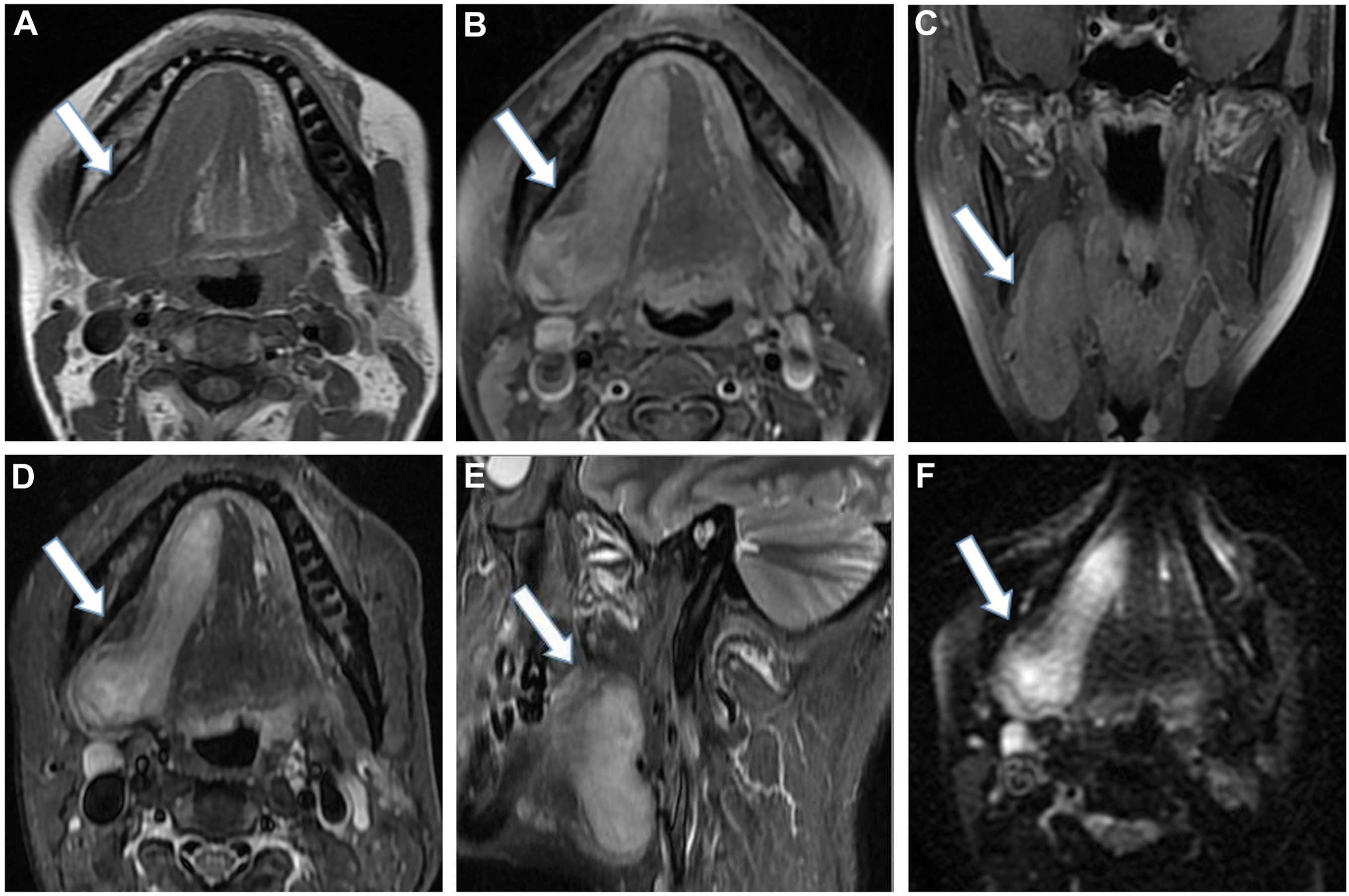
A plain MRI scan confirmed the presence of a well-circumscribed, irregular, soft-tissue mass measuring 71×29 mm, located in the right salivary area, associated with the right salivary gland (Fig. 1). The tumor exhibited marked and homogeneous enhancement following intravenous injection of contrast material. The adjacent bone was not involved and the lymph nodes in the carotid sheath area were observed. A benign tumor was suspected and subsequently removed by local excision. The surgical specimen was examined and an irregular-shaped mass with a complete capsule was detected. 
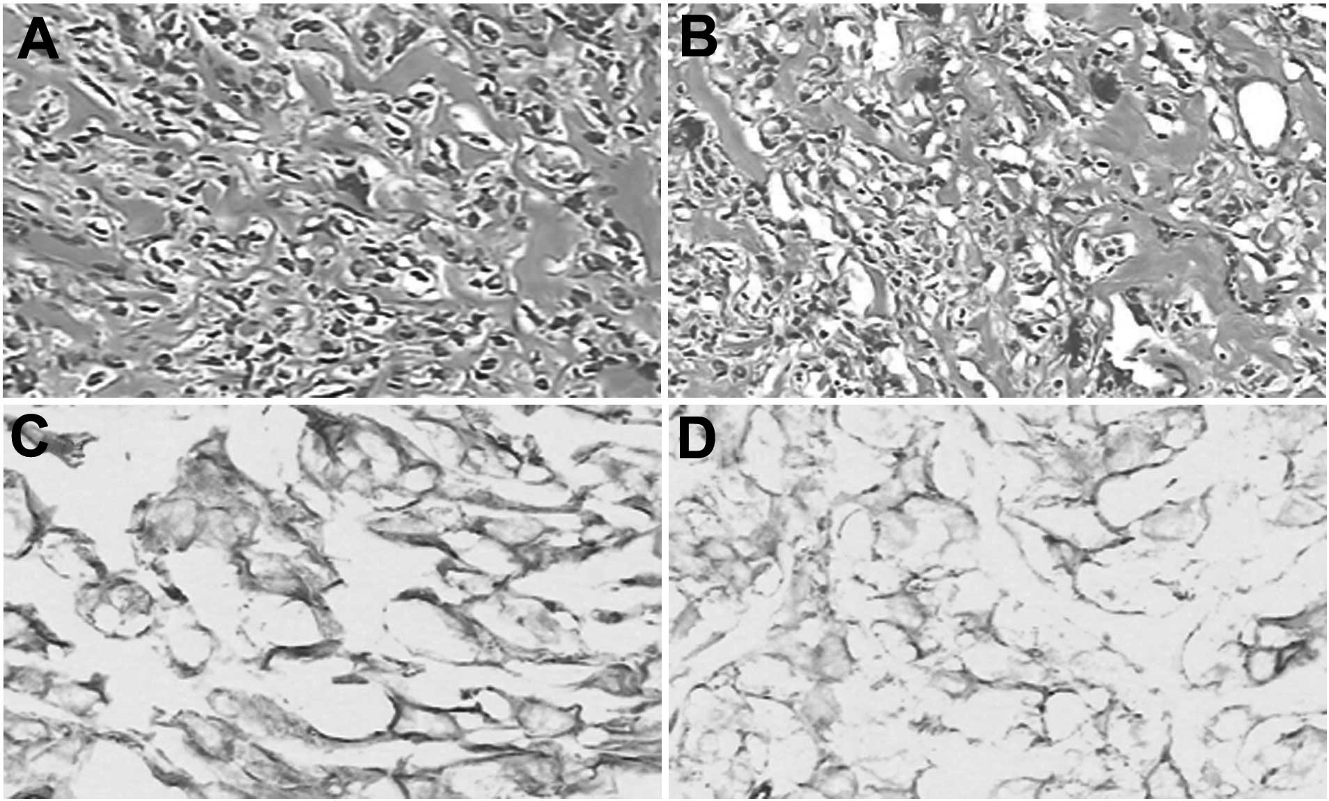
Microscopic examination demonstrated spindle cells and a collagen component in the mesenchyme, with reduced cytoplasm (Fig. 2A and B). Immunohistochemical analysis indicated neoplastic cells that were strongly positive for cluster of differentiation (CD)34 monoclonal antibody (Fig. 2C and D). The cytoplasm (brown) was identified as strongly positive for vimentin monoclonal antibody. Thus, the case was diagnosed as an SFT. Written informed consent was obtained from the patient for publication of this study.

Figure 1.

MRI of the tumor measuring 71×29 mm in the right salivary area. (A) Axial T1-weighted MRI demonstrating a well-defined mass invading the adjacent salivary gland, which is hypointense compared with the adjacent muscle. (B) Contrast-enhanced axial image scan illustrating homogeneous enhancement of the mass (arrow). (C) Contrast-enhanced coronal image scan demonstrating homogeneous enhancement of the mass. (D) Axial fat-suppressed T2-weighted MRI demonstrating a predominantly hyperintense mass. (E) Sagittal fat-suppressed T2-weighted MRI exhibiting isointense to hyperintense signal intensities in the mass. (F) Diffusion-weighted imaging illustrating the hyperintensified lesion. Arrows indicate the tumor mass. MRI, magnetic resonance imaging.

Figure 2.

(A and B) Photomicrograph demonstrating a solitary fibrous tumor composed of spindle cells (hematoxylin and eosin staining). (C and D) Immunohistochemical staining of neoplastic cells positive for cluster of differentiation 34 monoclonal antibody in the membrane and cytoplasm (brown), and cells positive for vimentin monoclonal antibody in the cytoplasm (brown). Magnification, ×400.

Discussion

SFTs primarily occur in adults ranging from 30 to 64 years old (6). SFTs are rare lesions that usually occur in the pleura and were first described by Klemperer and Rabin in 1931 (8). Previous studies have also reported such lesions in the head and neck regions, and ~80 cases have been described regarding SFTs located in the oral cavity (9). The risks of local recurrence and metastasis are associated with tumor size and histological grade of surgical resection margins (3,5,10).

In general, 79–100% positivity for CD34 on immunohistochemistry is considered a highly specific marker of SFTs (11). In addition, malignant SFTs tend to exhibit decreased CD34 immunoreactivity and overexpress p53, S-100, keratin and vimentin (12–14). However, separating SFTs arising in the head and neck region from those arising at other sites by histopathological or immunophenotypical features is not straightforward (15).

In the present case, the salivary gland SFT presented as a well-circumscribed, submucosal mass that was asymptomatic and slow-growing, with normal staining. SFTs can often be confused with other salivary gland conditions, including submandibular gland inflammation and sublingual gland cysts (1).

In the head and neck regions, the radiological findings of SFTs are not specific. However, a number of SFTs are detected incidentally through radiological examinations, or when the symptoms appear associated with a mass in the surrounding tissues (6). The components of the tumor itself may cause the variable intensity observed on MRI images (6). However, radiological differentiation of SFTs from other salivary gland tumors, including pleomorphic adenoma, is difficult. Therefore, in clinical practice, imaging techniques combined with immunohistochemical and pathological results may be advantageous for diagnosis.

SFTs are often observed as a soft-tissue attenuation on imaging, comprised of a well-marginated, lobulated mass when the tumor has prominent collateral feeding vessels or a visible fatty component. Such features alert the radiologist to a possible SFT diagnosis. When the tumor is ≥10 cm in size with central necrosis and effusion, it is likely to be a malignant tumor (5,16,17).

The most important prognostic factor in treating SFTs is treatment with a complete resection (18,19). Due to the high probability of late recurrence, long-term follow-ups are required (18).

In conclusion, the present study findings suggest that an SFT should be considered in the differential diagnosis of a well-marginated lesion with salivary gland association, a soft-tissue component, isointensity to hyperintensity on T2-weighted images and a high signal intensity on diffusion-weighted images, including homogeneous contrast-enhancement.

References

1 

Ferreiro JA and Nascimento AG: Solitary fibrous tumor of the major salivary glands. Histopathology. 28:261–264. 1996. View Article : Google Scholar : PubMed/NCBI

2 

Cho KJ, Ro JY, Choi J, Choi SH, Nam SY and Kim SY: Mesenchymal neoplasms of the major salivary glands: Clinicopathologcal features of 18 cases. Eur Arch Otorhinolaryngol. 265(Suppl 1): S47–S56. 2008. View Article : Google Scholar : PubMed/NCBI

3 

Garcia-Bennett J, Olivé CS, Rivas A, Domínguez-Oronoz R and Huguet P: Soft tissue solitary fibrous tumor. Imaging findings in a series of nine cases. Skeletal Radiol. 41:1427–1433. 2012. View Article : Google Scholar : PubMed/NCBI

4 

Yamada H, Hamada Y, Fujihara H, Fukami K, Mishima K, Nakaoka K, Kumagai K and Imamura E: Solitary fibrous tumor of the buccal space resected in combination with coronoidectomy. Oral Surg Oral Med Oral Pathol Oral Radiol. 114:e9–e14. 2012. View Article : Google Scholar : PubMed/NCBI

5 

Wignall OJ, Moskovic EC, Thway K and Thomas JM: Solitary fibrous tumors of the soft tissues: Review of the imaging and clinical features with histopathologic correlation. AJR Am J Roentgenol. 195:W55–W62. 2010. View Article : Google Scholar : PubMed/NCBI

6 

Kim HJ, Lee HK, Seo JJ, Kim HJ, Shin JH, Jeong AK, Lee JH and Cho KJ: MR imaging of solitary fibrous tumors in the head and neck. Korean J Radiol. 6:136–142. 2005. View Article : Google Scholar : PubMed/NCBI

7 

Jeong AK, Lee HK, Kim SY and Cho KJ: Solitary fibrous tumor of the parapharyngeal space: MR imaging findings. AJNR Am J Neuroradiol. 23:473–475. 2002.PubMed/NCBI

8 

Klemperer P and Rabin CB: Primary neoplasm of the pleura: A report of 5 cases. Arch Pathol (Chic). 1:11–28. 1931.

9 

Amico P, Colella G, Rossiello R, Maria Vecchio G, Leocata P and Magro G: Solitary fibrous tumor of the oral cavity with a predominant leiomyomatous-like pattern: A potential diagnostic pitfall. Pathol Res Pract. 206:499–503. 2010. View Article : Google Scholar : PubMed/NCBI

10 

Daigeler A, Lehnhardt M, Langer S, Steinstraesser L, Steinau HU, Mentzel T and Kuhnen C: Clinicopathological findings in a case series of extrathoracic solitary fibrous tumors of soft tissues. BMC Surg. 6:102006. View Article : Google Scholar : PubMed/NCBI

11 

Ali SZ, Hoon V, Hoda S, Heelan R and Zakowski MF: Solitary fibrous tumor. A cytologic-histologic study with clinical, radiologic, and immunohistochemical correlations. Cancer. 81:116–121. 1997. View Article : Google Scholar : PubMed/NCBI

12 

Civin CI, Strauss LC, Brovall C, Fackler MJ, Schwartz JF and Shaper JH: Antigenic analysis of hematopoiesis. III. A hematopoietic progenitor cell surface antigen defined by a monoclonal antibody raised against KG-1a cells. J Immunol. 133:157–165. 1984.PubMed/NCBI

13 

de Perrot M, Kurt AM, Robert JH, Borisch B and Spiliopoulos A: Clinical behavior of solitary fibrous tumors of the pleura. Ann Thorac Surg. 67:1456–1459. 1999. View Article : Google Scholar : PubMed/NCBI

14 

Yokoi T, Tsuzuki T, Yatabe Y, Suzuki M, Kurumaya H, Koshikawa T, Kuhara H, Kuroda M, Nakamura N, Nakatani Y and Kakudo K: Solitary fibrous tumour: Significance of p53 and CD34 immunoreactivity in its malignant transformation. Histopathology. 32:423–432. 1998. View Article : Google Scholar : PubMed/NCBI

15 

de Oliveira DH, Albuquerque AF, de Araújo Barreto MD, Nonaka CF, da Silva JS, Germano AR and Queiroz LM: Large solitary fibrous tumor of the oral cavity - report of a case. Pathol Res Pract. 210:1064–1067. 2014. View Article : Google Scholar : PubMed/NCBI

16 

Ferretti GR, Chiles C, Choplin RH and Coulomb M: Localized benign fibrous tumors of the pleura. AJR Am J Roentgenol. 169:683–686. 1997. View Article : Google Scholar : PubMed/NCBI

17 

Lee SC, Tzao C, Ou SM, Hsu HH, Yu CP and Cheng YL: Solitary fibrous tumors of the pleura: Clinical, radiological, surgical and pathological evaluation. Eur J Surg Oncol. 31:84–87. 2005. View Article : Google Scholar : PubMed/NCBI

18 

Magdeleinat P, Alifano M, Petino A, Le Rochais JP, Dulmet E, Galateau F, Icard P and Regnard JF: Solitary fibrous tumors of the pleura: Clinical characteristics, surgical treatment and outcome. Eur J Cardiothorac Surg. 21:1087–1093. 2002. View Article : Google Scholar : PubMed/NCBI

19 

Altinok T, Topçu S, Tastepe AI, Yazici U and Cetin G: Localized fibrous tumors of the pleura: Clinical and surgical evaluation. Ann Thorac Surg. 76:892–895. 2003. View Article : Google Scholar : PubMed/NCBI

Related Articles

  • Abstract
  • View
  • Download
  • Twitter
Copy and paste a formatted citation
Spandidos Publications style
Zhou Y, Zheng J, Zhu Q, Xia W and Bhagat SK: Solitary fibrous tumor of the salivary gland: A case report. Oncol Lett 11: 901-903, 2016.
APA
Zhou, Y., Zheng, J., Zhu, Q., Xia, W., & Bhagat, S.K. (2016). Solitary fibrous tumor of the salivary gland: A case report. Oncology Letters, 11, 901-903. https://doi.org/10.3892/ol.2015.3948
MLA
Zhou, Y., Zheng, J., Zhu, Q., Xia, W., Bhagat, S. K."Solitary fibrous tumor of the salivary gland: A case report". Oncology Letters 11.1 (2016): 901-903.
Chicago
Zhou, Y., Zheng, J., Zhu, Q., Xia, W., Bhagat, S. K."Solitary fibrous tumor of the salivary gland: A case report". Oncology Letters 11, no. 1 (2016): 901-903. https://doi.org/10.3892/ol.2015.3948
Copy and paste a formatted citation
x
Spandidos Publications style
Zhou Y, Zheng J, Zhu Q, Xia W and Bhagat SK: Solitary fibrous tumor of the salivary gland: A case report. Oncol Lett 11: 901-903, 2016.
APA
Zhou, Y., Zheng, J., Zhu, Q., Xia, W., & Bhagat, S.K. (2016). Solitary fibrous tumor of the salivary gland: A case report. Oncology Letters, 11, 901-903. https://doi.org/10.3892/ol.2015.3948
MLA
Zhou, Y., Zheng, J., Zhu, Q., Xia, W., Bhagat, S. K."Solitary fibrous tumor of the salivary gland: A case report". Oncology Letters 11.1 (2016): 901-903.
Chicago
Zhou, Y., Zheng, J., Zhu, Q., Xia, W., Bhagat, S. K."Solitary fibrous tumor of the salivary gland: A case report". Oncology Letters 11, no. 1 (2016): 901-903. https://doi.org/10.3892/ol.2015.3948
Follow us
  • Twitter
  • LinkedIn
  • Facebook
About
  • Spandidos Publications
  • Careers
  • Cookie Policy
  • Privacy Policy
How can we help?
  • Help
  • Live Chat
  • Contact
  • Email to our Support Team