Introduction
Hepatoid adenocarcinoma is an extrahepatic tumor
with an incidence of 0.38–0.73% (1).
The occurrence of this malignancy has been described in several
organs, including the lungs, gallbladder, esophagus, uterus and
stomach (2). Hepatoid adenocarcinoma
of stomach (HAS) refers to a rare type of gastric carcinoma
characterized by a distinct morphology and elevated α-fetoprotein
(AFP) levels (3). The diagnosis of
HAS is largely depend on the pathological analysis. In 1981, Kodama
et al (4) initially described
two histologic types of AFP-producing gastric carcinoma with
medullary or papillotubular arrangements. Subsequently, Ishikura
et al (5) proposed the term
‘hepatoid adenocarcinoma of the stomach’ for primary gastric
carcinomas characterized by hepatoid differentiation and the
production of large amounts of AFP. At present, the prognosis of
HAS is rather poor and according to a literature search of
PubMed/Medline between January 2001 and December 2013, only a few
cases of this disease have been reported (Table I) (6–8). The
current study reports a case of HAS, and summarizes the treatment
and outcome for the disease.
 | Table I.Literature review from January 2001 to
December 2013a. |
Table I.
Literature review from January 2001 to
December 2013a.
| Author (reference
no.) | Gender | Age (years) | AFP | Treatment | Follow-up |
|---|
| Gálvez-Muñoz et
al (7) | Male | 75 | Positive | Palliative total
gastrectomy; palliative chemotherapy with cisplatin and
capecitabine (6 cycles) | Alive at 8 months
post surgery |
| Ahn et al
(6) | Male | 68 | Positive | Billroth type II
subtotal gastrectomy; palliative chemotherapy with cisplatin and
capecitabine; second-line palliative chemotherapy with fluororacil,
leucovorin and irinotecan | Alive during 21
months of follow-up |
| Ye et al
(8) | Male | 58 | Positive | Distal gastrectomy;
epirubicin, oxaliplatin and fluorouracil (6 cycles) | Alive at 20 months
post surgery |
|
| Male | 54 | Positive | Total gastrectomy
with lymph node dissection; oxaliplatin plus fluorouracil adjuvant
chemotherapy (6 cycles); paclitaxel and capecitabine (2
cycles) | Deceased at 18 months
post surgery |
|
| Female | 61 | Positive | Chemotherapy with
oxaliplatin plus S-1 | Deceased at 8 months
after treatment |
| Present study | Male | 70 | Positive | Expanded gastrectomy
and radical resection of left lung lobe; chemotherapy using
oxaliplatin and capecitabine | Alive during 7 months
of follow-up |
Case report
A 70-year-old male patient presented to Shaoxing
People's Hospital (Shaoxing, China) due to muscle weakness and
palpitations lasting for 2 months. No abnormalities were noted
during the physical examination. During laboratory tests, fecal
occult blood was noted, and the patient's AFP level was 14,399.9
ng/ml (normal range, 0–13.4 ng/ml). Ultrasound examination
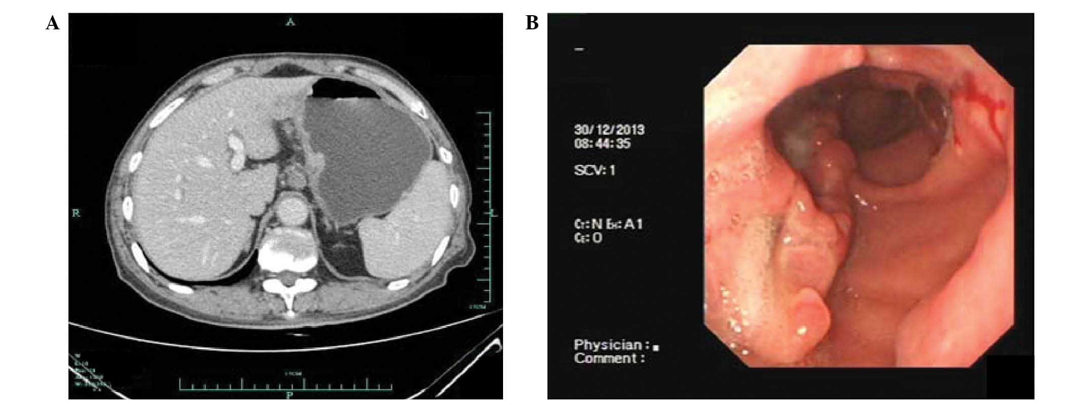
indicated an occupying lesion in right upper quadrant. Computed
tomography imaging was performed, revealing gastric cancer and
infiltration of cancer cells into the left lobe of the liver, as
well as lymphadenectasis of the group 1 and 3 nodes (Fig. 1A). Gastroscopy and pathological tests
revealed irregular bulges in the gastric antrum and oedema in the
peripheral mucous membrane. In addition, highly-differentiated
adenocarcinoma cells were observed. On this basis, the patient was
diagnosed with adenocarcinoma of the gastric antrum (Figs. 1B and 2A). The diagnosis of HAS was primarily based
on the presence of the following aspects: papillary adenocarcinoma
of high, moderate or low grade; mucinous adenocarcinoma;
undifferentiated carcinoma and poorly differentiated hepatocellular
carcinoma combined with presence of hyaline bodies; poorly
differentiated hepatocellular carcinoma and hyaline body-like
structures in the liver cancer specimen. Two cycles of chemotherapy
with oxaliplatin (130 mg; day 1) and capecitabine (2 mg, twice
daily; days 1–14) were administered.
At 7 weeks after chemotherapy, resection of the
stomach and external lobe of the left liver were conducted, during
which a tumor mass measuring ~2.0×1.5×1.0 cm was observed in
gastric corpus. Meanwhile, a gastric biopsy revealed an ulcerating
tumor (4.0×3.0×1.5 cm) from the gastric angle to the gastric
antrum, and infiltration of cancer cells into left liver
(4.0×3.0×3.0 cm). No postoperative complications were reported.
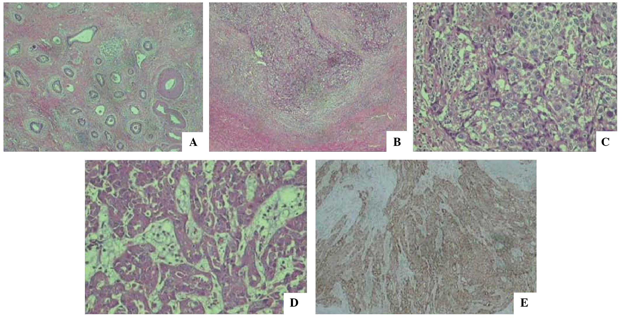
Biopsy of liver indicated adenoid carcinoma with no infiltration of
cancer cells in the incisal margins, and no lymph node metastasis
(Fig. 2B and C). The
immunohistochemistry results were AFP(+), cytokeratin (CK) 19(+),
CK7(−) and CK20(−). Biopsy of gastric samples indicated
adenocarcinoma (Fig. 2D); the
immunohistochemistry results were AFP(−), c-erbB-2(+),
E-cadherin(+), epidermal growth factor receptor(+), Ki-67 (+65%),
CK7(+) and CK20(+). AFP levels were measured on postsurgical days 9
and 30 at 116.7 and 17.3 ng/ml, respectively. Subsequently,
chemotherapy was performed 3 weeks after the after surgery. The
chemptherapy regimen, was 130 mg oxaliplatin on day 1 plus 2 mg
capecitabine, twice daily, on days 1–14, for two cycles. The
patient survived during 7 months of follow-up, at which point the
AFP levels were 10.4 ng/ml.
Discussion
For patients with HAS, the initial symptoms are
usually upper abdominal pain (9);
more rarely, individuals may also exhibit melena. On pathological
evaluation, elevated AFP is commonly noted in these patients. In
such cases, the AFP is typically significantly reduced compared
with the baseline levels following surgery and chemotherapy.
The diagnosis of HAS is largely dependent upon
hematoxylin and eosin (H&E) staining and immunohistochemical
staining. When stained with H&E, HAS typically exhibits similar
features to hepatocellular carcinoma. In addition, proliferation of
polygonal tumor cells is observed in trabecular and intestinal-like
structures. For the differential diagnosis, immunohistochemical
staining is required. In the current case, AFP expression was
confirmed using immunohistochemical staining, as shown in Fig. 2E.
All articles cited in the Medline/PubMed database
between January 2001 and December 2013 were searched using the
terms ‘hepatoid adenocarcinomas of stomach’, ‘AFP-producing tumor’
and ‘AFP-producing gastric cancer’. Simultaneously, a manual search
of all relevant articles was performed. The search was limited to
English language articles. A total of 6 patients (including the
current case) were included in the literature review (Table I) (6–8).
To date, no consensus has been reached regarding the
treatment of HAS, as little data are available in the literature
(10,11). Usually, the disease is treated using
similar strategies as those used to treat gastric adenocarcinoma.
Radical surgery is considered to be the optimal treatment option
and, at the same time, adjuvant chemotherapy and radiotherapy
should be performed according to the indications of the gastric
cancer (12). In the literature
review, 4 patients (66.7%) received gastrectomy and chemotherapy,
while 2 patients (33.3%) received only chemotherapy. For patients
with metastasis, including metastasis to the liver, simultaneous
resections are necessary (2). In the
present case, the patient received oxaliplatin (130 mg; day 1) and
capecitabine (2 mg, twice daily; days 1–14), and radical surgery
was performed to remove the lesions.
The literature review indicates that men are more
prone to developing HAS compared with women. In addition, the mean
age of the patients was 64.3 years, demonstrating that individuals
of advanced age are more prone to developing the disease. The rate
of positive AFP expression was 100.0%. Following surgery, AFP
expression was also used as a marker for the subsequent treatment.
In the present study, AFP expression levels were significantly
reduced on postoperative day 30 compared with that on day 9. In
addition, AFP expression levels were stable during the 7-month
follow-up.
In summary, HAS is a rare type of carcinoma with
poor prognosis. The current study presented a case of HAS occurring
in a 70-year-old male, and summarized the treatment outcomes of the
chemotherapy and/or gastrectomy in previous studies of this
disease. The present results indicated surgery and chemotherapy may
positively affect the outcome of similar patients. As the number of
cases is still limited, further randomized controlled trials are
necessary to confirm the efficacy of this treatment for HAS.
References
|
1
|
Gao YB, Zhang DF, Jin XL and Xiao JC:
Preliminary study on the clinical and pathological relevance of
gastric hepatoid adenocarcinoma. J Dig Dis. 8:23–28. 2007.
View Article : Google Scholar : PubMed/NCBI
|
|
2
|
Baek SK, Han SW, Oh DY, Im SA, Kim TY and
Bang YJ: Clinicopathologic characteristics and treatment outcomes
of hepatoid adenocarcinoma of the stomach, a rare but unique
subtype of gastric cancer. BMC Gastroenterol. 11:562011. View Article : Google Scholar : PubMed/NCBI
|
|
3
|
Gitlin D, Perricelli A and Gitlin GM:
Synthesis of -fetoprotein by liver, yolk sac, and gastrointestinal
tract of the human conceptus. Cancer Res. 32:979–982.
1972.PubMed/NCBI
|
|
4
|
Kodama T, Kameya T, Hirota T, Shimosato Y,
Ohkura H, Mukojima T and Kitaoka H: Production of α-fetoprotein,
normal serum proteins, and human chorionic gonadotropin in stomach
cancer, Histologic and inmunohistochemical analysis of 35 cases.
Cancer. 4:1647–1655. 1981. View Article : Google Scholar
|
|
5
|
Ishikura H, Fukasawa Y, Ogasawara K,
Natori T, Tsukada Y and Aizawa M: An AFP-producing gastric
carcinoma with features of hepatic differentiation, A case report.
Cancer. 56:840–848. 1985. View Article : Google Scholar : PubMed/NCBI
|
|
6
|
Ahn JS, Jeon JR, Yoo HS, Park TK, Park CK,
Sinn DH and Paik SW: Hepatoid adenocarcinoma of the stomach, An
unusual case of elevated alpha-fetoprotein with prior treatment for
hepatocellular carcinoma. Clin Mol Hepatol. 19:173–178. 2013.
View Article : Google Scholar : PubMed/NCBI
|
|
7
|
Gálvez-Muñoz E, Gallego-Plazas J,
Gonzalez-Orozco V, Menarguez-Pina F, Ruiz-Maciá JA and Morcillo MA:
Hepatoid adenocarcinoma of the stomach - a different histology for
not so different gastric adenocarcinoma, A case report. Int Semin
Surg Oncol. 6:132009. View Article : Google Scholar : PubMed/NCBI
|
|
8
|
Ye MF, Tao F, Liu F and Sun AJ: Hepatoid
adenocarcinoma of the stomach, A report of three cases. World J
Gastroenterol. 19:4437–4442. 2013. View Article : Google Scholar : PubMed/NCBI
|
|
9
|
Liu X, Cheng Y, Sheng W, Lu H, Xu X, Xu Y,
Long Z, Zhu H and Wang Y: Analysis of clinicopathologic features
and prognostic factors in hepatoid adenocarcinoma of the stomach.
Am J Surg Pathol. 34:1465–1471. 2010. View Article : Google Scholar : PubMed/NCBI
|
|
10
|
Inagawa S, Shimazaki J, Hori M, Yoshimi F,
Adachi S, Kawamoto T, Fukao K and Itabashi M: Hepatoid
adenocarcinoma of the stomach. Gastric Cancer. 4:43–52. 2001.
View Article : Google Scholar : PubMed/NCBI
|
|
11
|
Trompetas V, Varsamidakis N, Frangia K,
Polimeropoulos V and Kalokairinos E: Gastric hepatoid
adenocarcinoma and familial investigation, Does it always produce
alpha-fetoprotein? Eur J Gastroenterol Hepatol. 15:1241–1244. 2003.
View Article : Google Scholar : PubMed/NCBI
|
|
12
|
Xie Y, Zhao Z, Li P, Wang Y, Guo C, Wang
X, Tang W, Liu Q, Lu N, Xue L and Zhao D: Hepatoid adenocarcinoma
of the stomach is a special and easily misdiagnosed or missed
diagnosed subtype of gastric cancer with poor prognosis but
curative for patients of pN0/1: The experience of a single center.
Int J Clin Exp Med. 8:6762–6772. 2015.PubMed/NCBI
|