Introduction
One of the key characteristics of cancer cells is
the upregulated telomerase activity. This enzyme is active in
~80–90% of all cases of cancer (1,2). A lack of
telomerase activity in the majority of normal cells (or a
significantly lower activity level) is associated with the end
replication problem, resulting from the inability of DNA polymerase
to complete replication of the terminal end of the chromosome.
Thus, chromosomes in normal cells lose 50–100 base pairs at the 5′
end during each cell division (3).
This mechanism functions as a biological clock, eliminating cells
that have lived for too long or long enough to accumulate
aberrations or mutations (4,5). The unlimited number of cell divisions
may lead to the loss of genetic information; therefore, telomere
shortening is a signal for the induction of cell death by apoptosis
or p53-dependent aging (6).
Consequently, it prevents the transformation of cells. In several
types of eukaryotic cells, including cancer cells, telomeres are
rebuilt using telomerase (6,7).
Telomerase regulation and its association with
carcinogenesis is a complex process. However, it is suspected that
changes in the human body, even on a local scale (initiation of
neoplasia in a single cell), may be reflected at the molecular
level in the whole organism (for example, leukocytes), and vice
versa (8,9). Thus, changes in telomerase activity
and/or consequent telomere length alterations in peripheral blood
cells, including leukocytes and migrating cancer cells, are
perceived as potentially promising prediction markers of the
carcinogenic process (10). It is
understood that cancer cells first go through numerous divisions,
resulting in significant telomere shortening and telomerase
activity subsequently being restored (11). Consequently, cancer cells typically
have short telomeres. It is believed that the shortening of
telomeres in cancer cells is also associated with characteristics
and prognosis of cancer. Recently, telomere length has been
reported to be associated with tumor size, lymph node metastasis,
histological grade and specific breast tumor subtypes (12,13).
Shorter telomeres in tumor tissue have been associated with more
aggressive subtypes, including luminal B, human epidermal growth
factor 2 (HER2+) and triple-negative tumors (14). Although it is not yet fully
understood, these processes may also be reflected in telomere
length alteration in peripheral blood leukocytes (15). Therefore, the present study
investigated whether telomere length in peripheral blood leukocytes
serves as a useful parameter in breast cancer diagnostics.
Materials and methods
Patients and controls
The study group consisted of 52 women (mean age,
58±12 years; BMI, 25.9±3.53) with histologically diagnosed breast
cancer at various stages, based on the World Health Organization
criteria. The histopathological classification of breast carcinoma
is based on the diversity of the morphological features of the
tumor. It includes 20 major tumor types and 18 minor subtypes. A
major drawback of this classification is that 70%–80% of the all
breast cancer cases will eventually belong to one of the two major
histopathological classes, namely invasive ductal carcinomas not
otherwise specified or invasive lobular carcinoma (10). All of the patients were recruited from
the Department of Oncological Surgery, Poznan University of Medical
Sciences (Poznan, Poland). The control group comprised of 47
healthy volunteers and blood donors (all female; mean age, 55±16
years; BMI, 25.1±4.54). The patients and controls were Caucasians
from the same region of Poland (Poznan). The study protocol was
approved by the Ethics Committee of Poznan University of Medical
Sciences (146/11) and written consent was provided by the
participating individuals. Tumors were characterized according to
histological grade, tumor size, metastasis, lymph node and receptor
status [including the estrogen receptor (ER), progesterone receptor
(PgR) and HER2] (Table I).
 | Table I.Study group characteristics. |
Table I.
Study group characteristics.
|
| Group data |
|---|
|
|
|
|---|
| Feature | n | % |
|---|
| Total patients
studied | 52 | 100.0 |
| Tumor size |
|
|
| T1 | 31 | 59.6 |
| T2 | 17 | 32.7 |
| T3 | 4 | 7.7 |
| Metastasis |
|
|
| M0 | 43 | 82.7 |
| M1 | 1 | 1.9 |
| Mx | 8 | 15.4 |
| Lymph nodes |
|
|
| N0 | 13 | 25.0 |
| N1 | 30 | 57.7 |
| N2 | 0 |
0.0 |
| N3 | 1 |
1.9 |
| Nx | 8 | 15.4 |
| Histological
grade |
|
|
| G1 | 13 | 25.0 |
| G2 | 23 | 44.2 |
| G3 | 13 | 25.0 |
| Gx | 3 |
5.8 |
| ER status |
|
|
|
ER+ | 45 | 86.5 |
|
ER− | 7 | 13.5 |
| PgR status |
|
|
|
PgR+ | 39 | 75.0 |
|
PgR− | 13 | 25.0 |
| HER2 status |
|
|
|
HER2+ | 26 | 50.0 |
|
HER2− | 26 | 50.0 |
DNA isolation
DNA was extracted from peripheral blood leukocytes
as previously described (16), using
a Blood Mini DNA Isolation kit (A&A Biotechnology, Gdynia,
Poland) and was stored at −20°C. A high concentration sample of
genomic DNA was prepared in decimal concentrations that were used
to run as a standard curve.
Quantitative polymerase chain reaction
(qPCR)
Telomere length was assessed using two pairs of
primers, consisting of telomere-specific and single copy
gene-specific (albumin) primers (17,18).
Primers were selected (Table II)
that had previously been demonstrated to work specifically
(17), with conditions providing
efficiency close to 100% (2.5 mM MgCl2; 0.5 µM primers).
Albumin primers were designed using Universal ProbeLibrary (Roche
Diagnostics, Indianapolis, IN, USA). Initial denaturation and
polymerase activation (hot start) was performed at 95°C for 10 min,
followed by two cycles of 94°C for 15 sec and 49°C for 15 sec
without fluorescence acquisition. The signal was detected during a
further 40 cycles (94°C for 10 sec, 66°C for 10 sec and 72°C for 10
sec). Melting analysis (range, 65–95°C; resolution, 0.2°C) was
performed subsequent to qPCR to verify the specificity of the
product, and indicated that the melting temperature (Tm) was
81.7°C. The efficiency of the reaction was no lower than 97.8%.
Notably, this result was repeated for all the samples that were
analyzed (in serial dilutions). Similarly, the reaction conditions
for albumin were as follows: Denaturation at 95°C for 10 min (hot
start); followed by 45 cycles at 94°C for 10 sec, 61°C for 10 sec
and 72°C for 10 sec. The Tm of the products (analysis performed as
aforementioned) was 80.7°C and the efficiency was 99.6%. The
primers and MgCl2 were utilized at 0.5 µM and 2.5 mM,
respectively. Telomere length was assessed using the LightCycler®
2.0 Instrument and the LightCycler® FastStart DNA Master SYBR Green
I kit (Roche Diagnostics).
| Table II.Sequence of primers used in
quantitative polymerase chain reaction assays to assess relative
telomere length. |
Table II.
Sequence of primers used in
quantitative polymerase chain reaction assays to assess relative
telomere length.
| Primer | Sequence | Reference no. |
|---|
| Telg |
ACACTAAGGTTTGGGTTTGGGTTTGGGTTTGGGTTAGTGT | (12) |
| Telc |
TGTTAGGTATCCCTATCCCTATCCCTATCCCTATCCCTAACA | (12) |
| ALBF |
TTTGCAGATGTCAGTGAAAGAGA |
|
| ALBR |
TGGGGAGGCTATAGAAAATAAGG |
|
Southern blot analysis
Telomere length assessment obtained using qPCR was
verified by Southern blot (19), with
certain modifications as previously described (20). Briefly, 1.5 µg DNA obtained from two
samples was digested with the restriction enzymes HinfI and
RsaI (Roche Diagnostics); following this, the DNA fragments
were separated by gel electrophoresis (Sigma-Aldrich, St. Louis,
MO, USA), which was run at 5 V/cm in 1X
Tris-acetate-ethylenediaminetetraacetic acid buffer (TeloTAGGG
Telomere Length assay kit; Roche Diagnostics), and transferred to a
nylon membrane (Roche Diagnostics), according to the manufacturer's
protocols. Subsequently, a digoxigenin (DIG)-labeled probe
(TeloTAGGG Telomere Length assay kit; Roche Diagnostics) specific
to the telomeric sequence was applied and detected using anti-DIG
antibody conjugated to horseradish peroxidase (TeloTAGGG Telomere
Length assay kit; Roche Diagnostics). Chemiluminesce visualization
of telomeric DNA was conducted using the ChemiDoc XRS+ system
(BioRad Laboratories, Inc., Hercules, CA, USA) and was analyzed
using ImageJ (www.imagej.nih.gov/ij/), which evaluated the mean
length of the telomeres for each studied sample (terTRF).
Statistical analysis
Statistical analysis was performed using the
Mann-Whitney U test and Student's t-test calculated via Graph Pad
Prism 5 (GraphPad Software, Inc., La Jolla, CA, USA). P<0.05 was
considered to indicate a statistically significant difference.
Results
Telomere length
The average telomere lengths in the study and
control groups were analyzed and no significant statistical
difference was observed (P=0.9358; Fig.
1A). Additionally, the association between the telomere lengths
in leukocytes and the tumor stage [according to the tumor node
metastasis (TNM) classification] was investigated (10). As presented in Fig. 1B, it was demonstrated that the average
length of the telomeric sequences was significantly shorter
(P=0.0207) in leukocytes derived from individuals with breast
cancer of a higher stage (T2N1M0) than in leukocytes from patients
in the early stages of the disease (T1N0M0). The correlation of ER,
PgR and HER status with telomere length was also assessed. As
demonstrated in samples derived from HER2+ patients,
telomeres were significantly longer compared with samples from
HER2− patients (Fig. 1C;
P=0.0347). Telomere length was not significantly different between
patients that were positive and patients that were negative for ER
or PgR (P=0.1312 and P=0.1145, respectively; Fig. 1D and E). Also, no association was
identified between the histological cancer grade and alterations of
telomere length in the peripheral blood cells (G1 vs. G2, P=0.7381;
G1 vs. G3, P=0.3232; G2 vs. G3, P=0.1878; Fig. 1F).
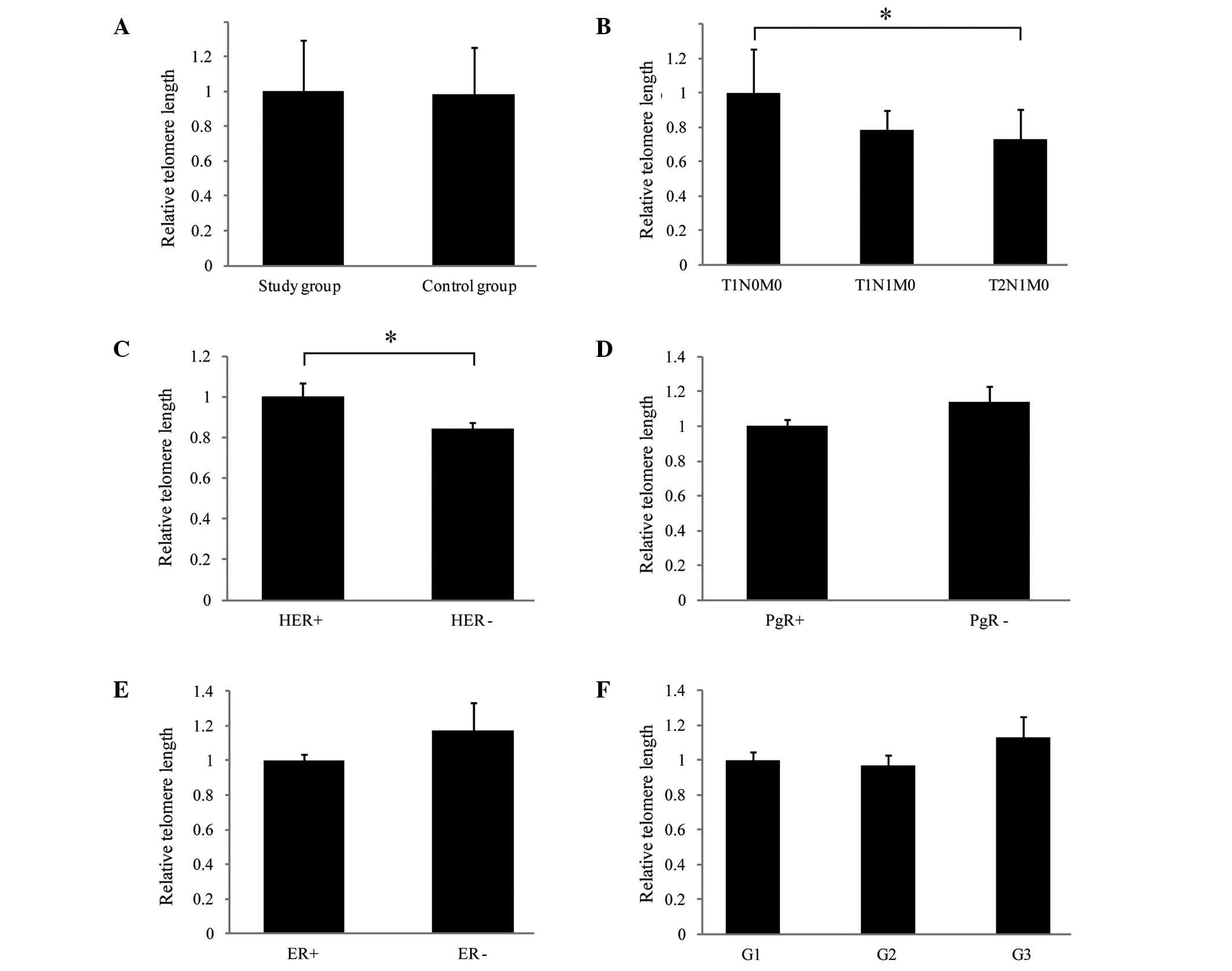 | Figure 1.Quantitative analysis of telomere
length in peripheral leukocytes from patients with breast cancer
(study group) and control subjects, and the correlation with
clinical parameters. Relative telomere length was assessed
according to protocol previously described by Cawthon (with
modifications) (12) using
quantitative polymerase chain reaction. Telomere length was
presented relative to a single copy gene albumin. Relative length
of telomeres, comparing the (A) study and control groups; (B) tumor
stage (according to the TNM classification as follows: T1N0M0,
small primary tumor and no lymph node involvement or distant
metastases; T1N1M0, small primary tumor, sentinel node metastasis
with no distant metastases; T2N1M0, larger primary tumor, sentinel
node metastasis with no distant metastases); (C) HER status; (D)
PgR status; (E) ER status; and (F) tumor grade. *P<0.05,
comparison shown by brackets. T, tumor; N, node; M, metastasis;
HER2, human epidermal growth factor receptor 2; PgR, progesterone
receptor; ER, estrogen receptor; G1/2/3, grades 1/2/3. |
Discussion
In terms of telomerase and telomeres as suitable
markers to aid cancer therapy and diagnostics, further research is
required. Telomerase activity is recognized as a unique feature of
cancer cells; however, the specificity of this marker has been
questioned, as the activity of this enzyme was previously observed
in stem cells, and in certain somatic cells (21–24).
Despite this, telomerase activity continues to be recognized as a
potential therapeutic target for cancer, primarily due to the
likely low number of side effects observed to be associated with
such treatment. It is established, however, that the success of any
therapy is predominantly reliant on an early and accurate
diagnosis. Telomerase activity and telomere length assessment
remain to be the most promising factors in early diagnostics. The
detection of increased telomerase activity and shortening of
telomeres are hypothesized to serve as specific signals of
carcinogenesis. According to a number of studies (25–27), the
detection of telomerase activity or telomere length alterations may
be observed either in migrating cancer cells or in peripheral blood
leukocytes. Such results may provide an opportunity to distinguish
or qualify patients with differential cancer stages or grades.
In terms of telomere length as a diagnostic factor,
certain studies have demonstrated that changes in telomere length
are linked to the risk of cancer (28–31).
Identification of telomere length is considered to be useful as a
prognostic factor, particularly when considering assays based on
telomere length analysis in blood cells. Such tests should be
efficient and non-invasive.
In the present study, no significant difference in
average telomere length between the control subjects and patients
with breast cancer was observed. However, significant differences
were identified between the telomere length in cells derived from
patients with different TNM stages of breast cancer and varying HER
statuses. A number of studies observed no association between
telomere length and breast cancer risk (32), whilst studies of small telomere length
variation (TLV; defined as the coefficient variation of telomere
lengths among examined cells) in normal epithelial cells adjacent
to the tumor demonstrated that TLV was significantly associated
with a 5-fold (95% confidence interval = 1.2–22.2) increased risk
of breast cancer local recurrence (33). Meta-analyses conducted on bladder,
esophageal, gastric, head and neck, ovarian, renal and overall
cancer identified significant associations between short telomeres
and the aforementioned types of cancer (assessed in surrogate and
cancer tissues) (34).
The results of the present study are noteworthy,
particularly since HER2 overexpression is considered to be an
important biological marker of poor prognosis and increased disease
aggressiveness, in addition to being a useful indicator of response
to anti-HER2 therapy in breast cancer (35). Previous studies have demonstrated that
HER2 amplification is associated with increased telomerase reverse
transcriptase (hTERT) expression and telomerase activity in cancer
cells (36). Similarly, Vageli et
al (37) indicated that the
mechanism of hTERT transcriptional activation may involve HER2-ER81
interactions; and possible connections between pathways that
regulate the expression of HER2 and hTERT provide an explanation
for telomere lengthening as a result of increased telomerase
activity. By contrast, Sugishita et al (38) demonstrated that the telomeres in cases
of HER2+ thyroid cancer are shorter than those of
HER2− cases, and suggested that short telomeres in
cancer tissues may serve as a further negative prognostic
indicator. However, Shen et al (39) observed increased telomere length among
cases of HER2+ breast cancer, and a significant
association between longer telomeres and all-cause mortality among
HER2− cases. Such results suggest that simultaneous
monitoring of HER2 status and telomere length may provide important
information regarding breast cancer development, malignancy and
prognosis.
In conclusion, the analysis of telomere length by
qPCR may serve as a diagnostic tool reflecting changes in telomere
length in leukocytes. This may subsequently result from the genome
instability, driving the mechanisms that eventually lead to
carcinogenesis. However, it should be noted that, despite the
evidence that malignant transformation contributes to telomere
length alterations, there are additional factors influencing this
parameter, including varying age, gender, xenobiotics/hormones
exposition, genetic profile and telomere length at birth. As
telomere length in breast cancer patients demonstrates a level of
prognostic value, future studies should be conducted in larger
groups of patients. If this trend is confirmed, measurement of
telomere length may enable a quick and noninvasive assessment of
potential risk associated with advanced cancer.
Acknowledgements
The current study was supported by The National
Science Centre (grant no. 2011/03/B/NZ7/00512) and the Poznan
University of Medical Sciences (grant no.
502-05-03318432-50736).
References
|
1
|
Chen CH and Chen RJ: Prevalence of
telomerase activity in human cancer. J Formos Med Assoc.
110:275–289. 2011. View Article : Google Scholar : PubMed/NCBI
|
|
2
|
Shtessel L and Ahmed S: Telomere
dysfunction in human bone marrow failure syndromes. Nucleus.
2:24–29. 2011. View Article : Google Scholar : PubMed/NCBI
|
|
3
|
Harley CB, Futcher AB and Greider CW:
Telomeres shorten during ageing of human fibroblasts. Nature.
345:458–460. 1990. View
Article : Google Scholar : PubMed/NCBI
|
|
4
|
Oeseburg H, de Boer RA, van Gilst WH and
van der Harst P: Telomere biology in healthy aging and disease.
Pflugers Arch. 459:259–268. 2010. View Article : Google Scholar : PubMed/NCBI
|
|
5
|
Watson JM and Riha K: Telomeres, aging,
and plants: From weeds to Methuselah - a mini-review. Gerontology.
57:129–136. 2011. View Article : Google Scholar : PubMed/NCBI
|
|
6
|
Chan SR and Blackburn EH: Telomeres and
telomerase. Philos Trans R Soc Lond B Biol Sci. 359:109–121. 2004.
View Article : Google Scholar : PubMed/NCBI
|
|
7
|
de Lange T: How telomeres solve the
end-protection problem. Science. 326:948–952. 2009. View Article : Google Scholar : PubMed/NCBI
|
|
8
|
Barczak W and Rubiś B: Telomere length as
a prognostic marker in breast and lung cancer. Nowotwory.
62:376–384. 2012.
|
|
9
|
Yao YG, Ogasawara Y, Kajigaya S, Molldrem
JJ, Falcão RP, Pintão MC, McCoy JP Jr, Rizzatti EG and Young NS:
Mitochondrial DNA sequence variation in single cells from leukemia
patients. Blood. 109:756–762. 2007. View Article : Google Scholar : PubMed/NCBI
|
|
10
|
Lakhani SR, Ellis IO, Schnitt SJ, Tan PH
and van de Vijver MJ: World Health Organization Classification of
Tumours of the Breast. World Health Organization Classification of
Tumours. 4:(Fourth). (Lyon). IARC Press. 2012.
|
|
11
|
Hackett JA and Greider CW: Balancing
instability: Dual roles for telomerase and telomere dysfunction in
tumorigenesis. Oncogene. 21:619–626. 2002. View Article : Google Scholar : PubMed/NCBI
|
|
12
|
Gramades MM, Telli ML, Balise R and Ford
JM: Longer relative telomere length in blood from women with
sporadic and familiar breast cancer compared with healthy controls.
Cancer Epidemiol Biomarkers Prev. 19:605–613. 2010. View Article : Google Scholar : PubMed/NCBI
|
|
13
|
Martinez-Delgado B, Yanowsky K,
Inglada-Perez L, Domingo S, Urioste M, Osorio A and Benitez J:
Genetic anticipation is associated with telomere shortening in
hereditary breast cancer. PLoS Genet. 7:e10021822011. View Article : Google Scholar : PubMed/NCBI
|
|
14
|
Heaphy CM, Subhawong AP, Gross AL, Konishi
Y, Kouprina N, Argani P, Visvanathan K and Meeker AK: Shorter
telomeres in luminal B, HER-2 and triple-negative breast cancer
subtypes. Mod Pathol. 24:194–200. 2011. View Article : Google Scholar : PubMed/NCBI
|
|
15
|
Shao L, Wood CG, Zhang D, Tannir NM, Matin
S, Dinney CP and Wu X: Telomere dysfunction in peripheral
lymphocytes as a potential predisposition factor for renal cancer.
J Urol. 178:1492–1496. 2007. View Article : Google Scholar : PubMed/NCBI
|
|
16
|
Rubiś B, Hołysz H, Barczak W, Gryczka R,
Łaciński M, Jagielski P, Czernikiewicz A, Półrolniczak A, Wojewoda
A, Perz K, et al: Study of ABCB1 polymorphism frequency in breast
cancer patients from Poland. Pharmacol Rep. 64:1560–1566. 2012.
View Article : Google Scholar : PubMed/NCBI
|
|
17
|
Cawthon RM: Telomere length measurement by
a novel monochrome multiplex quantitative PCR method. Nucleic Acids
Res. 37:e212009. View Article : Google Scholar : PubMed/NCBI
|
|
18
|
O'Callaghan NJ and Fenech M: A
quantitative PCR method for measuring absolute telomere length.
Biol Proced Online. 13:32011. View Article : Google Scholar : PubMed/NCBI
|
|
19
|
Kimura M, Stone RC, Hunt SC, Skurnick J,
Lu X, Cao X, Harley CB and Aviv A: Measurement of telomere length
by the Southern blot analysis of terminal restriction fragment
lengths. Nat Protoc. 5:1596–1607. 2010. View Article : Google Scholar : PubMed/NCBI
|
|
20
|
Gruszecka A, Kopczyński P, Cudziło D,
Lipińska N, Romaniuk A, Barczak W, Rozwadowska N, Totoń E and Rubiś
B: Telomere shortening in Down syndrome patients - when does it
start? DNA Cell Biol. 34:412–417. 2015. View Article : Google Scholar : PubMed/NCBI
|
|
21
|
Härle-Bachor C and Boukamp P: Telomerase
activity in the regenerative basal layer of the epidermis inhuman
skin and in immortal and carcinoma-derived skin keratinocytes. Proc
Natl Acad Sci USA. 93:6476–6481. 1996. View Article : Google Scholar : PubMed/NCBI
|
|
22
|
Hiyama E, Tatsumoto N, Kodama T, Hiyama K,
Shay J and Yokoyama T: Telomerase activity in human intestine. Int
J Oncol. 9:453–458. 1996.PubMed/NCBI
|
|
23
|
Yasumoto S, Kunimura C, Kikuchi K, Tahara
H, Ohji H, Yamamoto H, Ide T and Utakoji T: Telomerase activity in
normal human epithelial cells. Oncogene. 13:433–439.
1996.PubMed/NCBI
|
|
24
|
Yui J, Chiu CP and Lansdorp PM: Telomerase
activity in candidate stem cells from fetal liver and adult bone
marrow. Blood. 91:3255–3262. 1998.PubMed/NCBI
|
|
25
|
Huang YK, Fan XG, Qiu F and Wang ZM:
Combined detection of mRNA expression of Alpha-fetoprotein in
peripheral blood and telomerase activity of monocytes in
hepatocellular carcinoma patients. Hepatogastroenterology. 60:1–5.
2013.PubMed/NCBI
|
|
26
|
Lu L, Zhang C, Zhu G, Irwin M, Risch H,
Menato G, Mitidieri M, Katsaros D and Yu H: Telomerase expression
and telomere length in breast cancer and their associations with
adjuvant treatment and disease outcome. Breast Cancer Res.
13:R562011. View
Article : Google Scholar : PubMed/NCBI
|
|
27
|
Merchant NB, Dutta SK, Girotra M, Arora M
and Meltzer SJ: Evidence for enhanced telomerase activity in
Barrett's esophagus with dysplasia and adenocarcinoma. Asian Pac J
Cancer Prev. 14:679–683. 2013. View Article : Google Scholar : PubMed/NCBI
|
|
28
|
Bau DT, Lippman SM, Xu E, Gong Y, Lee JJ,
Wu X and Gu J: Short telomere lengths in peripheral blood
leukocytes are associated with an increased risk of oral
premalignant lesion and oral squamous cell carcinoma. Cancer.
119:4277–4283. 2013. View Article : Google Scholar : PubMed/NCBI
|
|
29
|
Lan Q, Cawthon R, Gao Y, Hu W, Hosgood HD
III, Barone-Adesi F, Ji BT, Bassig B, Chow WH, Shu X, et al: Longer
telomere length in peripheral white blood cells is associated with
risk of lung cancer and the rs2736100 (CLPTM1L-TERT) polymorphism
in a prospective cohort study among women in China. PLoS One.
8:e592302013. View Article : Google Scholar : PubMed/NCBI
|
|
30
|
Qin Q, Sun J, Yin J, Liu L, Chen J, Zhang
Y, Li T, Shi Y, Wei S and Nie S: Telomere length in peripheral
blood leukocytes is associated with risk of colorectal cancer in
Chinese population. PLoS One. 9:e881352014. View Article : Google Scholar : PubMed/NCBI
|
|
31
|
Wang S, Chen Y, Qu F, He S, Huang X, Jiang
H, Jin T, Wan S and Xing J: Association between leukocyte telomere
length and glioma risk: A case-control study. Neuro Oncol.
16:505–512. 2014. View Article : Google Scholar : PubMed/NCBI
|
|
32
|
Kim S, Sandler DP, Carswell G, De Roo LA,
Parks CG, Cawthon R, Weinberg CR and Taylor JA: Telomere length in
peripheral blood and breast cancer risk in a prospective
case-cohort analysis: Results from the Sister Study. Cancer Causes
Control. 22:1061–1066. 2011. View Article : Google Scholar : PubMed/NCBI
|
|
33
|
Zhou X, Meeker AK, Makambi KH, Kosti O,
Kallakury BV, Sidawy MK, Loffredo CA and Zheng YL: Telomere length
variation in normal epithelial cells adjacent to tumor: Potential
biomarker for breast cancer local recurrence. Carcinogenesis.
33:113–118. 2012. View Article : Google Scholar : PubMed/NCBI
|
|
34
|
Wentzensen IM, Mirabello L, Pfeiffer RM
and Savage SA: The association of telomere length and cancer: A
meta-analysis. Cancer Epidemiol Biomarkers Prev. 20:1238–1250.
2011. View Article : Google Scholar : PubMed/NCBI
|
|
35
|
Martin V, Cappuzzo F, Mazzucchelli L and
Frattini M: HER2 in solid tumors: More than 10 years under the
microscope; where are we now? Future Oncol. 10:1469–1486. 2014.
View Article : Google Scholar : PubMed/NCBI
|
|
36
|
Papanikolaou V, Iliopoulos D, Dimou I,
Dubos S, Tsougos I, Theodorou K, Kitsiou-Tzeli S and Tsezou A: The
involvement of HER2 and p53 status in the regulation of telomerase
in irradiated breast cancer cells. Int J Oncol. 35:1141–1149.
2009.PubMed/NCBI
|
|
37
|
Vageli D, Ioannou MG and Koukoulis GK:
Transcriptional activation of hTERT in breast carcinomas by the
Her2-ER81-related pathway. Oncol Res. 17:413–423. 2009. View Article : Google Scholar : PubMed/NCBI
|
|
38
|
Sugishita Y, Kammori M, Yamada O, Poon SS,
Kobayashi M, Onoda N, Yamazaki K, Fukumori T, Yoshikawa K, Onose H,
et al: Amplification of the human epidermal growth factor receptor
2 gene in differentiated thyroid cancer correlates with telomere
shortening. Int J Oncol. 42:1589–1596. 2013.PubMed/NCBI
|
|
39
|
Shen J, Terry MB, Liao Y, Gurvich I, Wang
Q, Senie RT and Santella RM: Genetic variation in telomere
maintenance genes, telomere length and breast cancer risk. PLoS
One. 7:e443082012. View Article : Google Scholar : PubMed/NCBI
|


