18F‑fluoroethyl‑L‑tyrosine positron emission tomography for the differential diagnosis of tumefactive multiple sclerosis versus glioma: A case report

  • Authors:
    • Sied Kebir
    • Florian C. Gaertner
    • Marcus Mueller
    • Michael Nelles
    • Matthias Simon
    • Niklas Schäfer
    • Moritz Stuplich
    • Christina Schaub
    • Michael Niessen
    • Frederic Mack
    • Ralph Bundschuh
    • Susanne Greschus
    • Markus Essler
    • Martin Glas
    • Ulrich Herrlinger
  • View Affiliations

  • Published online on: February 4, 2016     https://doi.org/10.3892/ol.2016.4189
  • Pages: 2195-2198
Metrics: Total Views: 0 (Spandidos Publications: | PMC Statistics: )
Total PDF Downloads: 0 (Spandidos Publications: | PMC Statistics: )


Abstract

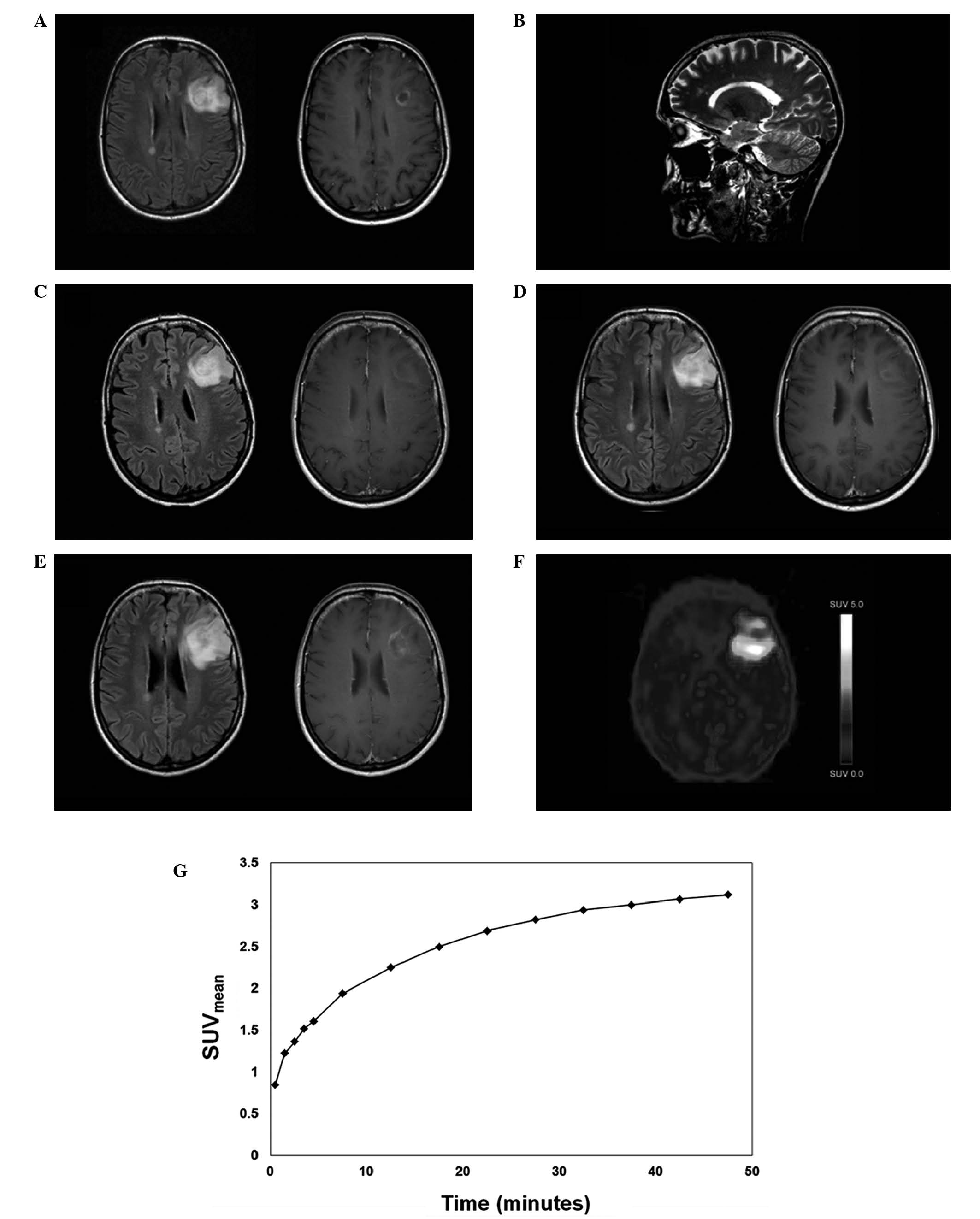
Large demyelinating inflammatory central nervous system (CNS) lesions may present with contrast enhancement on magnetic resonance imaging and may mimic CNS tumors such as glioma. In ambiguous cases, new diagnostic tools that may be helpful for distinguishing between demyelinating inflammatory and neoplastic CNS lesions are required. The current study presents the case of a patient with a large contrast‑enhanced frontal brain lesion, who was initially diagnosed with tumefactive multiple sclerosis. Following the progression of the brain lesion, an 18F‑fluoroethyl‑L‑tyrosine positron emission tomography (18F‑FET PET) was performed, revealing markedly elevated static 18F‑FET uptake parameters along with time activity‑curves consistent with glioma. Subsequently, a biopsy was undertaken, which confirmed the presence of anaplastic oligoastrocytoma. This case illustrates that 18F‑FET PET may provide useful diagnostic information in cases where distinction between neoplastic and demyelinating inflammatory CNS lesions is challenging. However, further systematic and prospective analyses are warranted to explore the value of this method in this setting.
View Figures
View References

Related Articles

Journal Cover

March-2016
Volume 11 Issue 3

Print ISSN: 1792-1074
Online ISSN:1792-1082

Sign up for eToc alerts

Recommend to Library

Copy and paste a formatted citation
x
Spandidos Publications style
Kebir S, Gaertner FC, Mueller M, Nelles M, Simon M, Schäfer N, Stuplich M, Schaub C, Niessen M, Mack F, Mack F, et al: 18F‑fluoroethyl‑L‑tyrosine positron emission tomography for the differential diagnosis of tumefactive multiple sclerosis versus glioma: A case report. Oncol Lett 11: 2195-2198, 2016
APA
Kebir, S., Gaertner, F.C., Mueller, M., Nelles, M., Simon, M., Schäfer, N. ... Herrlinger, U. (2016). 18F‑fluoroethyl‑L‑tyrosine positron emission tomography for the differential diagnosis of tumefactive multiple sclerosis versus glioma: A case report. Oncology Letters, 11, 2195-2198. https://doi.org/10.3892/ol.2016.4189
MLA
Kebir, S., Gaertner, F. C., Mueller, M., Nelles, M., Simon, M., Schäfer, N., Stuplich, M., Schaub, C., Niessen, M., Mack, F., Bundschuh, R., Greschus, S., Essler, M., Glas, M., Herrlinger, U."18F‑fluoroethyl‑L‑tyrosine positron emission tomography for the differential diagnosis of tumefactive multiple sclerosis versus glioma: A case report". Oncology Letters 11.3 (2016): 2195-2198.
Chicago
Kebir, S., Gaertner, F. C., Mueller, M., Nelles, M., Simon, M., Schäfer, N., Stuplich, M., Schaub, C., Niessen, M., Mack, F., Bundschuh, R., Greschus, S., Essler, M., Glas, M., Herrlinger, U."18F‑fluoroethyl‑L‑tyrosine positron emission tomography for the differential diagnosis of tumefactive multiple sclerosis versus glioma: A case report". Oncology Letters 11, no. 3 (2016): 2195-2198. https://doi.org/10.3892/ol.2016.4189