Spandidos Publications Logo
  • About
    • About Spandidos
    • Aims and Scopes
    • Abstracting and Indexing
    • Editorial Policies
    • Reprints and Permissions
    • Job Opportunities
    • Terms and Conditions
    • Contact
  • Journals
    • All Journals
    • Oncology Letters
      • Oncology Letters
      • Information for Authors
      • Editorial Policies
      • Editorial Board
      • Aims and Scope
      • Abstracting and Indexing
      • Bibliographic Information
      • Archive
    • International Journal of Oncology
      • International Journal of Oncology
      • Information for Authors
      • Editorial Policies
      • Editorial Board
      • Aims and Scope
      • Abstracting and Indexing
      • Bibliographic Information
      • Archive
    • Molecular and Clinical Oncology
      • Molecular and Clinical Oncology
      • Information for Authors
      • Editorial Policies
      • Editorial Board
      • Aims and Scope
      • Abstracting and Indexing
      • Bibliographic Information
      • Archive
    • Experimental and Therapeutic Medicine
      • Experimental and Therapeutic Medicine
      • Information for Authors
      • Editorial Policies
      • Editorial Board
      • Aims and Scope
      • Abstracting and Indexing
      • Bibliographic Information
      • Archive
    • International Journal of Molecular Medicine
      • International Journal of Molecular Medicine
      • Information for Authors
      • Editorial Policies
      • Editorial Board
      • Aims and Scope
      • Abstracting and Indexing
      • Bibliographic Information
      • Archive
    • Biomedical Reports
      • Biomedical Reports
      • Information for Authors
      • Editorial Policies
      • Editorial Board
      • Aims and Scope
      • Abstracting and Indexing
      • Bibliographic Information
      • Archive
    • Oncology Reports
      • Oncology Reports
      • Information for Authors
      • Editorial Policies
      • Editorial Board
      • Aims and Scope
      • Abstracting and Indexing
      • Bibliographic Information
      • Archive
    • Molecular Medicine Reports
      • Molecular Medicine Reports
      • Information for Authors
      • Editorial Policies
      • Editorial Board
      • Aims and Scope
      • Abstracting and Indexing
      • Bibliographic Information
      • Archive
    • World Academy of Sciences Journal
      • World Academy of Sciences Journal
      • Information for Authors
      • Editorial Policies
      • Editorial Board
      • Aims and Scope
      • Abstracting and Indexing
      • Bibliographic Information
      • Archive
    • International Journal of Functional Nutrition
      • International Journal of Functional Nutrition
      • Information for Authors
      • Editorial Policies
      • Editorial Board
      • Aims and Scope
      • Abstracting and Indexing
      • Bibliographic Information
      • Archive
    • International Journal of Epigenetics
      • International Journal of Epigenetics
      • Information for Authors
      • Editorial Policies
      • Editorial Board
      • Aims and Scope
      • Abstracting and Indexing
      • Bibliographic Information
      • Archive
    • Medicine International
      • Medicine International
      • Information for Authors
      • Editorial Policies
      • Editorial Board
      • Aims and Scope
      • Abstracting and Indexing
      • Bibliographic Information
      • Archive
  • Articles
  • Information
    • Information for Authors
    • Information for Reviewers
    • Information for Librarians
    • Information for Advertisers
    • Conferences
  • Language Editing
Spandidos Publications Logo
  • About
    • About Spandidos
    • Aims and Scopes
    • Abstracting and Indexing
    • Editorial Policies
    • Reprints and Permissions
    • Job Opportunities
    • Terms and Conditions
    • Contact
  • Journals
    • All Journals
    • Biomedical Reports
      • Information for Authors
      • Editorial Policies
      • Editorial Board
      • Aims and Scope
      • Abstracting and Indexing
      • Bibliographic Information
      • Archive
    • Experimental and Therapeutic Medicine
      • Information for Authors
      • Editorial Policies
      • Editorial Board
      • Aims and Scope
      • Abstracting and Indexing
      • Bibliographic Information
      • Archive
    • International Journal of Epigenetics
      • Information for Authors
      • Editorial Policies
      • Editorial Board
      • Aims and Scope
      • Abstracting and Indexing
      • Bibliographic Information
      • Archive
    • International Journal of Functional Nutrition
      • Information for Authors
      • Editorial Policies
      • Editorial Board
      • Aims and Scope
      • Abstracting and Indexing
      • Bibliographic Information
      • Archive
    • International Journal of Molecular Medicine
      • Information for Authors
      • Editorial Policies
      • Editorial Board
      • Aims and Scope
      • Abstracting and Indexing
      • Bibliographic Information
      • Archive
    • International Journal of Oncology
      • Information for Authors
      • Editorial Policies
      • Editorial Board
      • Aims and Scope
      • Abstracting and Indexing
      • Bibliographic Information
      • Archive
    • Medicine International
      • Information for Authors
      • Editorial Policies
      • Editorial Board
      • Aims and Scope
      • Abstracting and Indexing
      • Bibliographic Information
      • Archive
    • Molecular and Clinical Oncology
      • Information for Authors
      • Editorial Policies
      • Editorial Board
      • Aims and Scope
      • Abstracting and Indexing
      • Bibliographic Information
      • Archive
    • Molecular Medicine Reports
      • Information for Authors
      • Editorial Policies
      • Editorial Board
      • Aims and Scope
      • Abstracting and Indexing
      • Bibliographic Information
      • Archive
    • Oncology Letters
      • Information for Authors
      • Editorial Policies
      • Editorial Board
      • Aims and Scope
      • Abstracting and Indexing
      • Bibliographic Information
      • Archive
    • Oncology Reports
      • Information for Authors
      • Editorial Policies
      • Editorial Board
      • Aims and Scope
      • Abstracting and Indexing
      • Bibliographic Information
      • Archive
    • World Academy of Sciences Journal
      • Information for Authors
      • Editorial Policies
      • Editorial Board
      • Aims and Scope
      • Abstracting and Indexing
      • Bibliographic Information
      • Archive
  • Articles
  • Information
    • For Authors
    • For Reviewers
    • For Librarians
    • For Advertisers
    • Conferences
  • Language Editing
Login Register Submit
  • This site uses cookies
  • You can change your cookie settings at any time by following the instructions in our Cookie Policy. To find out more, you may read our Privacy Policy.

    I agree
Search articles by DOI, keyword, author or affiliation
Search
Advanced Search
presentation
Oncology Letters
Join Editorial Board Propose a Special Issue
Print ISSN: 1792-1074 Online ISSN: 1792-1082
Journal Cover
March-2016 Volume 11 Issue 3

Full Size Image

Sign up for eToc alerts
Recommend to Library

Journals

International Journal of Molecular Medicine

International Journal of Molecular Medicine

International Journal of Molecular Medicine is an international journal devoted to molecular mechanisms of human disease.

International Journal of Oncology

International Journal of Oncology

International Journal of Oncology is an international journal devoted to oncology research and cancer treatment.

Molecular Medicine Reports

Molecular Medicine Reports

Covers molecular medicine topics such as pharmacology, pathology, genetics, neuroscience, infectious diseases, molecular cardiology, and molecular surgery.

Oncology Reports

Oncology Reports

Oncology Reports is an international journal devoted to fundamental and applied research in Oncology.

Experimental and Therapeutic Medicine

Experimental and Therapeutic Medicine

Experimental and Therapeutic Medicine is an international journal devoted to laboratory and clinical medicine.

Oncology Letters

Oncology Letters

Oncology Letters is an international journal devoted to Experimental and Clinical Oncology.

Biomedical Reports

Biomedical Reports

Explores a wide range of biological and medical fields, including pharmacology, genetics, microbiology, neuroscience, and molecular cardiology.

Molecular and Clinical Oncology

Molecular and Clinical Oncology

International journal addressing all aspects of oncology research, from tumorigenesis and oncogenes to chemotherapy and metastasis.

World Academy of Sciences Journal

World Academy of Sciences Journal

Multidisciplinary open-access journal spanning biochemistry, genetics, neuroscience, environmental health, and synthetic biology.

International Journal of Functional Nutrition

International Journal of Functional Nutrition

Open-access journal combining biochemistry, pharmacology, immunology, and genetics to advance health through functional nutrition.

International Journal of Epigenetics

International Journal of Epigenetics

Publishes open-access research on using epigenetics to advance understanding and treatment of human disease.

Medicine International

Medicine International

An International Open Access Journal Devoted to General Medicine.

Journal Cover
March-2016 Volume 11 Issue 3

Full Size Image

Sign up for eToc alerts
Recommend to Library

  • Article
  • Citations
    • Cite This Article
    • Download Citation
    • Create Citation Alert
    • Remove Citation Alert
    • Cited By
  • Similar Articles
    • Related Articles (in Spandidos Publications)
    • Similar Articles (Google Scholar)
    • Similar Articles (PubMed)
  • Download PDF
  • Download XML
  • View XML
Case Report

Primary kidney parenchyma squamous cell carcinoma mimicking xanthogranulomatous pyelonephritis: A case report

  • Authors:
    • Zhao Wang
    • Bin Yan
    • Yong‑Bao Wei
    • Na Hu
    • Qin Shen
    • Duo Li
    • Jin‑Rui Yang
    • Xin Yang
  • View Affiliations / Copyright

    Affiliations: Department of Urology, The Second Xiangya Hospital, Central South University, Changsha, Hunan 410011, P.R. China, Department of PET/CT Center, The Second Xiangya Hospital, Central South University, Changsha, Hunan 410011, P.R. China, Department of Radiology, The Second Xiangya Hospital, Central South University, Changsha, Hunan 410011, P.R. China, Department of Pathology, The Second Xiangya Hospital, Central South University, Changsha, Hunan 410011, P.R. China, Department of Oncology, The Second Xiangya Hospital, Central South University, Changsha, Hunan 410011, P.R. China
  • Pages: 2179-2181
    |
    Published online on: February 8, 2016
       https://doi.org/10.3892/ol.2016.4200
  • Expand metrics +
Metrics: Total Views: 0 (Spandidos Publications: | PMC Statistics: )
Metrics: Total PDF Downloads: 0 (Spandidos Publications: | PMC Statistics: )
Cited By (CrossRef): 0 citations Loading Articles...

This article is mentioned in:


Abstract

Primary kidney parenchyma squamous cell carcinoma is extremely rare, and this is the forth case to be reported. In the present study, a case of a 61‑year old man is discussed. The man presented with recurrent lumbago, gross hematuria for nearly 2 months, and suspicious inflammatory kidney diseases on contrast‑enhanced computed tomography (CT) and fludeoxyglucose‑positron emission tomography (FDG‑PET)/CT, but a tumor can not be excluded completely prior to surgery. Finally, radical nephrectomy was performed, and histological analysis determined that the diagnosis was kidney parenchyma squamous cell carcinoma with inflammation invasion. The present case highlights the potential confusion of preoperative diagnosis of renal tumor with inflammation, and introduces the potential role of FDG‑PET in its diagnosis and survival evaluation in renal malignancies.

Introduction

Kidney squamous cell carcinomas are rare occurrences in renal malignancies, particularly for kidney parenchyma squamous cell carcinoma (1–5). To the best of our knowledge, only three cases of patients with kidney parenchyma cell carcinoma have been reported since December 2014 (2–5). The three cases consisted of a 73-year old man (2), a 51-year old man (5) and a 60-year old woman (4). All the patients underwent nephrectomies. The 73-year old man was followed up 3 months after surgery, the 51-year old man was followed up 6 and 12 months after surgery and the 60-year old woman was followed up 13 months after surgery; all patients remained alive with no evidence of disease at the time of follow-up (2–5). The present study reports a case of kidney parenchyma squamous cell carcinoma with inflammation invasion, which mimicked xanthogranulomatous pyelonephritis on radiological examination; the literature concerning kidney squamous cell carcinomas is also reviewed.

Case report

A 61-year old man presented to the Second Xiangya Hospital (Changsha, China) in October 2014 with right lumbago and gross hematuria that had been present for nearly 2 months. The patient had right renal parenchyma lithotomy ~14 years ago. Physical examination demonstrated positive percussion pain on the right kidney region. Palpation on the kidneys did not reveal any pathological symptoms.

On admission, routine urine tests demonstrated that the urine was clearly positive for white blood cells (WBCs) (WBC count, 1,187.50/µl; normal range, 0–25/µl). C-Reactive protein measured 72.20 mg/l (normal, <8 mg/l), erythrocyte sedimentation rate was 81 mm/h (normal, <15 mm/h), procalcitonin was 3.23 ng/ml (normal, <0.05 ng/ml). Renal and liver function tests results were within the normal ranges. Urine cytology examination demonstrated massive epithelial cells and neutrophils without 
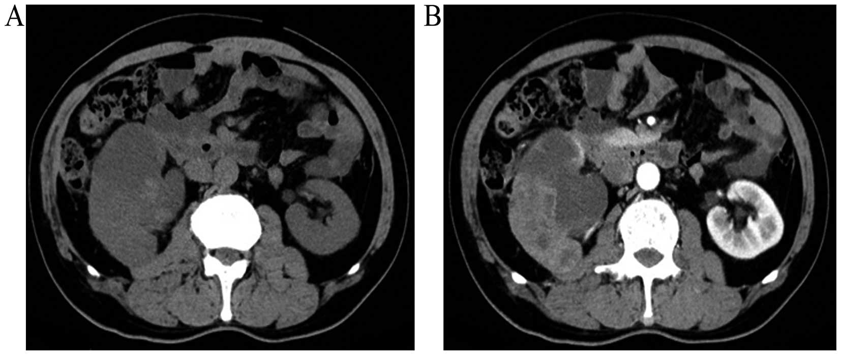
malignancies. Contrast-enhanced computed tomography (CT) was performed using a SOMATOM Sensation CT scanner (Siemens, Munich, Germany). It demonstrated enlargement of the right kidney with abnormal morphology and dilatation of the ureter, multiloculated cyst-like masses with soft tissues, and perirenal fatty space was fuzzy (Fig. 1A and B). The lesions observed on the CT scan were atypical and were considered as xanthogranulomatous pyelonephritis or tuberculosis; however, the presence of a rare renal tumor could not be completely excluded. Tuberculosis tests [purified protein derivative (PPD)-immunoglobulin (Ig)M, PPD-IgG and MycoDot] were performed and were negative.

Figure 1.

CT images of the patient. (A) Normal and (B) enhanced CT scans demonstrated the enlargement of the right kidney with abnormal morphology, multiple cyst-like masses with soft tissues and the perirenal fatty space was observed to be fuzzy. CT, computed tomography.

In total, 10 days following admission, the patient had a fever (highest temperature, 39.6°C) and chills, and blood rountine tests demonstrated a white blood cell count of 16.94×10⁹ cells/l (normal range, 3.50–9.50×10⁹ cells/l) and a neutrophil percentage of 92.34% (normal range, 40.00–75.00%). Therefore, physicians hypothesized that the patient had septicemia. Antibiotics were prescribed (latamoxef for 2 week, 1 g twice a day, intravenous combined with moxifloxacin for 5 days, 0.4 g once a day, intravenous), which were adjusted (imipenem cilastatin sodium, 0.5 g every 6 h, intravenous) according to a routine blood test (procalcitonin and C-reactive protein) and a routine urine examination, including color, glucose and protein concentration, pH and white and red blood cell count, urine culture and advice from clinical pharmacists. After septicemia was controlled in the 
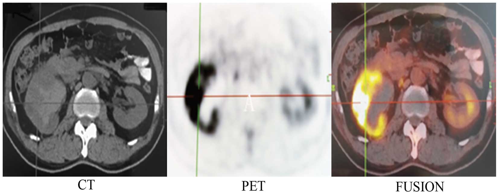
patient, cystoscopy was performed. Fludeoxyglucose-positron emission tomography (FDG-PET; Biography mCTx; Siemens) was also performed following the administration of 9.43 mCi FDG. Cystoscopy did not reveal the presence of carcinoma in the bladder and urethra. FDG-PET showed hydronephrosis with a fuzzy perirenal fatty space, and valid FDG was taken up by cystic-solid mixed masses in the right kidney (Fig. 2). The lesions were considered to be renal malignancies, but renal inflammation diseases could not be excluded completely.

Figure 2.

FDG-PET images. The images demonstrate hydronephrosis with fuzzy perirenal fatty space, and FDG was taken up by cystic-solid mixed masses in the right kidney. CT, computed tomography; PET, positron emission tomography; FDG, fludeoxyglucose.

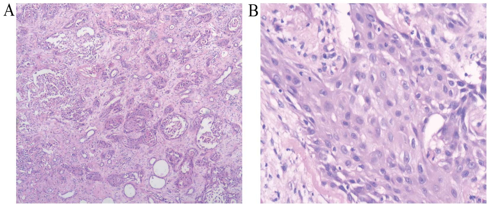
Right radical nephrectomy was performed. Histological examination using hematoxylin and eosin staining (Sinopharm Chemical Reagent Co., Ltd., Shanghai China) on paraffin-embedded tissues under a microscope (BX51TF; Olympus Corporation, Tokyo, Japan) demonstrated moderate-differentiated squamous cell carcinoma of the renal parenchyma with a massive invasion of inflammatory cells (Fig. 3A and B). The patient was discharged 10 days following surgery. Follow-up one month after surgery in December 2014 showed that the patient had no febrile, gross hematuria or abnormal abdominal signs. Written informed consent was obtained from the patient for the publication of the present study.

Figure 3.

Histological examination. Hematoxlyin and eosin staining at (A) ×40 and (B) ×200 magnification. A massive invasion of inflammatory cells was observed, which is a hallmark of squamous cell carcinoma of the renal parenchyma.

Discussion

Kidney squamous cell carcinomas are rare renal malignancies, and are classified as renal parenchyma and pelvic squamous cell carcinomas according to where they arise. Primary kidney pelvic squamous cell carcinomas account for 0.5–0.8% in kidney malignancies (1). Renal parenchyma squamous cell carcinomas are extremely rare; to the best of our knowledge only 3 cases have been reported until now (2–5).

Urinary calculi and chronic inflammation are some of the important factors associated with renal squamous cell carcinomas (4,5). In the present case, the patient has a history of kidney calculi for >14 years and surgery was performed to remove the stone. The patient exhibited chronic urinary inflammation and antibiotics were intermittently prescribed. These are two predisposing factors associated with the development of squamous cell carcinomas (4–5).

Ultrasound and CT are important tools to evaluate masses in renal malignancies. Xanthogranulomatous pyelonephritis, secondary malignancies should be taken into account in order to achieve a differential diagnosis for renal masses (1). In the present case, contrast-enhanced CT revealed the presence of lesions in the right kidney. According to the patient's history and primary blood and urine tests, xanthogranulomatous pyelonephritis was one of the most consistent diagnosis. Difficulties exist in evaluating primary renal masses and achieving differential diagnosis in radiological examinations prior to surgery due to the nonspecific features of these lesions (6).

FDG-PET/CT has been verified as an effective tool in diagnosis, preoperative and prognosis evaluation in renal tumors (7,8). A meta-analysis previously demonstrated that the sensitivity and specificity of FDG-PET for renal lesions are 62 and 88%, respectively; however, sensitivity and specificity increase to 84 and 91% for extra-renal lesions (9). FDG-PET is reportedly more consistent in detecting extra-renal lesions than renal lesions (9). In the present case, FDG-PET/CT was performed prior to surgery. Since the kidney has an abnormal structure and if invasive inflammation has occurred, it is difficult to make a definite diagnosis prior to surgery. Sometimes renal benign diseases resemble renal malignancies, such as acute pyelonephritis, xanthogranulomatous pyelonephritis or inflammatory pseudotumors on FDG-PET/CT (10–12).

Maximum standardized uptake value (SUVmax) in FDG-PET/CT is a potential measurement that may be used to evaluate patients' survival in renal cell carcinoma (8). For the patient in the present case, the SUVmax of the early image was 19.9, and 33.8 in the delayed image. According to Ferda et al (8) 12 month-mortality rate may be as high as 62.5%. Follow-up for the patient is therefore extremely important.

Primary kidney parenchyma squamous cell carcinoma is rare. The present study introduces a case of primary kidney parenchyma squamous cell carcinoma in a 61-year old man, which was initially diagnosed as xanthogranulomatous pyelonephritis on CT. In addition, FDG-PET/CT did not distinguish renal inflammation diseases from various types of tumors in the present case. Right radical nephrectomy was performed, and histological diagnosis determined a case of kidney parenchyma squamous cell carcinoma with inflammation invasion. The 1 year mortality for this case may reach as high as 62.5% according to the SUVmax. In conclusion, FDG-PET/CT is critical in assisting with the diagnosis and prognosis evaluation in kidney malignancies.

References

1 

Kalayci OT, Bozdag Z, Sonmezgoz F and Sahin N: Squamous cell carcinoma of the renal pelvis associated with kidney stones: Radiologic imaging features with gross and histopathological correlation. J Clin Imaging Sci. 3:142013. View Article : Google Scholar : PubMed/NCBI

2 

Terada T: Synchronous squamous cell carcinoma of the kidney, squamous cell carcinoma of the ureter and sarcomatoid carcinoma of the urinary bladder: A case report. Pathol Res Pract. 206:379–383. 2010. View Article : Google Scholar : PubMed/NCBI

3 

Pusiol T, Zorzi MG and Morini A: Comment on: Primary squamous cell carcinoma of the renal parenchyma. Indian J Pathol Microbiol. 56:702013. View Article : Google Scholar : PubMed/NCBI

4 

Kulshreshtha P, Kannan N, Bhardwaj R and Batra S: Primary squamous cell carcinoma of the renal parenchyma. Indian J Pathol Microbiol. 55:370–371. 2012. View Article : Google Scholar : PubMed/NCBI

5 

Ghosh P and Saha K: Primary intraparenchymal squamous cell carcinoma of the kidney: A rare and unique entity. Case Rep Pathol. 2014:2568132014.PubMed/NCBI

6 

Bhaijee F: Squamous cell carcinoma of the renal pelvis. Ann Diagn Pathol. 16:124–127. 2012. View Article : Google Scholar : PubMed/NCBI

7 

Oyama N, Ito H, Takahara N, Miwa Y, Akino H, Kudo T, Okazawa H, Fujibayashi Y, Komatsu K, Tsukahara K and Yokoyama O: Diagnosis of complex renal cystic masses and solid renal lesions using PET imaging: Comparison of 11C-acetate and 18F-FDG PET imaging. Clin Nucl Med. 39:e208–e214. 2014. View Article : Google Scholar : PubMed/NCBI

8 

Ferda J, Ferdova E, Hora M, Hes O, Finek J, Topolcan O and Kreuzberg B: 18F-FDG-PET/CT in potentially advanced renal cell carcinoma: A role in treatment decisions and prognosis estimation. Anticancer Res. 33:2665–2672. 2013.PubMed/NCBI

9 

Wang HY, Ding HJ, Chen JH, Chao CH, Lu YY, Lin WY and Kao CH: Meta-analysis of the diagnostic performance of [18F]FDG-PET and PET/CT in renal cell carcinoma. Cancer Imaging. 12:464–474. 2012. View Article : Google Scholar : PubMed/NCBI

10 

McCammack KC, Hawkes NC, Silverman ED and Paz DA: PET/CT appearance of acute pyelonephritis. Clin Nucl Med. 38:e299–e301. 2013. View Article : Google Scholar : PubMed/NCBI

11 

Cheng G, Torigian DA and Alavi A: FDG PET/CT and MRI findings in a patient with focal xanthogranulomatous pyelonephritis mimicking cystic renal malignancy. Clin Nephrol. 76:484–486. 2011. View Article : Google Scholar : PubMed/NCBI

12 

Lee JH, Lee KG, Park HK, Song SY, Kim JY, Kim YH, Choi YY, Jang KS and Park MH: Inflammatory pseudotumor of the kidney mimicking malignancy on 18F-FDG PET/CT in a patient with diabetes and hepatocellular carcinoma. Clin Nucl Med. 37:699–701. 2012. View Article : Google Scholar : PubMed/NCBI

Related Articles

  • Abstract
  • View
  • Download
  • Twitter
Copy and paste a formatted citation
Spandidos Publications style
Wang Z, Yan B, Wei YB, Hu N, Shen Q, Li D, Yang JR and Yang X: Primary kidney parenchyma squamous cell carcinoma mimicking xanthogranulomatous pyelonephritis: A case report. Oncol Lett 11: 2179-2181, 2016.
APA
Wang, Z., Yan, B., Wei, Y., Hu, N., Shen, Q., Li, D. ... Yang, X. (2016). Primary kidney parenchyma squamous cell carcinoma mimicking xanthogranulomatous pyelonephritis: A case report. Oncology Letters, 11, 2179-2181. https://doi.org/10.3892/ol.2016.4200
MLA
Wang, Z., Yan, B., Wei, Y., Hu, N., Shen, Q., Li, D., Yang, J., Yang, X."Primary kidney parenchyma squamous cell carcinoma mimicking xanthogranulomatous pyelonephritis: A case report". Oncology Letters 11.3 (2016): 2179-2181.
Chicago
Wang, Z., Yan, B., Wei, Y., Hu, N., Shen, Q., Li, D., Yang, J., Yang, X."Primary kidney parenchyma squamous cell carcinoma mimicking xanthogranulomatous pyelonephritis: A case report". Oncology Letters 11, no. 3 (2016): 2179-2181. https://doi.org/10.3892/ol.2016.4200
Copy and paste a formatted citation
x
Spandidos Publications style
Wang Z, Yan B, Wei YB, Hu N, Shen Q, Li D, Yang JR and Yang X: Primary kidney parenchyma squamous cell carcinoma mimicking xanthogranulomatous pyelonephritis: A case report. Oncol Lett 11: 2179-2181, 2016.
APA
Wang, Z., Yan, B., Wei, Y., Hu, N., Shen, Q., Li, D. ... Yang, X. (2016). Primary kidney parenchyma squamous cell carcinoma mimicking xanthogranulomatous pyelonephritis: A case report. Oncology Letters, 11, 2179-2181. https://doi.org/10.3892/ol.2016.4200
MLA
Wang, Z., Yan, B., Wei, Y., Hu, N., Shen, Q., Li, D., Yang, J., Yang, X."Primary kidney parenchyma squamous cell carcinoma mimicking xanthogranulomatous pyelonephritis: A case report". Oncology Letters 11.3 (2016): 2179-2181.
Chicago
Wang, Z., Yan, B., Wei, Y., Hu, N., Shen, Q., Li, D., Yang, J., Yang, X."Primary kidney parenchyma squamous cell carcinoma mimicking xanthogranulomatous pyelonephritis: A case report". Oncology Letters 11, no. 3 (2016): 2179-2181. https://doi.org/10.3892/ol.2016.4200
Follow us
  • Twitter
  • LinkedIn
  • Facebook
About
  • Spandidos Publications
  • Careers
  • Cookie Policy
  • Privacy Policy
How can we help?
  • Help
  • Live Chat
  • Contact
  • Email to our Support Team