Introduction
Ovarian cancer is the seventh most common cancer in
women worldwide, with ~239,000 cases and 151,000 mortalities
recorded in 2012 (1). Previous
studies have revealed that nulliparous women possess an increased
risk of developing epithelial ovarian cancer (EOC), while women who
have previously given birth, breastfed, undergone tubal ligation or
received oral contraceptives possess a reduced risk of developing
EOC (2). Patients exhibiting EOC have
been diagnosed with certain molecular abnormalities. However, the
role of these molecular abnormalities in early malignant
transformation remains to be elucidated (2). A previous study detected cytogenetic
abnormalities, mutations in the proto-oncogene p53 and
overexpression of pro-apoptotic genes (2). Lai et al (3) reported an association between epithelial
membrane protein 1 (EMP1) expression and tumor development and
progression. In particular, EMP1 was proposed to participate in the
development and progression of non-small cell lung cancer via
activation of the phosphoinositide 3-kinase/AKT signaling pathway
(3). To the best of our knowledge, no
previous studies describing the association between epithelial
ovarian tumors and EMP1 expression have been reported to date.
Serous tumors are the most frequently observed epithelial tumors of
the ovaries (2). In the present
study, the expression levels of EMP1 in patients with ovarian
serous tumors were investigated using immunohistochemistry, and the
association between EMP1 expression and certain clinical features
of these patients was analyzed.
Materials and methods
Patient samples
Following approval from the ethics committee of
Ondokuz Mayıs University (Samsun, Turkey; approval no. 568.2014),
informed consent was obtained from all patients enrolled in the
study. The present retrospective study included 84 cases of ovarian
serous tumor who had been diagnosed between 2005 and 2013 at the
Department of Pathology of the Faculty of Medicine of Ondokuz Mayıs
University. Each sample had undergone routine hematoxylin (cat. no.
105175) and eosin staining (cat. no. 115935; Merck KGaA, Darmstadt,
Germany), and all hematoxylin and eosin-stained slides were
re-examined to confirm the original diagnosis. A total of 82 serous
tumors were classified into three groups: i) Serous adenocarcinoma
(n=50); ii) borderline serous tumor (n=16); and iii) benign serous
tumor (n=18). All borderline serous tumors were categorized as
classical type, and micropapillary serous borderline tumors were
excluded from the study. All specimens were routinely fixed in
formalin (cat. no. 104003; Merck KGaA) and processed in paraffin
wax (cat. no. 107337; Merck KGaA). Representative samples were
selected for immunohistochemistry, and the association between EMP1
expression and tumor type, grade, stage and other prognostic
parameters in EMP1+ patients was investigated.
Immunohistochemical staining
Sections (4-µm) were prepared from routinely
processed paraffin blocks. Slides were placed into an oven at 56°C
for 1 h. All immunohistochemical stains were performed with
BOND-MAX autostainer (Leica Microsystems, Inc., Buffalo Grove, IL,
USA). Antigen retrieval was performed by incubating the slides in
citrate buffer (Thermo Fisher Scientific, Waltham, MA, USA) until
the temperature reached 95°C. Slides were then incubated for 30 min
at room temperature with primary rabbit anti-human EMP1 antibody
(cat. no. orb10588; polyclonal; 1:100 dilution; Biorbyt Ltd.,
Cambridge, UK), and revealed with the chromogen
3,3′-diaminobenzidine (cat. no. 901-DB801-082914; Biocare Medical,
Inc., Concord, CA, USA).
Assessment of immunohistochemical
staining
Pathologists assessed the stained slides blindly,
with no knowledge of the pathological diagnosis, using light
microscopy (Olympus BX51; Olympus Corporation, Tokyo, Japan).
Cytoplasmic staining was considered to indicate positivity for
EMP1. Each slide was evaluated according to the extent and
intensity of staining. Samples were categorized as negative if
<5% of tumor cells were positive for EMP1. Staining extent was
assessed as the percentage of EMP1+ cells present in the
sample, and scored semi-quantitatively using the following 0–3
scale: i) 0, <5%; ii) 1, 5–25%; iii) 2, 26–50%; and iv) 3,
51–100%. Staining intensity was categorized into three groups based
on the intensity of chromogen staining exhibited by the tumor
cells: i) 0, negative; ii) 1, weak staining (light yellow); iii) 2,
moderate staining (yellowish-brown); and iv) 3, strong staining
(brown). By adding the staining extent and intensity scores, a
final score for EMP1 expression was calculated. Samples were
divided into four groups, according to their final scores, as
follows: i) 0, negative; ii) 2, weak staining; iii) 3–4, moderate
staining; and iv) 5–6, strong staining. A final score of 1 was not
possible based on the scoring system used.
Statistical analyses
All statistical analyses were performed with SPSS
software version 16.0 (SPSS, Inc., Chicago, IL, USA). χ2
test was utilized to determine the P-values for the
clinicopathological features analyzed. P<0.05 indicated a
statistically significant difference.
Results
Demographic characteristics and
response rates
The characteristics of 50 malignant ovarian serous
adenocarcinoma patients, who were evaluated in the present study,
are summarized in Table I. The mean
age of the patients was 57.6 years, with a median CA125 level at
diagnosis of 719 U/ml (range, 18–5,000 U/ml). A total of 13
patients exhibited low-grade disease, while 37 patients exhibited
high-grade disease. Of the 50 patients with malignant ovarian
serous adenocarcinoma evaluated, the characteristics of 48 patients
with available demographic characteristics and chemotherapy
response rates were also summarized in Table I. A complete response was observed in
30 patients (62.5%), stable disease in 3 patients (6.2%),
progression in 2 patients (4.2%) and partial response in 13
patients (27.1%).
 | Table I.Clinicopathological features of
patients with ovarian serous tumors. |
Table I.
Clinicopathological features of
patients with ovarian serous tumors.
| Variable | Total patients, n
(%) |
|---|
| Age, years |
|
| Mean ±
standard deviation |
57.6±11.9 |
| CA125 levels at
diagnosis, U/ml |
|
| Median
(range) |
719
(18–5,000) |
| Surgery |
|
|
TAH-BSO | 40 (83.3) |
| Frozen
pelvis | 8
(16.7) |
| Surgery success |
|
|
Optimal | 16 (32.0) |
|
Suboptimal | 34 (68.0) |
| Stage |
|
| I | 11 (22.0) |
| II | 12 (24.0) |
| III | 15 (30.0) |
| IV | 12 (24.0) |
| Chemotherapy
response |
|
|
Complete | 30 (62.5) |
| Stable
disease | 3 (6.2) |
|
Progression | 2 (4.2) |
|
Partial | 13 (27.1) |
| Grade |
|
| Low | 13 (26.0) |
| High | 37 (74.0) |
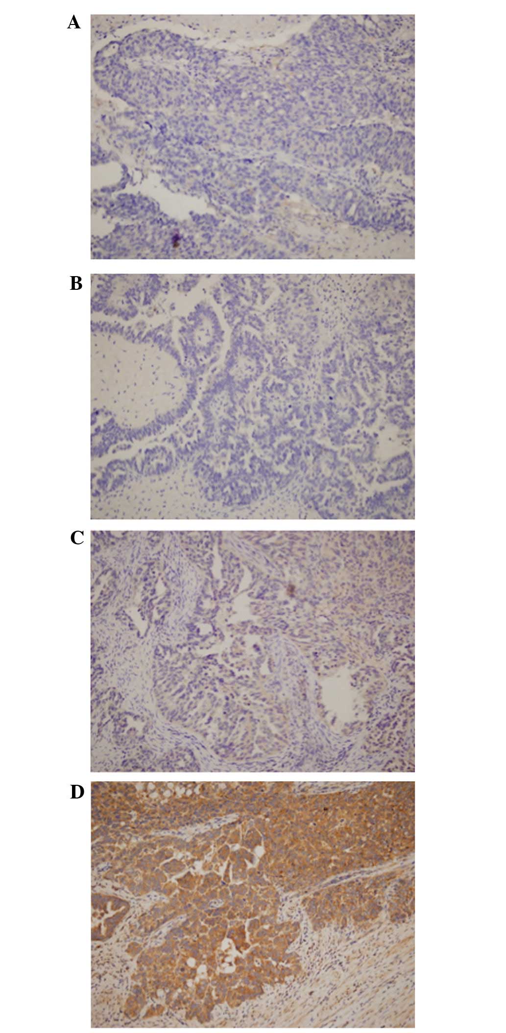
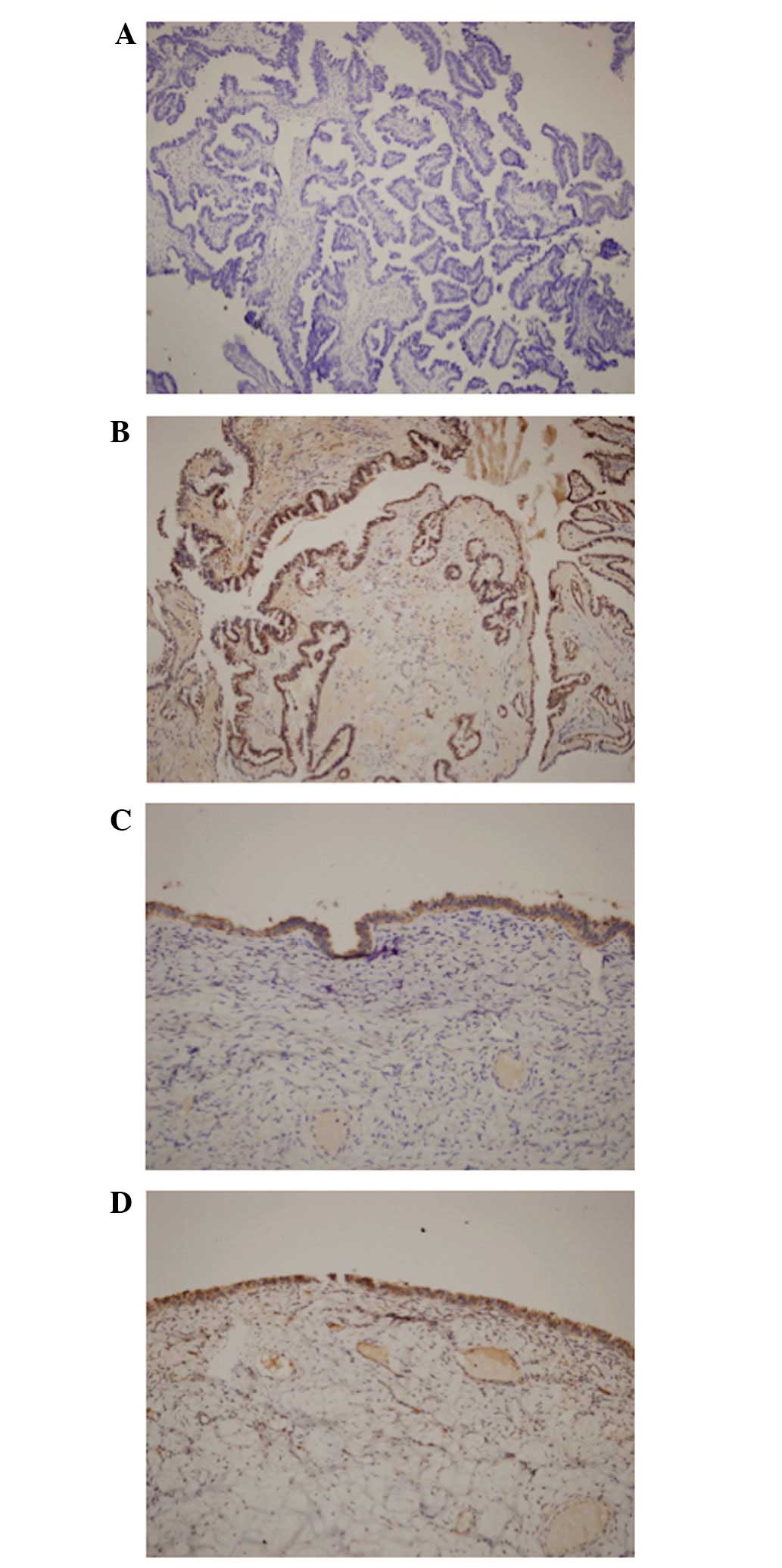
EMP1 expression
EMP1 was expressed in all the 50 cases of malignant
ovarian serous adenocarcinoma. However, the expression levels of
EMP1 in these patients were significantly reduced, compared with
those observed in the 34 borderline and benign serous tumor cases
(P<0.0001; Table II). Reduced
expression of EMP1 was correlated with high grade (P=0.009;
Table III) and stage (P<0.0001;
Table IV; Figs. 1 and 2)
of cancer. EMP1 expression was not correlated with any of the other
investigated parameters, including surgery, operation type or
chemotherapy response (P>0.005).
| Table II.Expression of epithelial membrane
protein 1 in patients with malignant vs. non-malignant ovarian
serous tumors. |
Table II.
Expression of epithelial membrane
protein 1 in patients with malignant vs. non-malignant ovarian
serous tumors.
| Score | Malignant, n (%) | Non-malignant, n
(%) | Total, n (%) |
|---|
| 0 | 22 (88.0) | 3
(12.0) | 25 (100.0) |
| 1 | 5
(83.3) | 1
(16.7) | 6
(100.0) |
| 2 | 14 (53.8) | 12 (46.2) | 26 (100.0) |
| 3 | 9
(33.3) | 18 (66.7) | 27 (100.0) |
| Totala | 50 (59.5) | 34 (40.5) | 84 (100.0) |
| Table III.Association between epithelial
membrane protein 1 expression and tumor grade in malignant ovarian
tissues. |
Table III.
Association between epithelial
membrane protein 1 expression and tumor grade in malignant ovarian
tissues.
| Score | Low grade, n (%) | High grade, n
(%) | Total, n (%) |
|---|
| 0 | 1
(4.5) | 21 (95.5) | 22 (100.0) |
| 1 |
1 (20.0) | 4
(80.0) | 5
(100.0) |
| 2 |
6 (42.9) | 8
(57.1) | 14 (100.0) |
| 3 |
5 (55.6) | 4
(44.4) | 9
(100.0) |
| Totala | 13
(26.0) | 37 (74.0) | 50 (100.0) |
| Table IV.Association between epithelial
membrane protein 1 expression and tumor stage in malignant ovarian
tissues. |
Table IV.
Association between epithelial
membrane protein 1 expression and tumor stage in malignant ovarian
tissues.
| Score | Stage I, n (%) | Stage II, n (%) | Stage III, n (%) | Stage IV, n (%) | Total, n (%) |
|---|
| 0 | 0
(0.0) | 1
(4.5) | 10
(45.5) | 11
(50.0) | 22 (100.0) |
| 1 |
1 (20.0) | 0
(0.0) |
3 (60.0) |
1 (20.0) | 5
(100.0) |
| 2 |
4 (28.6) |
8 (57.1) |
2 (14.3) | 0
(0.0) | 14 (100.0) |
| 3 |
6 (66.7) |
3 (33.3) | 0
(0.0) | 0
(0.0) | 9
(100.0) |
| Totala | 11
(22.0) | 12
(24.0) | 15
(30.0) | 12
(24.0) | 50 (100.0) |
Discussion
The EMP1 gene encodes four ~18 kDa transmembrane
domains (4). EMP1 differs from EMP2
and EMP3 in terms of hydrophobic groups (4). Zoidl et al (5) revealed that EMP1 was significantly
expressed in undifferentiated embryonic stem cells, while poorly
expressed in differentiated adult cells, and appeared to be
involved in prolonging the transition of Schwann cells from G to
S/G2/M phase. The EMP membrane glycoprotein family is
considered to be associated with cell proliferation and
differentiation (6). Previous studies
have demonstrated that the EMP1 gene is expressed in several normal
tissues, including the colon, lung, testis and ovary (7–9). Sun et
al (6–8), identified that the protein levels of
EMP1 were significantly reduced in nasopharyngeal carcinoma, breast
and prostate cancer, compared with normal tissue, and were
correlated with T stage, lymph node metastasis and clinical stage
in these tumors. In addition, Sun et al (9) identified that the expression levels of
EMP1 were significantly reduced in gastric cancer tissues, compared
with normal tissues, and observed that reduced EMP1 expression in
gastric cancer was associated with increased disease severity.
Similarly, the present study revealed that the expression levels of
EMP1 were significantly reduced in patients with malignant serous
tumors, compared with patients exhibiting benign and borderline
serous tumors. In addition, this decrease in EMP1 expression in
high-grade and advanced-stage tumors indicated that EMP1 expression
may be associated with tumor grade and stage. Gnirke and Weidle
(10) demonstrated that EMP1
expression was correlated with cell invasion and metastatic
characteristics in several human mammary carcinoma cell lines. Wang
et al (11) reported that the
EMP1 gene may act as a regulatory factor in cell signaling,
communication and adhesion. Zhang et al (12) observed that the downregulation of the
EMP1 gene was correlated with lymph node metastasis. Sun et
al (6) reported that the protein
levels of caspase-9 increased significantly with the levels of
EMP1, which indicated that the mitochondrial-dependent apoptosis
pathway may be involved in EMP1-induced apoptosis. Vascular
endothelial growth factor C (VEGFC) promotes the proliferation of
endothelial cells, increases vascular permeability, and acts as an
essential factor for tumor angiogenesis, invasion and metastasis
(6). Sun et al (6) demonstrated that VEGFC expression was
significantly decreased following transfection with EMP1, which
suggested that EMP1 was able to inhibit tumor angiogenesis by
suppressing VEGFC expression and tumor metastasis.
In conclusion, EMP1 may possess a significant role
in ovarian cancer cell proliferation, apoptosis, invasion and
metastasis, and may be involved in a number of biological
functions. Further extensive studies on EMP1 are required to aid
the development of novel therapy options for the treatment of
ovarian cancer, and the potential identification of novel
prognostic factors.
References
|
1
|
Ferlay J, Soerjomataram I, Ervik M,
Dikshit R, Eser S, Mathers C, Rebelo M, Parkin DM, Forman D and
Bray F: GLOBOCAN 2012 v1.0. Cancer Incidence and Mortality
Worldwide: IARC CancerBase No. 11. IARC (Lyon, France).
2013.http://globocan.iarc.frAccessed. December
31–2015
|
|
2
|
Cannistra SA, Gershenson DM and Recht A:
Ovarian cancer, fallopian tube carcinoma and peritoneal carcinoma.
DeVita, Hellman, and Rosenberg's Cancer: Principles and practice of
oncology. DeVita DT, Lawrence TS and Rosenberg SA: 2:(9th).
(Philadelphia, PA). Lippincott Williams & Wilkins. 1368–1391.
2011.
|
|
3
|
Lai S, Wang G, Cao X, Li Z, Hu J and Wang
J: EMP-1 promotes tumorigenesis of NSCLC through PI3K/AKT pathway.
J Huazhong Univ Sci Technolog Med Sci. 32:834–838. 2012. View Article : Google Scholar : PubMed/NCBI
|
|
4
|
Lobsiger CS, Magyar JP, Taylor V, et al:
Identification and characterization of a cDNA and the structural
gene encoding the mouse epithelial membrane protein-1. Genomics.
36:379–387. 1996. View Article : Google Scholar : PubMed/NCBI
|
|
5
|
Zoidl G, Blass-Kampmann S, D'Urso D,
Schmalenbach C and Müller HW: Retroviral-mediated gene transfer of
the peripheral myelin protein PMP22 in Schwann cells: Modulation of
cell growth. EMBO J. 14:1122–1128. 1995.PubMed/NCBI
|
|
6
|
Sun GG, Wang YD, Cui DW, Cheng YJ and Hu
WN: EMP1 regulates caspase-9 and VEGFC expression and suppresses
prostate cancer cell proliferation and invasion. Tumour Biol.
35:3455–3462. 2014. View Article : Google Scholar : PubMed/NCBI
|
|
7
|
Sun GG, Lu YF, Fu ZZ, Cheng YJ and Hu WN:
EMP-1 inhibitis nasopharyngeal cancer cell growth and metastasis
through induction apoptosis and angiogenesis. Tumour Biol.
35:3185–3193. 2014. View Article : Google Scholar : PubMed/NCBI
|
|
8
|
Sun GG, Wang YD, Lu YF and Hu WN: EMP-1, a
member of a new family of antiproliferative genes in breast
carcinoma. Tumour Biol. 35:3347–3354. 2014. View Article : Google Scholar : PubMed/NCBI
|
|
9
|
Sun G, Zhao G, Lu Y, Wang Y and Yang C:
Association of EMP1 with gastric carcinoma invasion, survival and
prognosis. Int J Oncol. 45:1091–1098. 2014.PubMed/NCBI
|
|
10
|
Gnirke AU and Weidle UH: Investigation of
prevalence and regulation of expression of progression associated
protein (PAP). Anticancer Res. 18:4363–4369. 1998.PubMed/NCBI
|
|
11
|
Wang HT, Kong JP, Ding F, Wang XQ, Wang
MR, Liu LX, Wu M and Liu ZH: Analysis of gene expression profile
induced by EMP-1 in esophageal cancer cells using cDNA microarray.
World J Gastroenterol. 9:392–398. 2003. View Article : Google Scholar : PubMed/NCBI
|
|
12
|
Zhang J, Cao W, Xu Q and Chen WT: The
expression of EMP1 is downregulated in oral squamous cell carcinoma
and possibly associated with tumour metastasis. J Clin Pathol.
64:25–29. 2011. View Article : Google Scholar : PubMed/NCBI
|