Introduction
High-grade gliomas are the most prevalent type of
primary tumor of the central nervous system in adults (1,2). Although
progress has been made in brain tumor therapies, the prognosis for
patients with malignant glioma remains extremely poor (1). The standard treatment for patients with
recently diagnosed glioblastoma, which comprises temozolomide and
radiotherapy, has increased the median overall survival (OS) time
by 15–20 months (1); however, tumor
recurrence remains inevitable. Salvage treatments for tumor
recurrence are palliative at best, and rarely provide the patient
with any notable survival benefit (1). The poor prognosis of the disease may be
attributed to the difficulty of early detection and to the high
rate of recurrence following initial treatment. Thus, it is
essential to further elucidate the biological features of malignant
gliomas in order to improve diagnosis and treatment of the
disease.
There are a number of histological grading schemes
for glioblastoma, of which the World Health Organization (WHO)
system (2) is the most commonly used
at present. A high WHO grade is associated with clinical
progression and a reduced survival rate (2); however, outcomes may vary between
individuals within diagnostic categories, even those with grade IV
glioma (1–3). This indicates the requirement for
additional markers of prognosis. The inadequacy of
histopathological grading is partially demonstrated by its
inability to prospectively recognize patients (3).
The S-phase index has been used as a prognostic
indicator for several types of tumor, and the S-phase fraction
estimated from tumor tissues has been previously demonstrated to be
the most important prognostic indicator (4–9). Numerous
studies that have utilized flow cytometric analysis of brain tumors
have also indicated that the DNA ploidy index or S-phase fraction
is associated with the grade of malignancy (10–15). These
previous studies focused on investigating the core of the tumor;
however, the predominant characteristic of malignant glioma is the
presence of invading tumor cells in the peritumoral zone. In
addition, distinguishing tumor cells from normal cells in the
peritumoral lesion is challenging. Therefore, the aim of the
present study was to investigate the cell-cycle phase measurements
of fixed paraffin-embedded specimens from the peritumoral invading
zone of high-grade gliomas using laser scanning cytometry
(LSC).
Materials and methods
Tumor specimens
Tumor specimens were obtained from patients who
underwent neurosurgery for primary brain tumors at the Department
of Neurosurgery, Brain Research Institute, Niigata University
(Niigata, Japan) between 1997 and 2000. Samples were specifically
re-reviewed according to the WHO grading system for tumors of the
central nervous system (2). The
research protocol was approved by the local ethics committee (Kyoto
Prefectural University of Medicine, Kyoto, Japan; approval no.
RBMR-G-123-1). Overall survival was measured between the date of
diagnosis at surgery, and the date of mortality.
Specimen analysis
A total of 4 specimens from 2 anaplastic
astrocytomas and 24 specimens from 10 glioblastomas were analyzed
at the tumor core and peritumoral invading zone, respectively.
Paraffin-embedded sections (50-µm thick) were deparaffinized in
xylene, rehydrated in decreasing ethanol concentrations (99.5, 90,
70 and 50%), and enzymatically disintegrated with trypsin and
protease (Sigma-Aldrich Co., LLC, Tokyo, Japan). The single cells
were subsequently stained with propidium iodide (Sigma-Aldrich Co.,
LLC) and placed on microscope slides.
LSC
The slides were scanned using an iCys LSC instrument
(CompuCyte, Cambridge, MA, USA) equipped with an Argon (Ar; 488 nm)
and a Helium-Neon (HeNe; 633 nm) laser and WinCyte® software
(CompuCyte). DNA staining based on propidium iodide served as the
trigger/contouring parameter. The following channels and settings
were used for data collection: Argon Green [photomultiplier tubes
(PMT), 22–32%; offset, 2,030; gain, 255], HeNe LongRed (LR) (PMT,
19–22; offset, 2,000–2,010; gain, 255), threshold on HeNe LR,
2,000–2,700 units; integration contour, +4 pixels; dynamic
background with inner/outer contour, 4/6 pixels; and minimum event
area, 25 µm2. Equal areas of 3,500×5,000 µm were scanned
in the middle of each well using a ×40 objective lens. DNA
histograms were exported and analyzed using ModFit LT™ (Verity,
Topsham, ME, USA). The histograms are presented with a smoothing
standard deviation of 2. The cell-cycle phase fraction was
calculated using a using the MultiCycle program (Phoenix Flow
Systems Inc, San Diego, CA, USA).
Statistical analysis
Analysis of the differences in cell-cycle phase
between the tumor core and the peritumoral invading zone were
performed based on three independent experiments, using the paired
two-tailed Student's t-test (two-tailed distribution or as
indicated). All analyses were completed using the JMP statistical
software package, version 10 (SAS Institute Japan Ltd. Tokyo,
Japan). Statistical tests were two-tailed with type one error of
0.05. Linear correlations between the results were estimated using
the product moment correlation coefficient.
Results
Patients
A total of 2 anaplastic astrocytoma (WHO grade III)
and 10 glioblastoma (WHO grade IV) specimens were obtained from
patients who underwent surgical resections (Table I). The median age of the patients was
61.5 years (range, 32–75 years), and the group comprised 8 male and
4 female patients. Following maximal surgical tumor resections,
patients received courses of external beam radiation therapy
(standard total dose, 60 Gy; applied to the tumor with a 2 cm
margin) and, for patients aged <65 years, first-line
chemotherapy (methyl chloroethyl nitroso urea, MCNU, 100
mg/m2, Vincristine 1.0 mg, intravenously, 3 cycles). The
patients were monitored for tumor recurrences during the initial
and maintenance therapy using magnetic resonance imaging (MRI) or
computed tomography scans. All treatments were performed at the
Department of Neurosurgery, Niigata University Hospital (Niigata,
Japan).
 | Table I.Patient characteristics. |
Table I.
Patient characteristics.
| Patient | Age, years | Gender | Histology |
Initial/Recurring | Overall survival,
days |
|---|
| 1 | 75 | Female | Glioblastoma | Initial | 193 |
| 2 | 51 | Male | Glioblastoma | Initial | 530 |
| 3 | 64 | Male | Glioblastoma | Initial | 85 |
| 4 | 74 | Male | Glioblastoma | Initial | 509 |
| 5 | 48 | Male | Glioblastoma | Recurring | 441 |
| 6 | 60 | Male | Glioblastoma | Initial | 372 |
| 7 | 32 | Male | Glioblastoma | Recurring | 300 |
| 8 | 50 | Female | Anaplastic
astrocytoma | Recurring | 527 |
| 9 | 56 | Male | Anaplastic
astrocytoma | Initial | 762 |
| 10 | 69 | Female | Glioblastoma | Initial | 568 |
| 11 | 70 | Male | Glioblastoma | Initial | 467 |
| 12 | 61 | Female | Glioblastoma | Initial | 815 |
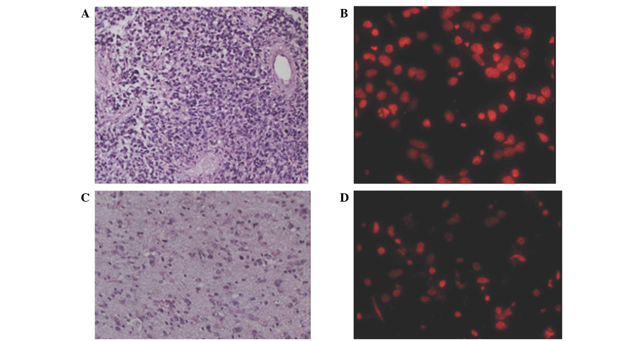
Measurement of cell cycle and
survival
The tumor core is defined as a densely cellular
lesion of neoplastic cells without normal brain tissue. By
contrast, the peritumoral invading zone is defined as a
low-cellular density lesion of neoplastic cells combined with
normal brain tissue close to the tumor core (Fig. 1). The S-phase fraction of the
peritumoral invading lesion was increased compared with the tumor
core in 9 of the patients. There was a slight trend for an
increased S-phase fraction of the peritumoral invading lesion
compared with the tumor core (P=0.24) (Table II). The regression coefficient of the
peritumoral invading zone S-phase fraction and the OS time was
−1,158.2 (P=0.12), indicating a correlation between the peritumoral
invading zone S-phase fraction and the OS time.
| Table II.Cell-cycle phase fraction of the tumor
core and peritumoral invading zone. |
Table II.
Cell-cycle phase fraction of the tumor
core and peritumoral invading zone.
| Tumor region | G0/G1 | S | G2 | M |
|---|
| Tumor core | 0.73±0.11 | 0.12±0.07 | 0.08±0.04 | 0.02±0.01 |
| Peritumoral invading
zone | 0.70±0.10 | 0.16±0.07 | 0.08±0.05 | 0.02±0.01 |
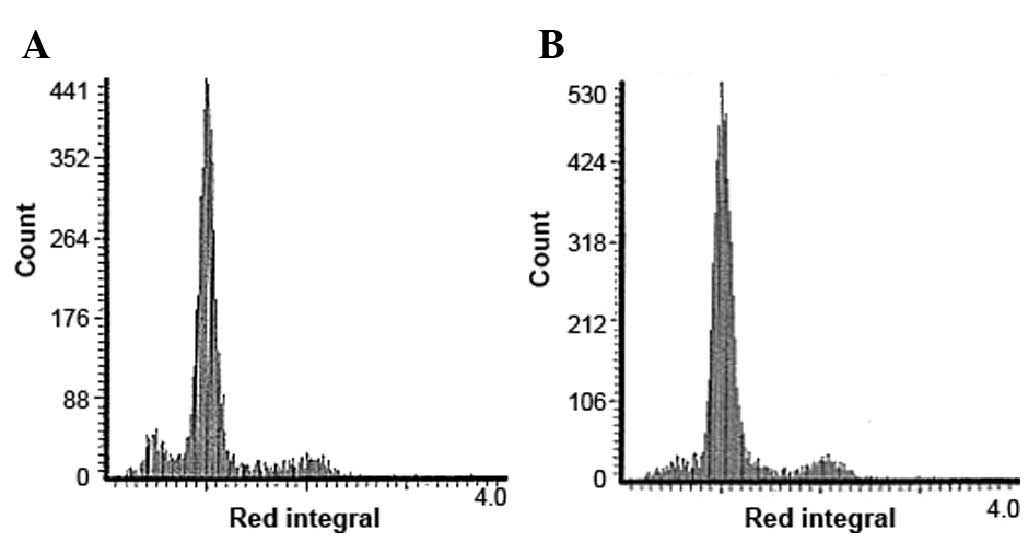
Illustrative case
A 60-year-old man presented with dizziness and right
hemicrania. An intra-axial contrast-enhanced mass was identified in
the right temporal lobe on MRI. A subtotal resection of the mass
was performed, and a diagnosis of glioblastoma was determined. A
total of 60-Gy of local radiotherapy and chemotherapy were
administered postoperatively. A cell-cycle analysis based on LSC is
shown in Fig. 2.
Discussion
LSC was developed to utilize the sensitivity of
fluorescence-based assays and the specificity of on-slide
measurements (16–18). LSC involves a microscope-based
cytofluorometer that is able to assess flow and image cytometry.
The laser-excited fluorescence emitted from individual
fluorochromed cells on a microscope slide is rapidly measured at
multiple wavelengths with high sensitivity and accuracy. LSC has a
number of applications, including the measurement of circulating
tumor cells, DNA damage, DNA ploidy and cell-cycle position
(19–27). The cells for the study are selected by
the operator in a manner that is similar to that for an image
analysis system. Alternatively, the entire cell population on the
slide may be measured automatically using small samples, including
biopsy specimens. Although flow cytometric analysis requires a
large number of cells, LSC may be performed with a relatively small
number (17–28).
Retrospective studies of stored paraffin-embedded
brain tumor samples may be conducted to investigate the interaction
between DNA parameters and patient prognosis. Intraoperative flow
cytometric analyses of glioma tissues, in which the differences
between the tumor margins and normal tissues were detected using
cell-cycle analysis, have been previously reported (29,30). The
malignancy index, defined as the ratio of the number of cells with
greater than normal DNA content to the total number of cells, was
observed to differ between neoplastic and perilesional tissue. An
optimal cut-off value of 6.8% was thus used to identify tumors in
the specimen (29).
Discriminating between tumor cells in the
peritumoral invading zone is challenging; therefore, the
quantitative assessment of tumor cells in glioma lesions is
important for predicting future recurrence. In addition, the
prognostic significance of changes in the malignancy index remains
to be determined in follow-up studies involving a greater number of
cases and more clinical data. In the present study, although the
results were above the usual threshold for statistical
significance, LSC was demonstrated to have potential clinical value
for the objective analysis of the cell cycle in gliomas, which may
provide important information for the characterization of tumor
margins. The profiling results of the present study may be useful
for the construction of a novel and improved classification scheme
for the assessment of clinical malignancies compared with the
conventional histological classification system. However, the
present study had several limitations. LSC analysis cannot
discriminate between glioma cells and normal cells. Therefore,
future studies which combine alternative modalities to complement
the investigation of the peritumoral invading zone are required.
Furthermore, although the assay used was simple, it did not provide
sufficient information regarding the molecular characterization of
gliomas. In future studies the assay may be expanded to provide
more information regarding glioma biology via the addition of
immunocytochemical markers, such as cyclins, inhibitors of
cyclin-dependent kinases, the tumor suppressor p53 and the Bcl-2
and Bax family of proteins, and by utilizing the full capabilities
of fluorescence excitation and emission measurements of LSC. In
addition, the results of the present study are based on a cohort of
12 patients and should therefore be considered as preliminary.
Thus, additional studies are required that assess a larger cohort
of patients in order to further evaluate the role of peritumoral
invading zone cell-cycle analysis for gliomas.
References
|
1
|
Stupp R, Hegi ME, Mason WP, van den Bent
MJ, Taphoorn MJ, Janzer RC, Ludwin SK, Allgeier A, Fisher B,
Belanger K, et al: European Organisation for Research and Treatment
of Cancer Brain Tumour and Radiation Oncology Groups; National
Cancer Institute of Canada Clinical Trials Group: Effects of
radiotherapy with concomitant and adjuvant temozolomide versus
radiotherapy alone on survival in glioblastoma in a randomised
phase III study: 5-year analysis of the EORTC-NCIC trial. Lancet
Oncol. 10:459–466. 2009. View Article : Google Scholar : PubMed/NCBI
|
|
2
|
Kleihues P, Louis DN, Wiestler OD, Burger
PC and Scheithauer BW: WHO grading of tumours of the central
nervous system. World Health Organization Classification of Tumours
of the Nervous System. Louis DN, Ohgaki H, Wiestler OD and Cavenee
WK: 1:(4th). (Lyon, France). IARC Press. 10–11. 2007.
|
|
3
|
Kawaguchi A, Yajima N, Tsuchiya N, Homma
J, Sano M, Natsumeda M, Takahashi H, Fujii Y, Kakuma T and Yamanaka
R: Gene expression signature-based prognostic risk score in
patients with glioblastoma. Cancer Sci. 104:1205–1210. 2013.
View Article : Google Scholar : PubMed/NCBI
|
|
4
|
Sun XF, Rütten S, Zhang H and Nordenskjöld
B: Expression of the deleted in colorectal cancer gene is related
to prognosis in DNA diploid and low proliferative colorectal
adenocarcinoma. J Clin Oncol. 17:1745–1750. 1999.PubMed/NCBI
|
|
5
|
Kolfschoten GM, Hulscher TM, Pinedo HM and
Boven E: Drug resistance features and S-phase fraction as possible
determinants for drug response in a panel of human ovarian cancer
xenografts. Br J Cancer. 83:921–927. 2000. View Article : Google Scholar : PubMed/NCBI
|
|
6
|
Russo A, Bazan V, Migliavacca M, Zanna I,
Tubiolo C, Tumminello FM, Dardanoni G, Cajozzo M, Bazan P, Modica
G, et al: Prognostic significance of DNA ploidy, S-phase fraction,
and tissue levels of aspartic, cysteine, and serine proteases in
operable gastric carcinoma. Clin Cancer Res. 6:178–184.
2000.PubMed/NCBI
|
|
7
|
Lin JK, Chang SC, Yang SH, Jiang JK, Chen
WC and Lin TC: Prognostic value of DNA ploidy patterns of
colorectal adenocarcinoma. Hepatogastroenterology. 50:1927–1932.
2003.PubMed/NCBI
|
|
8
|
Ross JS, Linette GP, Stec J, Ross MS,
Anwar S and Boguniewicz A: DNA ploidy and cell cycle analysis in
breast cancer. Am J Clin Pathol. 120(Suppl): S72–S84.
2003.PubMed/NCBI
|
|
9
|
Kim YT, Zhao M, Kim SH, Lee CS, Kim JH and
Kim JW: Prognostic significance of DNA quantification by flow
cytometry in ovarian tumors. Int J Gynaecol Obstet. 88:286–291.
2005. View Article : Google Scholar : PubMed/NCBI
|
|
10
|
Hoshino T, Prados M, Wilson CB, Cho KG,
Lee KS and Davis RL: Prognostic implications of the
bromodeoxyuridine labeling index of human gliomas. J Neurosurg.
71:335–341. 1989. View Article : Google Scholar : PubMed/NCBI
|
|
11
|
Vavruch L, Eneström S, Carstensen J,
Nordenskjöld B and Wingren S: DNA index and S-phase in primary
brain tumors. A comparison between fresh and deparaffinized
specimens studied by flow cytometry. J Neurosurg. 80:85–89. 1994.
View Article : Google Scholar : PubMed/NCBI
|
|
12
|
Gilbertson RJ, Jaros E, Perry RH, Kelly
PJ, Lunec J and Pearson AD: Mitotic percentage index: A new
prognostic factor for childhood medulloblastoma. Eur J Cancer.
33:609–615. 1997. View Article : Google Scholar : PubMed/NCBI
|
|
13
|
Struikmans H, Rutgers DH, Jansen GH,
Tulleken CA, van der Tweel I and Battermann JJ: Prognostic
relevance of cell proliferation markers and DNA-ploidy in gliomas.
Acta Neurochir (Wien). 140:140–147. 1998. View Article : Google Scholar : PubMed/NCBI
|
|
14
|
Weil RJ, Toms SA, Johnson MD and Mealer A:
Detection of proliferating S-phase brain tumor cells by in
situ DNA replication. J Neurosurg. 95:833–838. 2001. View Article : Google Scholar : PubMed/NCBI
|
|
15
|
Zamecnik J, Snuderl M, Eckschlager T,
Chanova M, Hladikova M, Tichy M and Kodet R: Pediatric intracranial
ependymomas: Prognostic relevance of histological,
immunohistochemical, and flow cytometric factors. Mod Pathol.
16:980–991. 2003. View Article : Google Scholar : PubMed/NCBI
|
|
16
|
Darzynkiewicz Z, Halicka HD and Zhao H:
Analysis of cellular DNA content by flow and laser scanning
cytometry. Adv Exp Med Biol. 676:137–147. 2010. View Article : Google Scholar : PubMed/NCBI
|
|
17
|
Mach WJ, Thimmesch AR, Orr JA, Slusser JG
and Pierce JD: Flow cytometry and laser scanning cytometry, a
comparison of techniques. J Clin Monit Comput. 24:251–259. 2010.
View Article : Google Scholar : PubMed/NCBI
|
|
18
|
Pozarowski P, Holden E and Darzynkiewicz
Z: Laser scanning cytometry: Principles and applications - an
update. Methods Mol Biol. 931:187–212. 2013. View Article : Google Scholar : PubMed/NCBI
|
|
19
|
Tsukazaki Y, Numa Y, Zhao S and Kawamoto
K: Analysis of DNA-ploidy using laser scanning cytometer in brain
tumors and its clinical application. Hum Cell. 13:221–228.
2000.PubMed/NCBI
|
|
20
|
Pollice AA, Smith CA, Brown K, Farkas DL,
Silverman JF and Shackney SE: Multiparameter analysis of human
epithelial tumor cell lines by laser scanning cytometry. Cytometry.
42:347–356. 2000. View Article : Google Scholar : PubMed/NCBI
|
|
21
|
Amirlak B and Couldwell WT: Apoptosis in
glioma cells: Review and analysis of techniques used for study with
focus on the laser scanning cytometer. J Neurooncol. 63:129–145.
2003. View Article : Google Scholar : PubMed/NCBI
|
|
22
|
Zabaglo L, Ormerod MG, Parton M, Ring A,
Smith IE and Dowsett M: Cell filtration-laser scanning cytometry
for the characterisation of circulating breast cancer cells.
Cytometry A. 55:102–108. 2003. View Article : Google Scholar : PubMed/NCBI
|
|
23
|
Foster SS, Leiman G, Schwarz JE, St John T
and Beatty BG: Laser scanning cytometry for the detection of
neoplasia in urologic cytology specimens. Cancer. 102:115–123.
2004. View Article : Google Scholar : PubMed/NCBI
|
|
24
|
Pozarowski P, Huang X, Gong RW, Priebe W
and Darzynkiewicz Z: Simple, semiautomatic assay of cytostatic and
cytotoxic effects of antitumor drugs by laser scanning cytometry:
Effects of the bis-intercalator WP631 on growth and cell cycle of
T-24 cells. Cytometry A. 57:113–119. 2004. View Article : Google Scholar : PubMed/NCBI
|
|
25
|
Liu XP, Sato T, Oga A, Ikemoto K, Kawauchi
S, Ikeda E and Sasaki K: Two subtypes of mucinous colorectal
carcinoma characterized by laser scanning cytometry and comparative
genomic hybridization. Int J Oncol. 25:615–621. 2004.PubMed/NCBI
|
|
26
|
Schwock J, Geddie WR and Hedley DW:
Analysis of hypoxia-inducible factor-1alpha accumulation and cell
cycle in geldanamycin-treated human cervical carcinoma cells by
laser scanning cytometry. Cytometry A. 68:59–70. 2005. View Article : Google Scholar : PubMed/NCBI
|
|
27
|
Fujiyoshi N, Ushijima K, Kawano K,
Fujiyoshi K, Yamaguchi T, Araki Y, Kakuma T, Watanabe S, Kaku T,
Nishida T and Kamura T: Radiation effects on DNA content of
cervical cancer cells: A rapid evaluation of radiation sensitivity
by laser scanning cytometry. Mol Clin Oncol. 3:51–54.
2015.PubMed/NCBI
|
|
28
|
Gerstner AO, Machlitt J, Laffers W, Tárnok
A and Bootz F: Analysis of minimal sample volumes from head and
neck cancer by laser scanning cytometry. Onkologie. 25:40–46. 2002.
View Article : Google Scholar : PubMed/NCBI
|
|
29
|
Shioyama T, Muragaki Y, Maruyama T, Komori
T and Iseki H: Intraoperative flow cytometry analysis of glioma
tissue for rapid determination of tumor presence and its
histopathological grade: Clinical article. J Neurosurg.
118:1232–1238. 2013. View Article : Google Scholar : PubMed/NCBI
|
|
30
|
Alexiou GA, Vartholomatos G, Goussia A,
Batistatou A, Tsamis K, Voulgaris S and Kyritsis AP: Fast cell
cycle analysis for intraoperative characterization of brain tumor
margins and malignancy. J Clin Neurosci. 22:129–132. 2015.
View Article : Google Scholar : PubMed/NCBI
|