Introduction
Glioma accounts for 40–60% of intracranial
malignancies (1). The average
survival time for individuals with glioma is 12–18 months (2) and the incidence of glioma has increased
continuously in the past 20 years (1). Glioma is characterized by invasive
growth and an undefined tumor edge; therefore, surgery cannot be
used to resect the whole tumor and as a result other treatments are
required post-surgery. Currently, adjuvant radiotherapy and
chemotherapy are the treatments used post-surgery (3). However, there are severe side effects
and complications associated with these treatments, including a
high recurrence rate of the tumor (3).
Photodynamic therapy (PDT) refers to a specific
wavelength of light that causes photosensitization of cells, which
leads to the production of reactive oxygen species that damage cell
structures and functions through numerous signaling pathways, and
eventually results in the death of cells (4). PDT has been an effective strategy in the
treatment of glioma in experimental and clinical studies (5,6).
Peritumoral edema is a key stage in the infiltration and recurrence
of glioma, and previous studies have demonstrated that PDT
increases the permeability of the blood-tumor barrier, which leads
to increased peritumoral edema (7).
The bilateral effects of PDT may lead to the spread of glioma and
decrease the effectiveness of PDT in halting the progression of
glioma.
Torasemide is a novel loop diuretic that acts on the
thick ascending limb of the kidney nephron, inhibits the
sodium-potassium-chloride carrier system and increases water
excretion (8). The present study
evaluated the effects of PDT combined with torasemide on the
expression of matrix metalloproteinase (MMP) 2 and
sodium-potassium-chloride cotransporter (NKCC) 1 in peritumoral
edema of rat glioma.
Material and methods
Cell culture
Rat glioma C6 cells were purchased from the Beijing
Institutes of Life Science, Chinese Academy of Sciences (Beijing,
China) and cultured as monolayers in Gibco® RPMI 1640 medium
(Thermo Fisher Scientific, Inc., Waltham, MA, USA) containing
Gibco® 10% Fetal Bovine Serum (Thermo Fisher Scientific, Inc.) in a
humidified incubator (NuAire, Inc., Caerphilly, UK) containing 5%
CO2 at 37°C. Cells in the exponential phase of growth
were used for the present study.
Rat C6 glioma model
All animal protocols in the present study were
approved by the Ethics Committee of the Institutional Research
Board of Harbin Medical University (Harbin, China; approval no.
HMUIRB20150051). Male pathogen-free Wistar rats weighing 220–250 g
(60 rats in total; age, 7–8 weeks) were purchased from the Animal
Experiment Center of Harbin Medical University, and were kept for
24 h at room temperature and with free access to water and standard
laboratory food prior to C6 cell injection. Injection of C6 cells
was performed as previously described (9). Briefly, the rats were anesthetized with
10% chloral hydrate (3 ml/kg; CAS no. 302-17-0; Yangzhou Aoxin
Chemical Factory, Yangzhou, China) and fixed in stereotaxic
apparatus (Motorized Lab Standard Stereotaxic Instrument; catalog
no. 51700, Stoelting Co., Wood Dale, IL, USA) for the facilitation
of the injection. Following sterilization and a skin incision in
the scalp of the rats, a hole was rendered in the skull 1.0 mm
anterior to the anterior fontanel and 1.0 mm lateral to the
sagittal suture. A syringe was inserted 5 mm into the cerebral
cortex and 10 µl C6 glioma cell suspension (1×10⁶ cells)
was injected, as described previously (7). The duration of the procedure was 10 min.
The syringe remained in the brain for 5 min followed by a slow
extraction. The hole was sealed with bone wax (catalog no. 1029754;
B. Braun Melsungen AG; Melsungen, Germany) and the scalp was
sutured. Following sterilization, the rats were returned to their
accommodation and monitored.
Study design
The presence of glioma was confirmed in the rats 7
days following injection with C6 cells using unenhanced and
enhanced magnetic resonance imaging (MRI; Signa HDxt 3.0T; GE
Healthcare Bio-Sciences, Pittsburgh, PA, USA), as previously
described (10). T1-weighted coronal
and axial images were acquired from unenhanced and enhanced scans.
Enhanced scans were achieved using an intravenous injection of
gadolinium-diethylene triamine pentaacetic acid (Gd-DTPA; 0.4
ml/kg; Magnevist®; Bayer AG, Leverkusen, Germany) through the tail
vein of the rats. The rats with glioma were randomly assigned to 4
groups (n=15): Control group, the rats received no treatment; PDT
group, the rats received PDT treatment on day 8 subsequent to
injection with C6 cells; torasemide group, the rats received 5mg/kg
torasemide intraperitoneally (Yoko Pharmaceutical Co., Ltd.,
Nanjing, China) on day 8 subsequent to injection for a duration of
3 days; and PDT + torasemide group, the rats received 5mg/kg
torasemide intraperitoneally following PDT on day 8 subsequent to
injection for a duration of 3 days. The rats in the control and PDT
groups received saline at volumes equivalent to the torasemide
received by the rats in the torasemide and PDT + torasemide groups.
On day 21 subsequent to injection, 5 rats in each group were
sacrificed. Glioma tissues were harvested and processed immediately
for wet-to-dry weight (W/D) ratio, histological examination,
immunohistochemistry and western blot analysis. The remaining 10
rats in each group were used to determine survival times.
PDT treatment
Hematoporphyrin monomethyl ether (HMME; Shanghai
Fudan-Zhangjiang Bio-Pharmaceutical Co., Ltd., Shanghai, China) was
administrated via the tail vein of the rats at a dose of 5 mg/kg on
day 8 subsequent to C6 cell injection, and were kept in the dark
for 3 h. Subsequently, the rats were anesthetized with 10% chloral
hydrate (3 ml/kg) and fixed in a stereotaxic apparatus to expand
the hole in the skull to 10 mm in diameter. The tumor was exposed
by a microneurosurgical method 5 min later. The optical fiber of
the PDT equipment (wavelength, 630 nm; DIOMED 630 PDT Laser; model,
T2USA; Diomed Ltd., Cambridge, UK) was placed on the tumor surface
and PDT was administered at 80 J/cm2 in a 30
mm2 region for 10 min, as previously described (8). Following PDT, the hole was sealed with
bone wax and the scalp was sutured.
W/D ratio of peritumoral tissues
Regions of peritumoral tissue 1–4 mm in diameter
were resected from glioma. The resected tissues were placed on an
ice plate and were immediately weighed. Subsequently, the tissues
were desiccated in a 105°C oven for 24 h until a stable dry weight
was achieved. The W/D ratio was calculated for water content of the
tumor.
Histological observation
Glioma tissues were harvested 21 days following C6
injection and were immediately fixed in 10% formalin (catalog no.
50-00-0; Dezhou Yun Xin Experimental Instrument Co., Ltd., Dezhou,
China), and embedded in paraffin. Tissue sections 6 µm thick were
cut and stained with hematoxylin and eosin (H&E; catalog no.
C0105, Beyotime Institute of Biotechnology, Shanghai, China) for
light microscopy (Eclipse 80i; Nikon Corporation, Tokyo,
Japan).
Immunohistochemistry
Paraffinized peritumoral edema tissues were cut into
6-µm thick sections for immunohistochemistry. The sections were
deparaffinized and hydrated followed by antigen retrieval; the
tissues were incubated with 3% hydrogen peroxide (catalog no.
7722-84-1; Shanghai Ziyi Reagent Factory, Shanghai, China) for 10
min and subsequently citric acid buffer (catalog no. CW0128;
Beijing Tianrui Technology Co., Ltd., Beijing, China) for 3 min.
The tissue sections were incubated overnight at 4°C with the
primary antibodies rabbit anti-rat NKCC-1 antibody (polyclonal;
dilution, 1:500; catalog no. ab59791, Abcam, Cambridge, UK), rabbit
anti-rat MMP2 antibody (polyclonal; dilution, 1:200; catalog no.
ab38898; Abcam) and mouse anti-rat glial fibrillary acidic protein
antibody (GFAP; polyclonal; dilution, 1:500; catalog no. ab7260;
Abcam). Subsequently the tissues were washed with
phosphate-buffered saline three times prior to the addition of the
goat anti-rabbit secondary antibody (monoclonal; dilution, 1:2,000;
catalog no. TA130024; OriGene Technologies, Inc., Beijing, China)
for 30 min at 37°C. The positive index of a tissue section was the
number of cells out of 50 cells that expressed stained cells in 5
fields of view. The tissue sections were blindly examined by a
pathologist (Harbin Medical University).
Western blot analysis
Peritumoral edema tissue was ground in liquid
nitrogen and dissolved in a RIPA Lysis Buffer (Beyotime Institute
of Biotechnology), which contained the protease inhibitor
phenylmethanesulfonyl fluoride. The total protein was extracted
from the tissue, and the protein concentration was determined using
the BCA Protein Assay Kit (catalog no. P0012; Beyotime Institute of
Biotechnology) and stored at −80°C for additional analysis. Equal
amounts of protein (>20 µg) from each group were loaded and
resolved using 10% sodium dodecyl sulfate-polyacrylamide gel
electrophoresis and transferred to polyvinylidene difluoride
membranes (0.45 µm; EMD Millipore, Boston, MA, USA). The membranes
were blocked with 5% non-fat milk in Tris-buffered saline, and
subsequently incubated with rabbit anti-rat MMP2 and rabbit
anti-rat NKCC1 antibodies at 4°C overnight. The membranes were
incubated with goat anti-rabbit secondary antibody (monoclonal;
dilution, 1:2,000; catalog no. TA130024; OriGene Technologies,
Inc.) for 2 h at room temperature. The optical density of bands was
determined using ImageJ software (version 1.48; National Institutes
of Health, Bethesda, MA, USA) and the data were quantified by
normalization to the density of β-actin (OriGene Technologies,
Inc.).
Reverse transcription-quantitative
polymerase chain reaction (RT-qPCR)
Total RNA of each tissue sample was collected using
the RNAsimple Total RNA kit (catalog no. DP419; Tiangen Biotech,
Co., Ltd., Beijing, China), according to the manufacturer's
protocol. A final volume of 20 µl cDNA was synthesized from 500 ng
total RNA using Super M-MLV Reverse Transcriptase (BioTek China,
Beijing, China) and the AffinityScript cDNA Synthesis kit (Agilent
Technologies, Inc., Santa Clara, CA, USA), according to the
manufacturer's protocol. The qPCR was performed on standardized
quantities of cDNA using Exicycler™ 96 (Bioneer Corporation,
Daejeon, Korea). The following primers (Shanghai Kehua
Bio-engineering Co., Ltd., Shanghai, China) were used: MMP2,
forward 5′-GTGGCAATGGAGATGGACA-3′ and reverse
5′-GGTCATAATCCTCGGTGGTG-3′; NKCC1, forward
5′-CCTGGGAGAGTTCCACGAT-3′ and reverse 5′-TTCGGCAGTGTATGTGACCA-3′;
and β-actin, forward 5′-TCAGGTCATCACTATCGGCAAT-3′ and reverse
5′-AAAGAAAGGGTGTAAAACGCA-3′. All samples were run in duplicate.
Relative mRNA expression levels were calculated as
2−∆∆Cq (11).
Statistical analysis
All data were represented as the mean ± standard
deviation. Statistical analyses were performed using the SPSS
version 10.0 software (SPSS, Inc., Chicago, IL, USA). The
difference in results between the various groups was analyzed using
one-way analysis of variance. Survival data were analyzed by
Kaplan-Meier survival analysis, and log rank test was used to
compare the four groups. P<0.05 was considered to indicate a
statistically significant difference.
Results
H&E staining, GFAP expression and
MRI
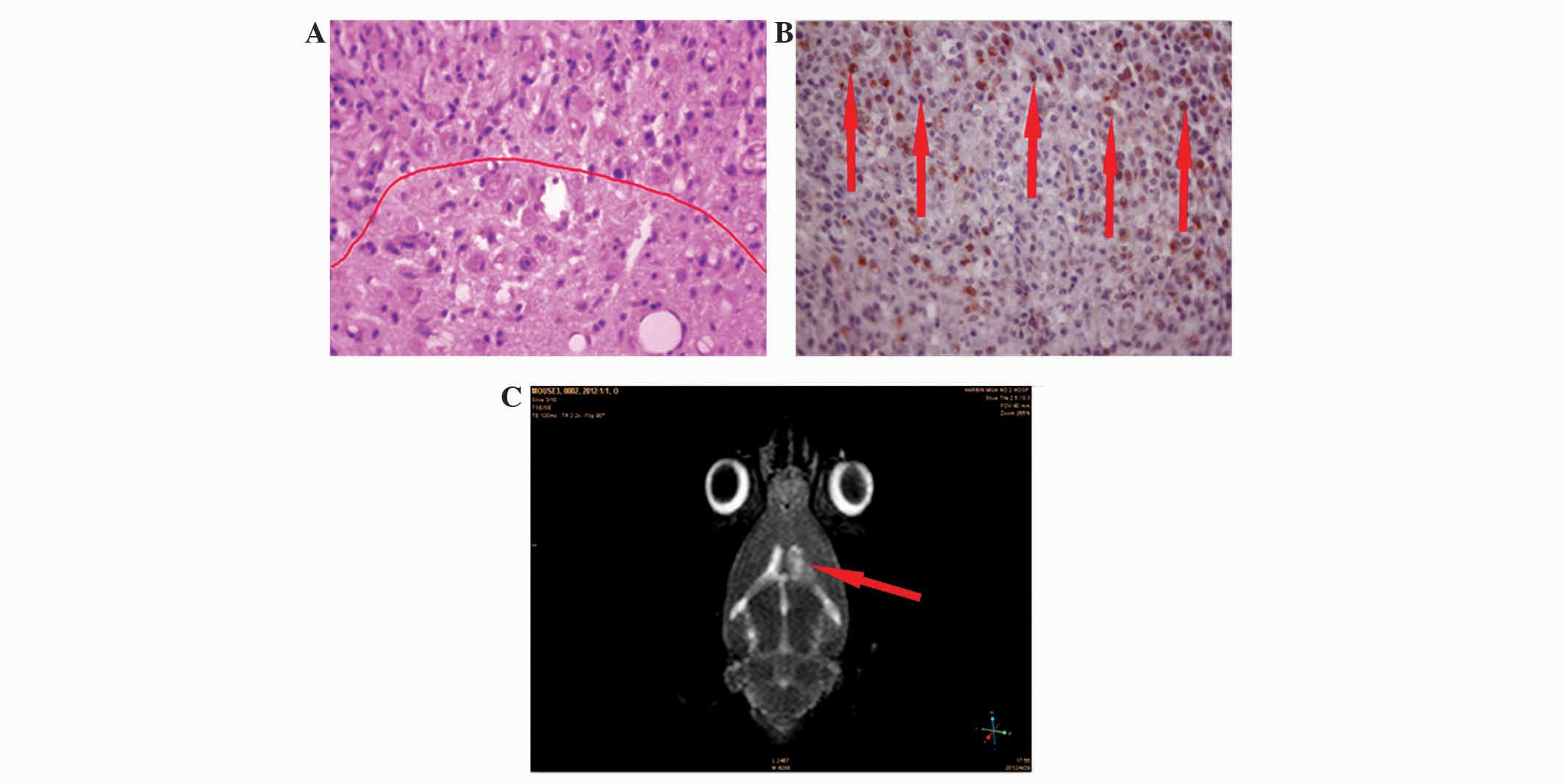
H&E staining of glioma sections are shown in
Fig. 1A. The tissues exhibited
invasive growth of glioma and disintegration of the nuclei of the
cells. Peritumoral edema tissues exhibited fewer venous lakes
compared with normal tissues. The cancer cells invaded normal brain
tissues without a defined margin between normal and cancerous brain
tissue.
Immunohistochemistry revealed that numerous cells
expressed GFAP, which presented as brown granules, and this
suggests the presence of glioma (Fig.
1B). A representative MRI scan of a rat with glioma is
presented in Fig. 1C. MRI scans were
performed 7 days subsequent to injection of C6 cells in all groups
to confirm the presence of glioma.
Water content of peritumoral edema
tissues
Water content of peritumoral edema tissues was
determined by the W/D ratio. The W/D ratio was significantly
increased in the PDT group (5.12±0.73) and decreased in the
torasemide group (4.03±1.17)compared with the control group
(4.76±0.57) (P<0.05). Compared with PDT group, the W/D ratio
decreased significantly in the PDT + torasemide group (4.79±0.65;
P<0.05).
Analysis of MMP2 and NKCC1 expression
using immunohistochemistry
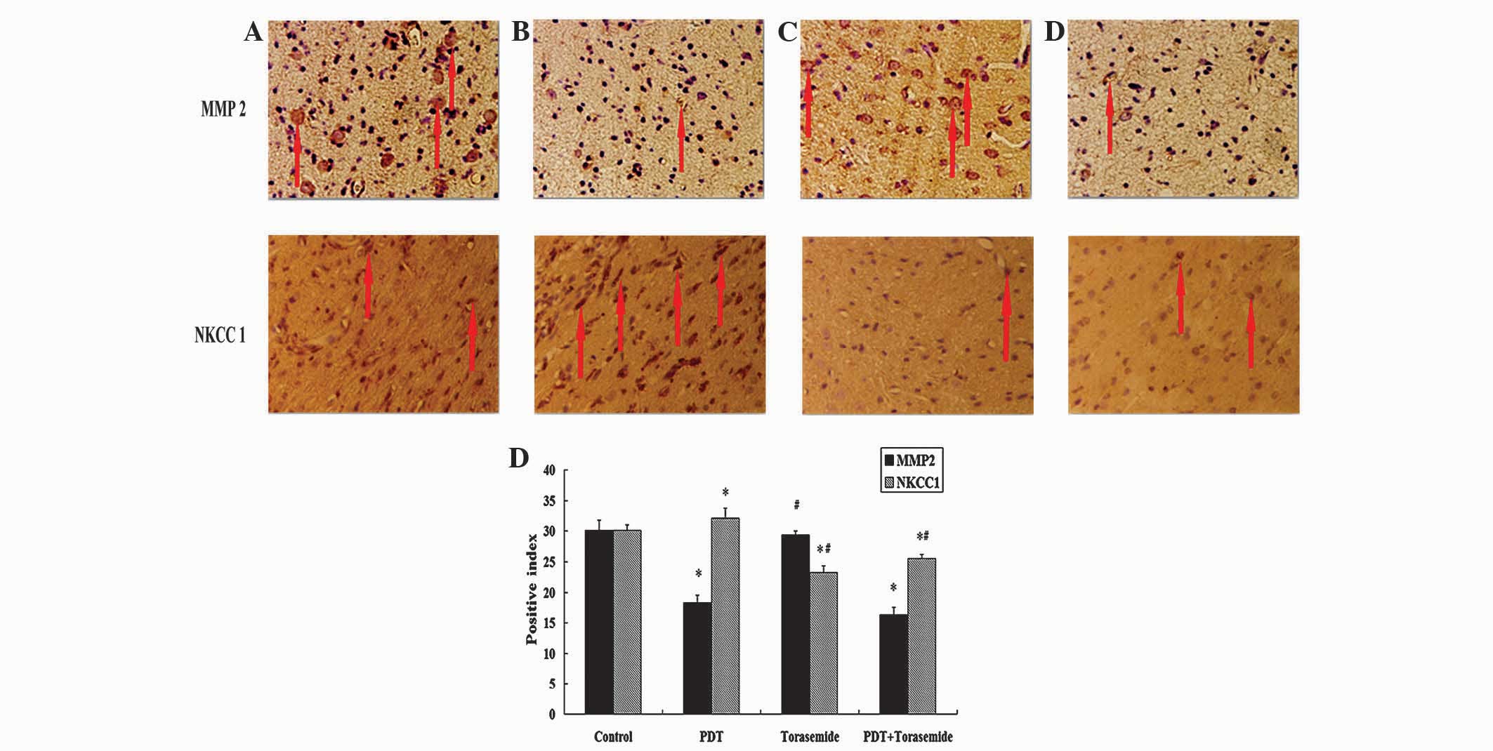
The results of MMP2 immunohistochemistry are
presented in Fig. 2. There was a
decrease in the number of MMP2-positive cells in the PDT group and
PDT + torasemide group compared with the control group. However,
the number of MMP2-positive cells between the PDT group and PDT +
torasemide group did not vary. The number of MMP2-positive cells in
the control group and torasemide group also did not vary.
Similarly, the positive index in the PDT group (18.3±1.3) and PDT +
torasemide group (16.4±1.2) was decreased compared to the control
group (30.2±1.6) and torasemide group (29.4±0.6) (P<0.05).
However, no significant difference was observed in the positive
index in the PDT group and PDT + torasemide group.
The results of NKCC1 immunohistochemical analysis
are also presented in Fig. 2.
Compared with the control group, the number of NKCC1-positive cells
was increased in the PDT group and decreased in the torasemide
group. Compared with the PDT group, the number of NKCC1-positive
cells was decreased in the PDT + torasemide group. Similarly,
compared with the control group (30.2±0.9), the positive index was
increased in the PDT group (32.1±1.6) and significantly decreased
in the torasemide group (23.2±1.1; P<0.05). Compared with the
PDT group, the positive index in the PDT + torasemide group was
significantly decreased (25.5±1.2; P<0.05).
Protein expression of MMP2 and NKCC1
using western blot analysis
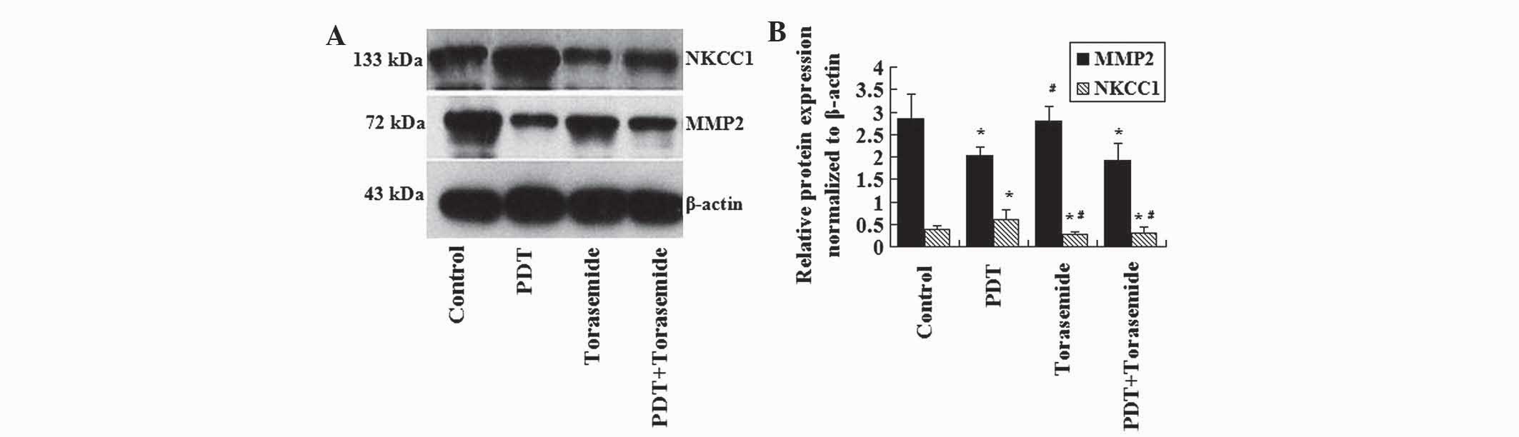
The protein expression of MMP2 and NKCC1 was
determined using western blotting and the results are presented as
the ratio of optical density to β-actin (control). The protein
expression of MMP2 was decreased in the PDT group and PDT +
torasemide group compared with the control group (Fig. 3; P<0.05). The protein expression of
MMP2 between the PDT and PDT + torasemide group had no significant
difference. The protein expression of NKCC1 in the PDT group was
significantly increased compared with the control group, and the
protein expression of NKCC1 in the torasemide group was
significantly decreased compared with the control group (Fig. 3; P<0.05). The NKCC1 protein
expression in the PDT + torasemide group was significantly
decreased compared with the PDT group (P<0.05).
Expression of MMP2 and NKCC1 mRNA
using RT-qPCR
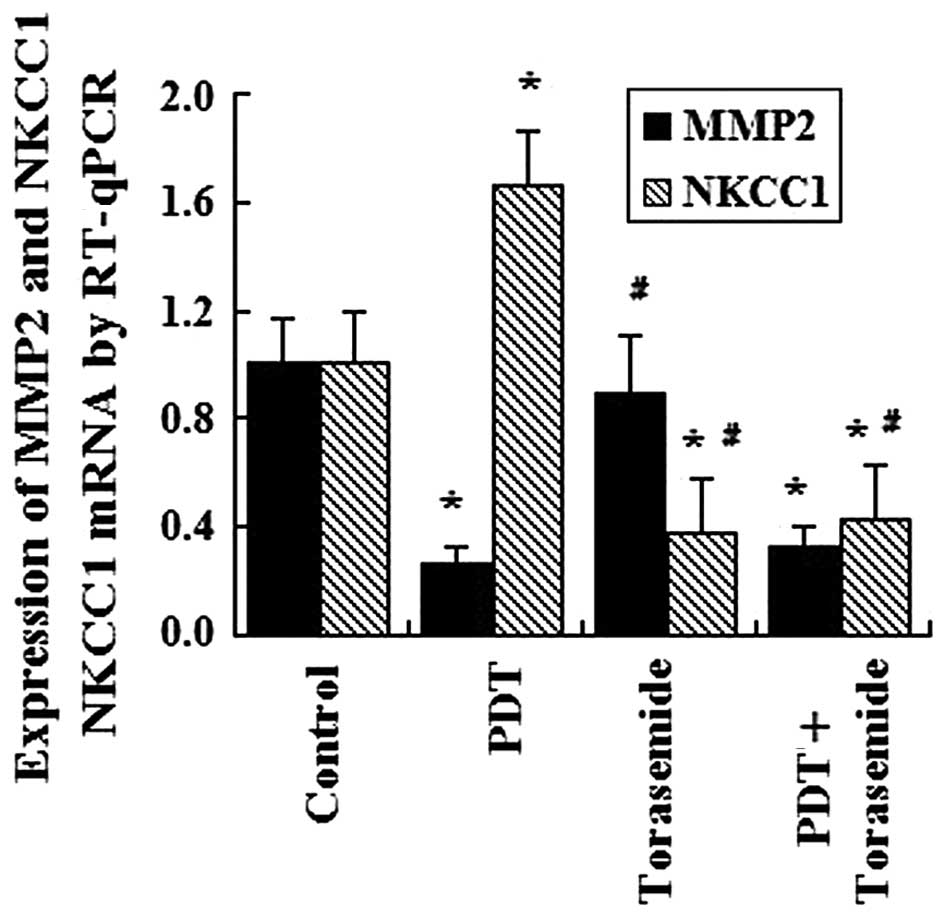
The mRNA expression of MMP2 and NKCC1 in the
peritumoral edema tissues was determined using RT-qPCR (Fig. 4). The mRNA expression levels of MMP2
in the PDT group and PDT + torasemide group were decreased compared
with the control group (P<0.05). A statistical diference was not
observed between the mRNA expression levels of MMP2 in the PDT
group and PDT + torasemide group. The mRNA expression levels of
NKCC1 in the PDT group increased significantly compared with the
control group, and the value in the torasemide group decreased
significantly compared with the control group (P<0.05). Compared
with the PDT group, the mRNA expression levels of NKCC1 in the PDT
+ torasemide group were significantly decreased (P<0.05).
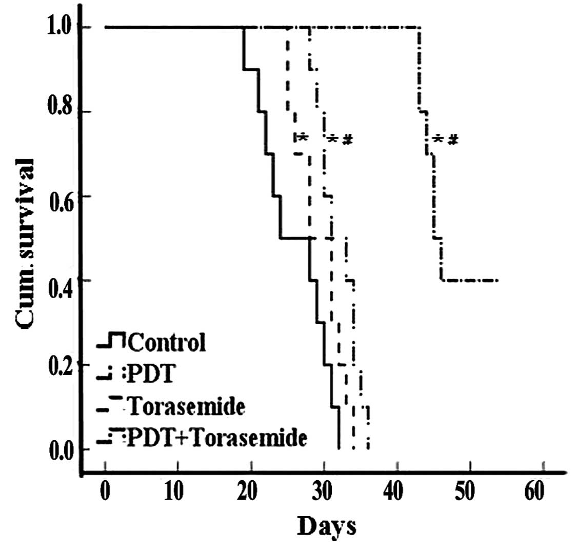
Survival time
Survival time was analyzed using Kaplan-Meier
analysis. The mean survival times for the PDT and torasemide groups
were 30.6±2.4 and 28.3±2.0 days, respectively, which were
significantly increased compared with the control group (24.5±2.2)
(P<0.05). The mean survival time in the PDT + torasemide group
was 47.7±2.5 days, which was significantly increased compared with
the PDT and torasemide groups (Fig.
5; P<0.05).
Discussion
Peritumoral edema is located between the gray, soft
glioma tissue and the white, moist normal tissue, which is
relatively easy to identify under a light microscope (6). Peritumoral edema contains glioma cells
and is a key stage during the invasive growth and recurrence of
glioma. A previous study demonstrated that PDT treatment leads to
increased permeability of the blood-tumor barrier, which increases
the extent of peritumoral edema (12). However, whether the invasion ability
of glioma cells is caused by a water and salt metabolic imbalance
requires investigation. Torasemide is a novel loop diuretic
(13), and has a role in the
medullary thick ascending limb of the kidney where it inhibits the
sodium-potassium-chloride carrier system, which leads to an
increase in the excretion of sodium, potassium, chloride and water
in the urine that may in theory reduce peritumoral edema.
Currently, the mechanism that causes peritumoral
edema is unknown; however, the invasion abilities and mechanisms
that are associated with cerebral edema may be involved (14), and the invasion of glioma is
considered to be an important factor (14,15). Gene
products produced as a result of cerebral edema have been
demonstrated to transport tumor cells, water, macromolecules and
plasma through the basement membranes of vessel walls to the
peritumoral region, which leads to the development of peritumoral
edema and glioma invasion; this process is closely associated with
the MMP family (16–18). The growth of malignant glioma
compresses normal tissue and releases a large number of
neurotransmitters, which alters the environment of the tissue to
provide beneficial anatomical and physicochemical alterations that
increase the growth of glioma.
MMPs are secreted by connective tissue cells,
macrophages and certain tumor cells and degrade extracellular
matrix proteolytic enzymes associated with gliomas in a
zinc-dependent manner (17). MMP2 and
peritumoral edema have been demonstrated to be increased with a
more severe glioma grade (6). The
degradation of the extracellular matrix and basement membrane is a
result of an imbalance between MMPs and tissue inhibitors of MMPs
(19). An increased expression of
MMP2 in glioma degrades the extracellular matrix and basement
membrane of blood vessels directly and indirectly, which leads to
the invasion of glioma and the development of peritumoral edema.
NKCC1 induces sodium, chloride and potassium ions to enter a cell
(20,21), while the potassium and chlorine
cotransporter (KCC2) induces chloride and potassium ions to leave a
cell. The balance between NKCC1 and KCC2 mediates the
electrochemical gradient of the membrane (22,23). NKCC1
has an increased expression in peritumoral edema, which leads to an
imbalance in ions that causes the cell to swell and results in cell
dysfunction (22). Therefore,
vasogenic edema and cytotoxicity of cells coexist in peritumoral
edema.
PDT uses 630 nm wavelengths of visible light to
cause photosensitization of cells, which aggregates tumor tissue
and eventually leads to tumor cell death (24). HMME is a novel stable photosensitizer
with a high specificity to tumor tissue, and has been used
clinically for numerous years (25).
The present study demonstrated that HMME-PDT is a useful method to
induce tumor cell apoptosis and necrosis effectively. Cell
apoptosis induced by PDT may be caused by the release of a variety
of mitochondrial apoptotic factors, including cytochrome C,
apoptosis inducing factor and procaspases that activate caspase-3
(25). The present results
demonstrate that PDT decreased the protein and mRNA expression of
MMP2, but increased the protein and mRNA expression of NKCC1, which
may affect the effectiveness of PDT. In addition, PDT increased the
water content of peritumoral edema tissues and destroyed blood
vessels, which was confirmed by the development of ischemia and
hypoxia in the peritumoral edema region, and aggravated ischemia
and hypoxia of peritumoral tissue. This may cause high levels of
intracellular chloride due to an increased expression of NKCC1.
Torasemide causes an increase in the excretion of
sodium, potassium, chloride and water in the urine. It has a long
half-life and a high plasma protein binding and bioavailability
rate. Torasemide inhibits NKCC1 in peritumoral edema tissue, and is
located at the cancerous tissue boundary. It may then relocate to
the blood due to the osmotic pressure gradient, which leads to an
attenuation of peritumoral edema (26). In the present study, torasemide
inhibited the protein and mRNA expression of NKCC1 and decreased
the W/D ratio. This suggests that torasemide affects NKCC1 directly
and indirectly and decreases peritumoral edema.
In the present study, torasemide treatment following
PDT decreased the protein and mRNA expression of NKCC1 and W/D
ratio. There was no significant increase in the protein and mRNA
expression of MMP2. In addition, torasemide treatment following PDT
greatly increased the survival time of rats with glioma.
In conclusion, PDT combined with torasemide
treatment significantly prolonged the survival time of rats with
glioma, and reduced the invasion of glioma and peritumoral edema.
PDT combined with torasemide may be more effective if it affects
MMP2 and NKCC1; however, even though this treatment strategy did
not affect MMP2 it may be a beneficial treatment strategy for
patients with glioma.
References
|
1
|
Davis FG, Kupelian V, Freels S, McCarthy B
and Surawicz T: Prevalence estimates for primary brain tumors in
the United States by behavior and major histology groups. Neuro
Oncol. 3:152–158. 2001. View Article : Google Scholar : PubMed/NCBI
|
|
2
|
Li JH, Song DY, Xu YG, Huang Z and Yue W:
In vitro study of haematoporphyrin monomethyl ether-mediated
sonodynamic effects on C6 glioma cells. Neurol Sci. 29:229–235.
2008. View Article : Google Scholar : PubMed/NCBI
|
|
3
|
Chibbaro S, Benvenuti L, Caprio A,
Carnesecchi S, Pulerà F, Faggionato F, Serino D, Galli C,
Andreuccetti M, Buxton N and Gagliardi R: Temozolomide as
first-line agent in treating high-grade gliomas: Phase II study. J
Neurooncol. 67:77–81. 2004. View Article : Google Scholar : PubMed/NCBI
|
|
4
|
Choudhary S, Nouri K and Elsaie ML:
Photodynamic therapy in dermatology: A review. Lasers Med Sci.
24:971–980. 2009. View Article : Google Scholar : PubMed/NCBI
|
|
5
|
Tsutsumi M, Miki Y, Akimoto J, Haraoka J,
Aizawa K, Hirano K and Beppu M: Photodynamic therapy with
talaporfin sodium induces dose-dependent apoptotic cell death in
human glioma cell lines. Photodiagn Photodyn Ther. 10:103–110.
2013. View Article : Google Scholar
|
|
6
|
Zhan Q, Yue W and Hu S: Effect of
photodynamic therapy and endostatin on human glioma xenografts in
nude mice. Photodiagn Photodyn Ther. 8:314–320. 2011. View Article : Google Scholar
|
|
7
|
Zhang X, Li X and Wu J: Experimental study
on the invasion of glioma in vivo. Zhonghua Yi Xue Za Zhi.
81:150–153. 2001.(In Chinese). PubMed/NCBI
|
|
8
|
Schreiber S, Gross S, Brandis A, Harmelin
A, Rosenbach-Belkin V, Scherz A and Salomon Y: Local photodynamic
therapy (PDT) of rat C6 glioma xenografts with
Pd-bacteriopheophorbide leads to decreased metastases and increase
of animal cure compared with surgery. Int J Cancer. 99:279–285.
2002. View Article : Google Scholar : PubMed/NCBI
|
|
9
|
Zelenkov P, Baumgartner R, Bise K, Heide
M, Meier R, Stocker S, Sroka R, Goldbrunner R and Stummer W: Acute
morphological sequelae of photodynamic therapy with
5-aminolevulinic acid in the C6 spheroid model. J Neurooncol.
82:49–60. 2007. View Article : Google Scholar : PubMed/NCBI
|
|
10
|
Ulmer S, Reeh M, Krause J, Herdegen T,
Heldt-Feindt J, Jansen O and Rohr A: Dynamic contrast-enhanced
susceptibility-weighted perfusion MRI (DSC-MRI) in a glioma model
of the rat brain using a conventional receive-only surface coil
with a inner diameter of 47 mm at a clinical 1.5 T scanner. J
Neurosci Methods. 172:168–172. 2008. View Article : Google Scholar : PubMed/NCBI
|
|
11
|
Livak KJ and Schmittgen TD: Analysis of
relative gene expression data using real-time quantitative PCR and
the 2(−Delta Delta C(T)) Method. Methods. 25:402–408. 2001.
View Article : Google Scholar : PubMed/NCBI
|
|
12
|
Zhang X, Cong D, Shen D, Gao X, Chen L and
Hu S: The effect of bumetanide on photodynamic therapy-induced
peri-tumor edema of C6 glioma xenografts. Lasers Surg Med.
46:422–430. 2014. View Article : Google Scholar : PubMed/NCBI
|
|
13
|
Fowler SF and Murray KM: Torsemide: A new
loop diuretic. Am J Health Syst Pharm. 52:1771–1780; quiz
1814–1815. 1995.PubMed/NCBI
|
|
14
|
Nase G, Helm PJ, Enger R and Ottersen OP:
Water entry into astrocytes during brain edema formation. Glia.
56:895–902. 2008. View Article : Google Scholar : PubMed/NCBI
|
|
15
|
Kiyatkin EA, Brown PL and Sharma HS: Brain
edema and breakdown of the blood-brain barrier during
methamphetamine intoxication: Critical role of brain hyperthermia.
Eur J Neurosci. 26:1242–1253. 2007. View Article : Google Scholar : PubMed/NCBI
|
|
16
|
Scott JG, Bauchet L, Fraum TJ, Nayak L,
Cooper AR, Chao ST, Suh JH, Vogelbaum MA, Peereboom DM, Zouaoui S,
et al: Recursive partitioning analysis of prognostic factors for
glioblastoma patients aged 70 years or older. Cancer.
118:5595–5600. 2012. View Article : Google Scholar : PubMed/NCBI
|
|
17
|
Lebeau A, Müller-Aufdemkamp C, Allmacher
C, Sauer U, Nerlich A, Lichtinghagen R and Löhrs U: Cellular
protein and mRNA expression patterns of matrix
metalloproteinases-2, −3 and −9 in human breast cancer: Correlation
with tumour growth. J Mol Histol. 35:443–455. 2004. View Article : Google Scholar : PubMed/NCBI
|
|
18
|
VanMeter TE, Rooprai HK, Kibble MM,
Fillmore HL, Broaddus WC and Pilkington GJ: The role of matrix
metalloproteinase genes in glioma invasion: Co-dependent and
interactive proteolysis. J Neurooncol. 53:213–235. 2001. View Article : Google Scholar : PubMed/NCBI
|
|
19
|
Nelson AR, Fingleton B, Rothenberg ML and
Matrisian LM: Matrix metalloproteinases: Biologic activity and
clinical implications. J Clin Oncol. 18:1135–1149. 2000.PubMed/NCBI
|
|
20
|
Haas M and Forbush B III: The Na-K-Cl
cotransporters. J Bioenerg Biomembr. 30:161–172. 1998. View Article : Google Scholar : PubMed/NCBI
|
|
21
|
Kaplan MR, Mount DB, Delpire E, Gamba G
and Hebert SC: Molecular mechanisms of NaCl cotransport. Annu Rev
Physiol. 58:649–668. 1996. View Article : Google Scholar : PubMed/NCBI
|
|
22
|
Hartmann AM, Blaesse P, Kranz T, Wenz M,
Schindler J, Kaila K, Friauf E and Nothwang HG: Opposite effect of
membrane raft perturbation on transport activity of KCC2 and NKCC1.
J Neurochem. 111:321–331. 2009. View Article : Google Scholar : PubMed/NCBI
|
|
23
|
Lang F, Ritter M, Gamper N, Huber S,
Fillon S, Tanneur V, Lepple-Wienhues A, Szabo I and Gulbins E: Cell
volume in the regulation of cell proliferation and apoptotic cell
death. Cell Physiol Biochem. 10:417–428. 2000. View Article : Google Scholar : PubMed/NCBI
|
|
24
|
Li JH, Chen ZQ, Huang Z, Zhan Q, Ren FB,
Liu JY, Yue W and Wang Z: In vitro study of low intensity
ultrasound combined with different doses of PDT. Effects on C6
glioma cells. Oncol Lett. 5:702–706. 2013.PubMed/NCBI
|
|
25
|
Devi DG, Cibin TR and Abraham A: Bis
(3,5-diiodo-2,4,6-trihydroxyphenyl) squaraine photodynamic therapy
induces in vivo tumor ablation by triggering cytochrome c dependent
mitochondria mediated apoptosis. Photodiagn Photodyn Ther.
10:510–517. 2013. View Article : Google Scholar
|
|
26
|
Kahle KT, Staley KJ, Nahed BV, Gamba G,
Hebert SC, Lifton RP and Mount DB: Roles of the cation-chloride
cotransporters in neurological disease. Nat Clin Pract Neurol.
4:490–503. 2008. View Article : Google Scholar : PubMed/NCBI
|