Spandidos Publications Logo
  • About
    • About Spandidos
    • Aims and Scopes
    • Abstracting and Indexing
    • Editorial Policies
    • Reprints and Permissions
    • Job Opportunities
    • Terms and Conditions
    • Contact
  • Journals
    • All Journals
    • Oncology Letters
      • Oncology Letters
      • Information for Authors
      • Editorial Policies
      • Editorial Board
      • Aims and Scope
      • Abstracting and Indexing
      • Bibliographic Information
      • Archive
    • International Journal of Oncology
      • International Journal of Oncology
      • Information for Authors
      • Editorial Policies
      • Editorial Board
      • Aims and Scope
      • Abstracting and Indexing
      • Bibliographic Information
      • Archive
    • Molecular and Clinical Oncology
      • Molecular and Clinical Oncology
      • Information for Authors
      • Editorial Policies
      • Editorial Board
      • Aims and Scope
      • Abstracting and Indexing
      • Bibliographic Information
      • Archive
    • Experimental and Therapeutic Medicine
      • Experimental and Therapeutic Medicine
      • Information for Authors
      • Editorial Policies
      • Editorial Board
      • Aims and Scope
      • Abstracting and Indexing
      • Bibliographic Information
      • Archive
    • International Journal of Molecular Medicine
      • International Journal of Molecular Medicine
      • Information for Authors
      • Editorial Policies
      • Editorial Board
      • Aims and Scope
      • Abstracting and Indexing
      • Bibliographic Information
      • Archive
    • Biomedical Reports
      • Biomedical Reports
      • Information for Authors
      • Editorial Policies
      • Editorial Board
      • Aims and Scope
      • Abstracting and Indexing
      • Bibliographic Information
      • Archive
    • Oncology Reports
      • Oncology Reports
      • Information for Authors
      • Editorial Policies
      • Editorial Board
      • Aims and Scope
      • Abstracting and Indexing
      • Bibliographic Information
      • Archive
    • Molecular Medicine Reports
      • Molecular Medicine Reports
      • Information for Authors
      • Editorial Policies
      • Editorial Board
      • Aims and Scope
      • Abstracting and Indexing
      • Bibliographic Information
      • Archive
    • World Academy of Sciences Journal
      • World Academy of Sciences Journal
      • Information for Authors
      • Editorial Policies
      • Editorial Board
      • Aims and Scope
      • Abstracting and Indexing
      • Bibliographic Information
      • Archive
    • International Journal of Functional Nutrition
      • International Journal of Functional Nutrition
      • Information for Authors
      • Editorial Policies
      • Editorial Board
      • Aims and Scope
      • Abstracting and Indexing
      • Bibliographic Information
      • Archive
    • International Journal of Epigenetics
      • International Journal of Epigenetics
      • Information for Authors
      • Editorial Policies
      • Editorial Board
      • Aims and Scope
      • Abstracting and Indexing
      • Bibliographic Information
      • Archive
    • Medicine International
      • Medicine International
      • Information for Authors
      • Editorial Policies
      • Editorial Board
      • Aims and Scope
      • Abstracting and Indexing
      • Bibliographic Information
      • Archive
  • Articles
  • Information
    • Information for Authors
    • Information for Reviewers
    • Information for Librarians
    • Information for Advertisers
    • Conferences
  • Language Editing
Spandidos Publications Logo
  • About
    • About Spandidos
    • Aims and Scopes
    • Abstracting and Indexing
    • Editorial Policies
    • Reprints and Permissions
    • Job Opportunities
    • Terms and Conditions
    • Contact
  • Journals
    • All Journals
    • Biomedical Reports
      • Information for Authors
      • Editorial Policies
      • Editorial Board
      • Aims and Scope
      • Abstracting and Indexing
      • Bibliographic Information
      • Archive
    • Experimental and Therapeutic Medicine
      • Information for Authors
      • Editorial Policies
      • Editorial Board
      • Aims and Scope
      • Abstracting and Indexing
      • Bibliographic Information
      • Archive
    • International Journal of Epigenetics
      • Information for Authors
      • Editorial Policies
      • Editorial Board
      • Aims and Scope
      • Abstracting and Indexing
      • Bibliographic Information
      • Archive
    • International Journal of Functional Nutrition
      • Information for Authors
      • Editorial Policies
      • Editorial Board
      • Aims and Scope
      • Abstracting and Indexing
      • Bibliographic Information
      • Archive
    • International Journal of Molecular Medicine
      • Information for Authors
      • Editorial Policies
      • Editorial Board
      • Aims and Scope
      • Abstracting and Indexing
      • Bibliographic Information
      • Archive
    • International Journal of Oncology
      • Information for Authors
      • Editorial Policies
      • Editorial Board
      • Aims and Scope
      • Abstracting and Indexing
      • Bibliographic Information
      • Archive
    • Medicine International
      • Information for Authors
      • Editorial Policies
      • Editorial Board
      • Aims and Scope
      • Abstracting and Indexing
      • Bibliographic Information
      • Archive
    • Molecular and Clinical Oncology
      • Information for Authors
      • Editorial Policies
      • Editorial Board
      • Aims and Scope
      • Abstracting and Indexing
      • Bibliographic Information
      • Archive
    • Molecular Medicine Reports
      • Information for Authors
      • Editorial Policies
      • Editorial Board
      • Aims and Scope
      • Abstracting and Indexing
      • Bibliographic Information
      • Archive
    • Oncology Letters
      • Information for Authors
      • Editorial Policies
      • Editorial Board
      • Aims and Scope
      • Abstracting and Indexing
      • Bibliographic Information
      • Archive
    • Oncology Reports
      • Information for Authors
      • Editorial Policies
      • Editorial Board
      • Aims and Scope
      • Abstracting and Indexing
      • Bibliographic Information
      • Archive
    • World Academy of Sciences Journal
      • Information for Authors
      • Editorial Policies
      • Editorial Board
      • Aims and Scope
      • Abstracting and Indexing
      • Bibliographic Information
      • Archive
  • Articles
  • Information
    • For Authors
    • For Reviewers
    • For Librarians
    • For Advertisers
    • Conferences
  • Language Editing
Login Register Submit
  • This site uses cookies
  • You can change your cookie settings at any time by following the instructions in our Cookie Policy. To find out more, you may read our Privacy Policy.

    I agree
Search articles by DOI, keyword, author or affiliation
Search
Advanced Search
presentation
Oncology Letters
Join Editorial Board Propose a Special Issue
Print ISSN: 1792-1074 Online ISSN: 1792-1082
Journal Cover
May-2016 Volume 11 Issue 5

Full Size Image

Sign up for eToc alerts
Recommend to Library

Journals

International Journal of Molecular Medicine

International Journal of Molecular Medicine

International Journal of Molecular Medicine is an international journal devoted to molecular mechanisms of human disease.

International Journal of Oncology

International Journal of Oncology

International Journal of Oncology is an international journal devoted to oncology research and cancer treatment.

Molecular Medicine Reports

Molecular Medicine Reports

Covers molecular medicine topics such as pharmacology, pathology, genetics, neuroscience, infectious diseases, molecular cardiology, and molecular surgery.

Oncology Reports

Oncology Reports

Oncology Reports is an international journal devoted to fundamental and applied research in Oncology.

Experimental and Therapeutic Medicine

Experimental and Therapeutic Medicine

Experimental and Therapeutic Medicine is an international journal devoted to laboratory and clinical medicine.

Oncology Letters

Oncology Letters

Oncology Letters is an international journal devoted to Experimental and Clinical Oncology.

Biomedical Reports

Biomedical Reports

Explores a wide range of biological and medical fields, including pharmacology, genetics, microbiology, neuroscience, and molecular cardiology.

Molecular and Clinical Oncology

Molecular and Clinical Oncology

International journal addressing all aspects of oncology research, from tumorigenesis and oncogenes to chemotherapy and metastasis.

World Academy of Sciences Journal

World Academy of Sciences Journal

Multidisciplinary open-access journal spanning biochemistry, genetics, neuroscience, environmental health, and synthetic biology.

International Journal of Functional Nutrition

International Journal of Functional Nutrition

Open-access journal combining biochemistry, pharmacology, immunology, and genetics to advance health through functional nutrition.

International Journal of Epigenetics

International Journal of Epigenetics

Publishes open-access research on using epigenetics to advance understanding and treatment of human disease.

Medicine International

Medicine International

An International Open Access Journal Devoted to General Medicine.

Journal Cover
May-2016 Volume 11 Issue 5

Full Size Image

Sign up for eToc alerts
Recommend to Library

  • Article
  • Citations
    • Cite This Article
    • Download Citation
    • Create Citation Alert
    • Remove Citation Alert
    • Cited By
  • Similar Articles
    • Related Articles (in Spandidos Publications)
    • Similar Articles (Google Scholar)
    • Similar Articles (PubMed)
  • Download PDF
  • Download XML
  • View XML
Case Report Open Access

Laparoscopic resection of benign schwannoma in the hepatoduodenal ligament: A case report and review of the literature

  • Authors:
    • Liang Tao
    • Shaoyan Xu
    • Zhigang Ren
    • Yiting Lu
    • Xiaowu Kong
    • Xiaohui Weng
    • Zhijie Xie
    • Zhiming Hu
  • View Affiliations / Copyright

    Affiliations: Department of General Surgery, The Third People's Hospital of Haining, Jiaxing, Zhejiang 314408, P.R. China, Department of Hepatobiliary and Pancreatic Surgery, The First Affiliated Hospital, Zhejiang University School of Medicine, Hangzhou, Zhejiang 310003, P.R. China, Department of Hepatobiliary and Pancreatic Surgery, Zhejiang Provincial People's Hospital, Hangzhou, Zhejiang 310014, P.R. China
    Copyright: © Tao et al. This is an open access article distributed under the terms of Creative Commons Attribution License.
  • Pages: 3349-3353
    |
    Published online on: April 5, 2016
       https://doi.org/10.3892/ol.2016.4410
  • Expand metrics +
Metrics: Total Views: 0 (Spandidos Publications: | PMC Statistics: )
Metrics: Total PDF Downloads: 0 (Spandidos Publications: | PMC Statistics: )
Cited By (CrossRef): 0 citations Loading Articles...

This article is mentioned in:


Abstract

Schwannomas are mesenchymal neoplasms with a low malignant potential, which arise from Schwann cells. The tumors can occur in most parts of the body; however, the head, neck and flexor surfaces of the extremities are the most common locations. Schwannomas occurring in the hepatoduodenal ligament are extremely rare. To the best of our knowledge, only two cases of schwannoma in the hepatoduodenal ligament have been reported in the literature, and treatment of such cases by laparoscopic surgery has not yet been reported. The present study reports a case of schwannoma in the hepatoduodenal ligament in a 50‑year‑old male patient. Physical and laboratory examinations showed no abnormal results. Ultrasound and computed tomography failed to definitively diagnose the mass and identify its location. During laparoscopic surgery, a mass was identified in the hepatoduodenal ligament and was completely removed. The gross specimen was a 4.5x2.5x2.5‑cm localized mass, yellowish‑white in color. Microscopic examination revealed that the tumor was mainly composed of spindle‑shaped cells and no atypical cells were identified. Immunohistochemical staining showed a strong positive S‑100 protein reaction, whereas cluster of differentiation 34 and epithelial membrane antigen were negative. The final diagnosis of the lesion was benign schwannoma of the hepatoduodenal ligament. The patient was followed‑up for 7 months and, at the time of writing, was healthy and without any complications. The aim of the present study was to describe a rare case of hepatoduodenal ligament schwannoma in a 50-year-old male patient, and present a review of the literature. To the best of our knowledge, this is the first case of hepatoduodenal ligament schwannoma treated by laparoscopic surgery.

Introduction

Schwannomas are neurogenic tumors that originate from Schwann cells, the cells of the peripheral nervous system (1). Schwannomas have been shown to occur in all ethnic groups and at all ages worldwide, with >90% of the tumors being benign (2). Schwannomas account for ~5% of benign soft-tissue neoplasms (3). Schwannomas may develop in any part of the body, but the most common sites include the head, neck and flexor surfaces of the extremities (4). Generally, schwannomas have a single place of origin, but 10% originate from multiple locations (5). Surgery is the most common treatment for schwannomas. Benign schwannomas rarely recur subsequent to complete surgical resection. Long-term survival can be expected in the majority of patients (4). Peripheral nerve sheath tumors account for 2–6% of all gastrointestinal stromal tumors; however, schwannomas occurring in the hepatoduodenal ligament are extremely rare. To the best of our knowledge, only two such cases have been reported in the literature (6,7). One patient was female and the other patient was male, aged 62 and 29 years old, respectively (6,7). As schwannomas of the hepatoduodenal ligament are normally asymptomatic and often discovered incidentally, preoperative diagnosis is considerably challenging. The two patients each accepted more than two imaging examinations prior to treatment by laparotomy. The lesions were solitary and found in the hepatoduodenal ligament in the two patients. Finally, the patients were definitively diagnosed with schwannoma by pathological examination (6,7). Following complete tumor excision, patients with benign schwannomas generally have a good prognosis. The present study describes the case of a hepatoduodenal ligament schwannoma in a 50-year-old male patient, and presents a review of the literature. To the best of our knowledge, this was the first time laparoscopic surgery was used for the treatment of a benign schwannoma of the hepatoduodenal ligament.

Case report

A 50-year-old male patient was referred to the Department of General Surgery, The Third People's Hospital of Haining (Jiaxing, Zhejiang, China), presenting with pain in the right abdomen following trauma, on July 11, 2014. Physical examination revealed a soft and flat abdomen, and no enlarged lymph nodes were identified in the examinable sites. The patient's family had no history of any specific disease; however, 3 years prior to admission, a mass was incidentally detected in the hepatogastric ligament of the patient by ultrasound (US; Philips IU22; Philips, Amsterdam, Netherlands), during a health examination. As no abnormal symptoms had presented at the time, the patient did not receive any treatment. During the period of hospitalization in July 
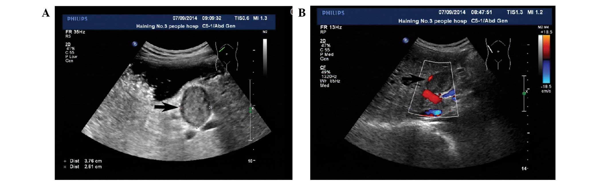
2014, an US revealed a well-defined hypodense lesion in the space between the porta hepatis and the stomach, which measured 3.6×2.4 cm (Fig. 1A). Color Doppler US (Philips IU22) showed no flow signals within the mass (Fig. 1B).

Figure 1.

US and Color Doppler US. (A) Conventional US showing a mass (arrow) measuring 3.8×2.6 cm in the space between the porta hepatis and the stomach. (B) Color Doppler US showing no blood supply in the mass (arrow). US, ultrasound.

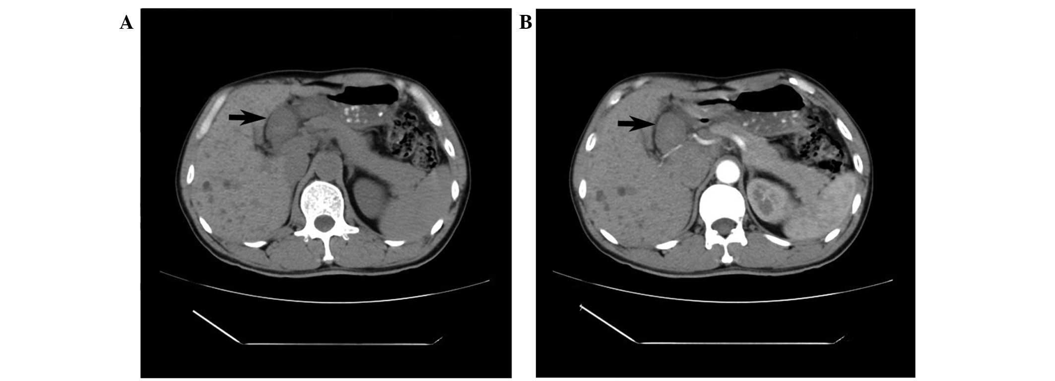
Subsequently, an abdominal computed tomography (CT) scan (GE Brightspeed Elite; Hangwei Tongyong Electric Medicine System Co., Ltd., Beijing, China) was performed to examine the region. The non-enhanced CT scan showed a well-defined round soft-tissue mass without internal calcification or liquefaction in the space between the porta hepatis and the stomach. The mass measured 3.8×3.0×2.5 cm, had a uniform density and was surrounded by a clear tunica (Fig. 2A). On the contrast-enhanced CT scan, the mass showed no evident enhancement in the arterial phase (Fig. 2B). Based on the findings of the CT scan, the diagnosis of gastric stromal tumor was primarily considered. However, since the mass had been growing slowly for 3 years and possessed a clear tunica, the mass was then considered to be a benign tumor, which may be removed.

Figure 2.

CT scans. (A) Non-enhanced CT scan showing a uniform density mass in the space between the porta hepatis and the stomach (arrow); (B) Enhanced CT scan showing no obvious enhancement in the arterial dominant phase (arrow). CT, computed tomography.

Laboratory results were as follows: Red blood cells, 4.38×10¹²/l (normal values, 4.30–5.80×10¹²/l); white blood cells, 5.60×10⁹/l (normal values, 4.00–10.00×10⁹/l); hemoglobin, 131 g/l (normal values, 130–175 g/l); lactate dehydrogenase, 146 international units (IU)/l (normal values, 80–240 IU/l); total protein, 58.10 g/l (normal values, 65.00–85.00 g/l); total bilirubin, 10.80 µmol/l (normal values, 0.00–21.00 µmol/l); conjugated bilirubin, 5.70 µmol/l (normal values, 0.00–10.00 µmol/l); bile acids, 2.40 µmol/l (normal values, 0.00–12.00 µmol/l); aspartate aminotransferase, 18 IU/l (normal values, 15–45 IU/l); alanine transaminase, 16 IU/l (normal values, 9–60 IU/l); γ-glutamyl transpeptidase, 34 U/l (normal values, 10–60 IU/l); alkaline phosphatase, 67 U/l (normal values, 45–125 IU/l); amylase, 77 U/l (normal values, <100 IU/l); antimitochondrial antibody, negative; α-fetoprotein, <20 ng/ml (normal values, <20 ng/ml); carcinoembryonic antigen, 0.67 ng/ml (normal values, <5.50 ng/ml); carbohydrate antigen 19–9, 4.93 U/ml (normal values, <37.00 U/ml); cancer antigen 125, <4.00 U/ml (normal values, <35.00 U/ml).

Subsequent to obtaining patient consent, laparoscopic surgery was performed (Laparoscope operating system, 22202011V110; Karl Storz GmbH & Co. KG, Tuttlingen, Germany). The patient was placed in a horizontal position. Following the successful induction of anesthesia, the skin of the surgery field was conventionally sterilized, and sterile drapes were whisked onto the patient's body. Subsequently, a ~2.0 cm long incision was made in the superior border of the umbilicus. A CO2 pneumoperitoneum was set up with 15 mmHg intra-abdominal pressure using a veress needle (26120J; Karl Storz GmbH & Co. KG, Tuttlingen, Germany), and a 1.1 cm Trocar puncture (30103MP; Karl Storz GmbH & Co. KG) was made to insert a laparoscopic lens (26003BA; Karl Storz GmbH & Co. KG). Under direct vision, 3 holes were made beneath the xiphoid process and left and right upper abdomen, and the ultrasonic scalpel (GEN04; Johnson & Johnson Medical (China) Ltd., Shanghai, China), dissecting forceps (33321ML; Karl Storz GmbH & Co. KG), scratch-free grasping forceps (33321R; Karl Storz GmbH & Co. KG) were inserted. 
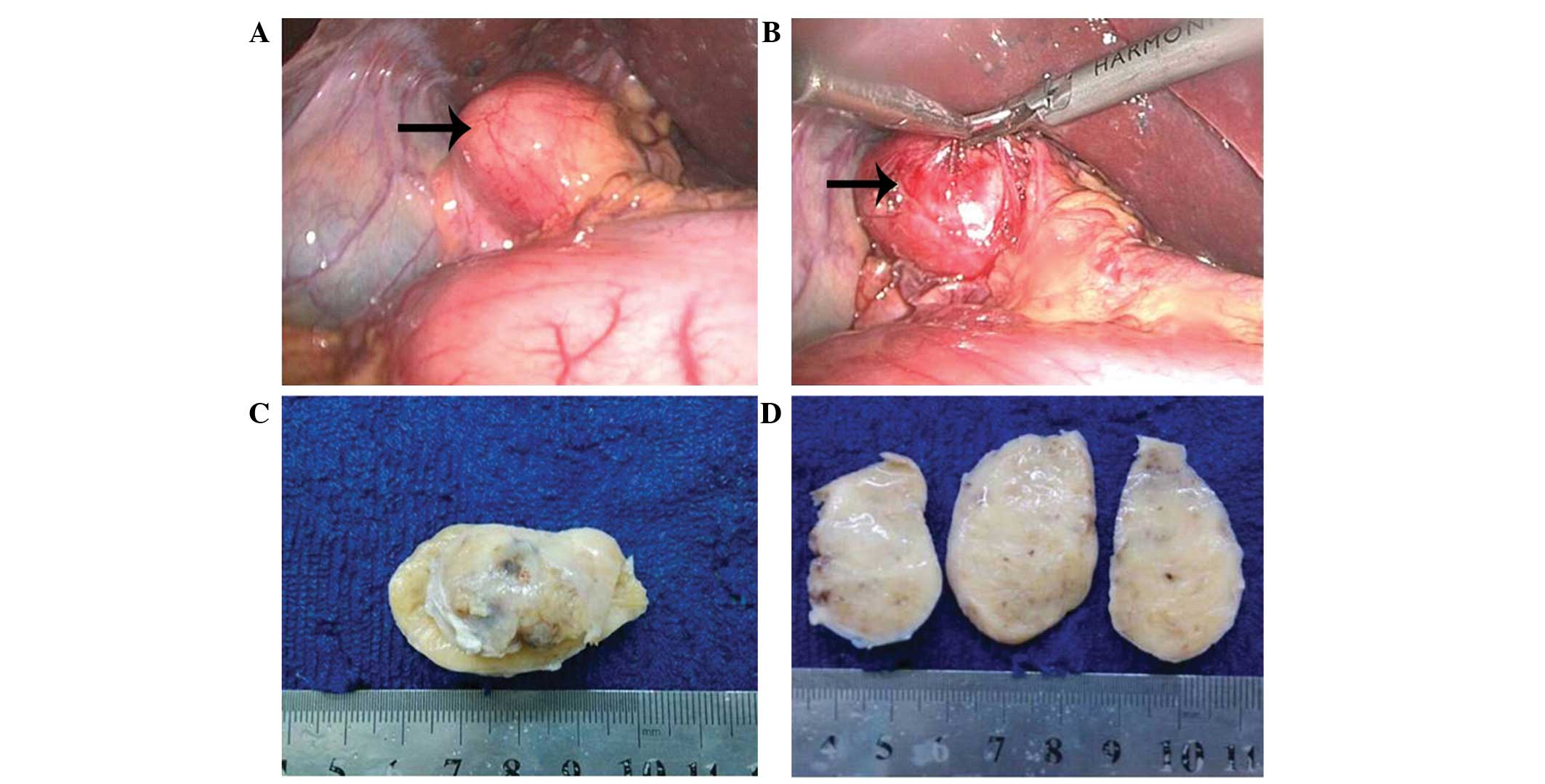
During laparoscopic exploration, a mass surrounded by a fibrous capsule was located in the hepatoduodenal ligament, medial to the gallbladder and near the upper antrum (Fig. 3A and B). No invasion of the surrounding tissue was observed and the biliary ducts were not dilated. The mass was completely resected. The capsule was then removed and the gross specimen was described as a 4.5×2.5×2.5-cm sized localized mass, yellowish-white in color (Fig. 3C and D). Following postoperative pathological analysis of an intraoperative frozen section, the mass was diagnosed as a schwannoma. Postoperatively, the patient had a smooth recovery and left the hospital 3 days later.

Figure 3.

Intraoperative and macroscopic findings. Intraoperatively, (A) the tumor was found to be located in the hepatoduodenal ligament, (B) with no invasion of the surrounding tissue (arrows). (C) Macroscopic findings showed a capsulated yellowish-white mass. (D) The capsule was then removed and the gross specimen was described as a 4.5×2.5×2.5-cm localized mass.

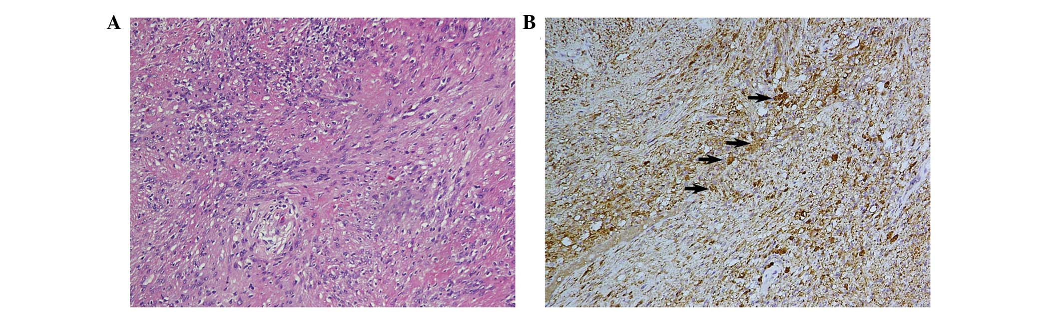
The tissue section was analyzed at the Department of Pathology, Zhejiang Provincial People's Hospital (Hangzhou, Zhejiang, China). Microscopically, the tumor mainly consisted of spindle-shaped cells, and no atypical cells or signs of malignancy were observed (Fig. 4A). Immunohistochemical analysis showed that the tumor cells were vimentin- and S-100 protein-positive (Fig. 4B), and epithelial membrane antigen- and cluster of differentiation 34-negative. The final diagnosis of the tumor was schwannoma. The patient was followed-up for 7 months and, at the time of writing, was healthy and without any complications.

Figure 4.

Pathological examination. (A) Microscopic examination showed that the tumor was mainly composed of spindle-shaped cells (stain, hematoxylin and eosin). (B) Immunohistochemical staining showed a strong tumor positivity for S-100 protein (arrows) (magnification, ×10; lens diameter, 20 mm).

The present study was approved by the Ethics Committee of the Third People's Hospital of Haining (Jiaxing, Zhejiang, China).

Discussion

Schwannomas originate from the sheaths of the peripheral nerves and have been shown to occur in all ethnic groups worldwide (2). Secondary degenerative changes of schwannomas, including cyst formation, calcification, hemorrhage, and hyalinization, sometimes appear (8). Schwannomas occur in most parts of the body, but the head, neck and flexor surfaces of the extremities are the most common sites (1). They rarely occur in the retroperitoneum, accounting for 6% of all primary retroperitoneal tumors (9,10). The occurrence of schwannomas in the gallbladder (11), gastroduodenal ligament (12), pancreas (13), bowel mesentery (14) or colon (15) is extremely rare. To the best of our knowledge, only two cases of schwannoma involving the hepatoduodenal ligament have been reported (6,7). The clinical characteristics of the aforementioned cases are listed in Table I. These tumors are usually asymptomatic and often incidentally discovered. Due to their low incidence and, therefore, lack of adequate understanding of the mechanism of this type of tumor, forming a preoperative diagnosis is challenging.

Table I.

Clinical characteristics of 3 patients with benign schwannomas in the hepatoduodenal ligament.

Table I.

Clinical characteristics of 3 patients with benign schwannomas in the hepatoduodenal ligament.

Author, yearGenderAge, yearsSymptomsImaging techniqueLocationNumberSize, cmPreoperative diagnosisTreatmentFollow-up, monthsStatusRef.
Nagafuchi et al, 1988F62Asymp-tomaticUS, CT, CA MRI, ERCHepato-duodenal ligamentSolitary9×5×4.5NALaparotomy26Survived(14)
Pinto et al, 2011M29Asymp-tomaticUS, MRI endoscopy, biopsyHepato-duodenal ligamentSolitary4.5×2.9Spindle cell neoplasia or stromal tumorLaparotomyNANA(15)
Present caseM50Right abdominal painUS, CT, CECTHepato-duodenal ligamentSolitary4.5×2.5×2.5Stromal tumorLaparoscopic surgery  7Survived

[i] F, female; M, male; NA, not available; US, ultrasound; CT, computed tomography; CECT, contrast-enhanced computed tomography; MRI, magnetic resonance imaging; ERC, endoscopic retrograde cholangiogram; CA, celiac angiography.

Prior to treatment, comprehensive imaging modalities, including US, CT and magnetic resonance imaging (MRI), should be used to establish a probable diagnosis and determine the lesion limits; however, these imaging techniques rarely provide a definitive diagnosis, due to a lack of distinguishing features of these tumors on imaging scans. Generally, schwannomas manifest as well-defined, hypoattenuating masses on non-enhanced CT scans, and on contrast-enhanced CT scans show peripheral enhancement with an irregular pattern. Delayed peripheral enhancement until the late venous phase reflects a fibrous capsule and an internal fibrillary element on CT scan (11). MRI comprises another useful tool for determining the nature of the tumor. On MRI scans, schwannomas are usually presented as masses with a low signal intensity on T1-weighted images, and inhomogeneous high signal intensity on T2-weighted images (16).

Pathology is important for a definitive diagnosis. Histologically, schwannomas consist of compact cellular lesions. Microscopically, schwannomas are encapsulated tumors with a biphasic growth pattern consisting of Antoni A (interlacing and cellular fascicles) and Antoni B (less cellular and myxoid) regions. In addition, typical Verocay bodies in the Antoni A areas may be detected using hematoxylin and eosin staining. In immunohistochemistry, positive uniform S-100 staining may be detected, which is an indicator of schwannoma (17,18). In the present case, the histological findings were predominantly cellular Antoni A areas and immunohistochemistry showed a strong positive staining of the tumor cells for S-100 protein, which supported the benign nature of the schwannoma in the present case.

In conclusion, surgical intervention may be the optimal treatment for schwannomas, as surgery may be used to determine the location and nature of the lesion and to successfully treat it. In the present case, a large mass had developed; however, no resultant symptoms were present prior to trauma. Definitively determining the nature of the tumor preoperatively was challenging, despite the use of imaging modalities. To the best of our knowledge, this was the first case of benign schwannoma of the hepatoduodenal ligament treated by laparoscopic surgery. Following complete tumor excision, patients with benign schwannomas generally have a good prognosis.

Acknowledgements

The authors would like to thank Dr Chen Yuan (Department of Pathology, Zhejiang Provincial People's Hospital for proofreading the pathological methods.

References

1 

Das Gupta TK and Brasfield RD: Tumors of peripheral nerve origin: Benign and malignant solitary schwannomas. CA Cancer J Clin. 20:228–233. 1970. View Article : Google Scholar : PubMed/NCBI

2 

Ariel IM: Tumors of the peripheral nervous system. CA Cancer J Clin. 33:282–299. 1983. View Article : Google Scholar : PubMed/NCBI

3 

Pilavaki M, Chourmouzi D, Kiziridou A, Skordalaki A, Zarampoukas T and Drevelengas A: Imaging of peripheral nerve sheath tumors with pathologic correlation: Pictorial review. Eur J Radiol. 52:229–239. 2004. View Article : Google Scholar : PubMed/NCBI

4 

Le Guellec S: Nerve sheath tumours. Ann Pathol. 35:54–70. 2015.(In French). View Article : Google Scholar : PubMed/NCBI

5 

Fenoglio L, Severini S, Cena P, Migliore E, Bracco C, Pomero F, Panzone S, Cavallero GB, Silvestri A, Brizio R and Borghi F: Common bile duct schwannoma: A case report and review of literature. World J Gastroenterol. 13:1275–1278. 2007. View Article : Google Scholar : PubMed/NCBI

6 

Honjo Y, Kobayashi Y, Nakamura T, Takehira Y, Kitagawa M, Ikematsu Y, Ozawa T and Nakamura H: Extrahepatic biliary schwannoma. Dig Dis Sci. 48:2221–2226. 2003. View Article : Google Scholar : PubMed/NCBI

7 

Lane RH, Stephens DH and Reiman HM: Primary retroperitoneal neoplasms: CT findings in 90 cases with clinical and pathologic correlation. AJR Am J Roentgenol. 152:83–89. 1989. View Article : Google Scholar : PubMed/NCBI

8 

Li ZQ, Wang HY, Li J and Teng L: Recurrent retroperitoneal Schwannomas displaying different differentiation from primary tumor: Case report and literature review. World J Surg Oncol. 8:662010. View Article : Google Scholar : PubMed/NCBI

9 

Liu LN, Xu HX, Zheng SG, Sun LP, Guo LH and Wu J: Solitary schwannoma of the gallbladder: A case report and literature review. World J Gastroenterol. 20:6685–6690. 2014. View Article : Google Scholar : PubMed/NCBI

10 

Bayraktutan U, Kantarci M, Ozgokce M, Aydinli B, Atamanalp SS and Sipal S: Education and Imaging. Gastrointestinal: Benign cystic schwannoma localized in the gastroduodenal ligament; a rare case. J Gastroenterol Hepatol. 27:9852012. View Article : Google Scholar : PubMed/NCBI

11 

Ciledag N, Arda K and Aksoy M: Pancreatic schwannoma: A case report and review of the literature. Oncol Lett. 8:2741–2743. 2014.PubMed/NCBI

12 

Tang SX, Sun YH, Zhou XR and Wang J: Bowel mesentery (meso-appendix) microcystic/reticular schwannoma: Case report and literature review. World J Gastroenterol. 20:1371–1376. 2014. View Article : Google Scholar : PubMed/NCBI

13 

Bugiantella W, Rondelli F, Mariani L, Peppoloni L, Cristallini E and Mariani E: Schwannoma of the colon: A case report. Oncol Lett. 8:2511–2512. 2014.PubMed/NCBI

14 

Nagafuchi Y, Mitsuo H, Takeda S, Ohsato K, Tsuneyoshi M and Enjoji M: Benign schwannoma in the hepatoduodenal ligament: Report of a case. Surg Today. 23:68–72. 1993. View Article : Google Scholar : PubMed/NCBI

15 

Pinto J, Afonso M, Veloso R, Tente D, Fernandes S, Proença L, Carvalho J, Pontes JM and Fraga J: Benign schwannoma of the hepatoduodenal ligament. Endoscopy. 43(Suppl 2): E195–E196. 2011. View Article : Google Scholar : PubMed/NCBI

16 

Rha SE, Byun JY, Jung SE, Chun HJ, Lee HG and Lee JM: Neurogenic tumors in the abdomen: Tumor types and imaging characteristics. Radiographics. 23:29–43. 2003. View Article : Google Scholar : PubMed/NCBI

17 

Das Gupta TK, Brasfield RD, Strong EW and Hajdu SI: Benign solitary Schwannomas (neurilemomas). Cancer. 24:355–366. 1969. View Article : Google Scholar : PubMed/NCBI

18 

Weiss SW, Langloss JM and Enzinger FM: Value of S-100 protein in the diagnosis of soft tissue tumors with particular reference to benign and malignant Schwann cell tumors. Lab Invest. 49:299–308. 1983.PubMed/NCBI

Related Articles

  • Abstract
  • View
  • Download
  • Twitter
Copy and paste a formatted citation
Spandidos Publications style
Tao L, Xu S, Ren Z, Lu Y, Kong X, Weng X, Xie Z and Hu Z: Laparoscopic resection of benign schwannoma in the hepatoduodenal ligament: A case report and review of the literature. Oncol Lett 11: 3349-3353, 2016.
APA
Tao, L., Xu, S., Ren, Z., Lu, Y., Kong, X., Weng, X. ... Hu, Z. (2016). Laparoscopic resection of benign schwannoma in the hepatoduodenal ligament: A case report and review of the literature. Oncology Letters, 11, 3349-3353. https://doi.org/10.3892/ol.2016.4410
MLA
Tao, L., Xu, S., Ren, Z., Lu, Y., Kong, X., Weng, X., Xie, Z., Hu, Z."Laparoscopic resection of benign schwannoma in the hepatoduodenal ligament: A case report and review of the literature". Oncology Letters 11.5 (2016): 3349-3353.
Chicago
Tao, L., Xu, S., Ren, Z., Lu, Y., Kong, X., Weng, X., Xie, Z., Hu, Z."Laparoscopic resection of benign schwannoma in the hepatoduodenal ligament: A case report and review of the literature". Oncology Letters 11, no. 5 (2016): 3349-3353. https://doi.org/10.3892/ol.2016.4410
Copy and paste a formatted citation
x
Spandidos Publications style
Tao L, Xu S, Ren Z, Lu Y, Kong X, Weng X, Xie Z and Hu Z: Laparoscopic resection of benign schwannoma in the hepatoduodenal ligament: A case report and review of the literature. Oncol Lett 11: 3349-3353, 2016.
APA
Tao, L., Xu, S., Ren, Z., Lu, Y., Kong, X., Weng, X. ... Hu, Z. (2016). Laparoscopic resection of benign schwannoma in the hepatoduodenal ligament: A case report and review of the literature. Oncology Letters, 11, 3349-3353. https://doi.org/10.3892/ol.2016.4410
MLA
Tao, L., Xu, S., Ren, Z., Lu, Y., Kong, X., Weng, X., Xie, Z., Hu, Z."Laparoscopic resection of benign schwannoma in the hepatoduodenal ligament: A case report and review of the literature". Oncology Letters 11.5 (2016): 3349-3353.
Chicago
Tao, L., Xu, S., Ren, Z., Lu, Y., Kong, X., Weng, X., Xie, Z., Hu, Z."Laparoscopic resection of benign schwannoma in the hepatoduodenal ligament: A case report and review of the literature". Oncology Letters 11, no. 5 (2016): 3349-3353. https://doi.org/10.3892/ol.2016.4410
Follow us
  • Twitter
  • LinkedIn
  • Facebook
About
  • Spandidos Publications
  • Careers
  • Cookie Policy
  • Privacy Policy
How can we help?
  • Help
  • Live Chat
  • Contact
  • Email to our Support Team