Introduction
Neurofibromatosis type 1 (NF1) (MIM no. 162200),
also known as von Recklinghausen disease, is clinically
characterized by the presence of simple, diffuse and plexiform
neurofibromas. Plexiform neurofibromas are unencapsulated,
poorly-circumscribed tumors infiltrating the nerves and adjacent
fat and muscles (1). The connective
overgrowth can be limited to a single nerve or a plexus; in the
latter case, when the plexus spreads to the epidermal and dermal
tissues, it is termed molluscum fibrosum. This can occur multiple
times, covering all body sites (including the forehead, temple,
eyes, nape and upper lip) with the exception of the palms and soles
(2). The plexiform neurofibroma
variant, mixoglioma gelatiniforme, is usually soft and is located
in the lower third of the leg, and when associated with
lymphangiomatosis, it can give rise to elephantiasis neuromatosa
(EN). EN is characterized by abnormal soft-tissue hypertrophy and
bone dysplasia together with early and excessive bone growth of the
affected leg compared with the contralateral leg (3,4).
Pachidermocele or dermatholysis may be associated with NF1, showing
an overlap of skin layers in the thorax, buttocks and roots of the
limbs.
The etiology of EN is not yet fully understood, but
the association of primary lymphatic dysplasia with a lymphatic
proliferative process has been proposed (5–7).
The current study presents a case of NF1-associated
EN with typical clinical manifestations. Written informed consent
was obtained from the patient.
Case report
Case history
In January 2014, the case of a female patient was
brought to our attention at the Department of Dermatology of
University of Modena and Reggio Emilia (Modena, Italy) due to a
29-year history of several neurofibromas and multiple (>6)
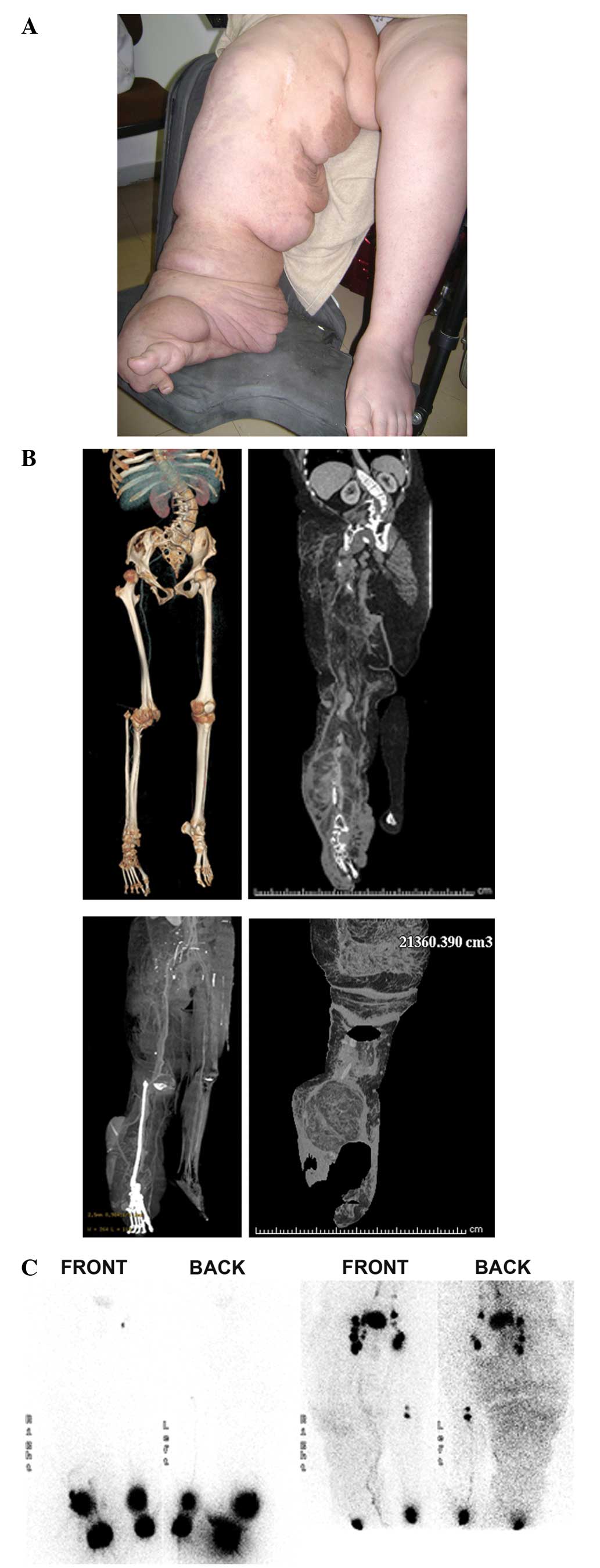
cafè-au-lait macules. The patient presented with giant
elephantiasis of the right leg, which had started to grow during
late childhood, and had since accelerated its expansion in the
following years (Fig. 1A). At birth,
the patient exhibited a cafè-au-lait macule on the right thigh. By
1 year old, a semi-liquid mass had developed at the same site. The
lesion was noticed by the parents and showed indolent growth with
no signs of bleeding or pain. Upon histopathological examination,
it presented with the aspects of a lymphangioma. Lymphedema of the
ipsilateral foot and discrepant leg lengths were noted
successively. In the following years, the uneven leg growth was
associated with bone proliferation, which required a number of
osteotomies with the aim of stopping the growth. Conventional
lymphoscintigraphy (LS) was performed to assess the lower limb
lymphatic drainage pathway.
The family history was suggestive of NF1, as the
patient's father exhibited macrocephaly, hypertelorism, and
multiple cafè-au-lait macules and neurofibromas.
Genetic testing, magnetic resonance imaging (MRI)
and three-dimensional computed tomography (3D-CT) were also
performed.
MRI and 3D-CT
The MRI and 3D-CT scan study showed a preponderance
of adipose tissue in the elephantiasic limb, corresponding to
three-quarters of its whole volume, in addition to a severe
dorso-lumbar-sacral scoliosis with convexity on the left. Two
central nervous system hamartomas of the pallidus nucleous were
also identified by MRI. 3D-CT revealed a mild lumbo-sacral
meningocele and giant L4 neurofibroma (Fig. 1B).
Germline mutation analysis of the NF1
gene
Genomic DNA was extracted from the peripheral blood
of the patient and the patient's father using the QIAamp DNA Blood
Mini kit (Qiagen Inc., Valencia, CA, USA), and stored at −20°C
until use. All NF1 exons were amplified by polymerase chain
reaction with intron spanning primers, as described previously
(8), and then analyzed with
denaturing high-performance liquid chromatography, as described
previously (9). For each abnormal
elution profile, genomic DNA was directly sequenced in each
direction using a CEQ Dye-Terminator Cycle Sequencing kit (Beckman
Coulter Inc., Miami, FL, USA) according to the manufacturer's
instructions.
Mutations were checked using the Mutalyzer program
(http://www.lovd.nl/mutalyzer). NF1
germline deletion g.129042_129043delAG; c.1541_1542del;
p.(Gln514Argfs*43) was found in the proband and the patient's
father. To the best of our knowledge, this type of mutation has not
previously been described.
Lymphedema and limb lymphatic
assessment
A common tape measure was used to assess the limb
circumferences of the patient. Reference circumferences were the
popliteal crease, point zero (K), +30 cm (A), +20 cm (B) and +10 cm
(C) in the thigh, −10 cm (D), −20 cm (E) and −30 cm (F) in the
lower leg, and 10 cm proximal from the tip of the first toe (G).
Leg volumes were calculated according to these measurements
(10).
Lower limb lymphatic function was assessed by LS,
using injections of 37-MBq 99mTc-labeled human serum
albumin. Injection points were in the first, second and fourth
interdigital and retromalleolar spaces of the affected and
contralateral foot. Image acquisition was obtained after 60 and 240
min using a dual head γ-camera (Philips Healthcare, Andover, MA,
USA) equipped with a low-energy and high-resolution collimator, and
an energy peak centered on 140 KeV (window of 20%).
The criteria to define lymphatic dysfunction include
delay, asymmetric or absent visualization of regional lymph nodes,
and the presence of dermal backflow. Other observations include the
visualization of asymmetric lymphatic channels, collateral
lymphatic channels, interrupted vascular structures, and lymph
nodes of the deep lymphatic system (i.e., popliteal lymph nodes
following web space injection in the lower extremities) (11).
In LS, the images obtained 60 min after the
injections showed bilateral lower limb lymph flow delay. Mild
dermal backflow in the absence of tracer migration was observed in
the affected lower limb, whilst one inguinal lymph node was
observed in the left limb. Posterior images confirmed the findings,
also visualizing a popliteal lymph node in the healthy (left) leg.
Images acquired at 240 min showed significant dermal backflow in
the right limb, and hyperplasia and hypertrophy of the inguinal and
external iliac lymph nodes, in comparison to those of the left leg
(Fig. 1C).
Treatment and patient outcome
In May 2014, the patient was treated with surgery to
reduce the volume of tissue in the leg region; tissue with a mass
of 4.3 kg was removed. However, the outcome was not as good as
expected due to hemorrhage during surgery, which prevented complete
exision. Following surgery, the patient exhibited limited
functional improvement and limb lightening. The patient was
subsequently administered anticoagulant therapy with enoxaparin
(8,000 IU, daily for one year) and underwent regular follow up
examinations every three months for the first year and every six
months subsequently. At the time of writing, the patient was well
with a good prognosis.
Discussion
EN is a rare clinical manifestation associated with
the NF1 phenotype. The condition should be defined as early and
excessive growth in the width and length of the affected limb due
to a neoplastic proliferation of the perineural connective tissue,
together with congenital lymphatic insufficiency and chronic
hyperemia. While no more than 30 cases are described in the
literature (Table I) (12–32), the
real incidence is estimated to be higher. The clinical expression
is characterized by plexiform neurofibromas located in the
superficial or deep nervous system associated with congenital
lymphangiomathosis. Signs usually appear during the first years of
life, due to lymphostasis and subsequent lymphedema causing
adipocyte metaplasia of the adjacent tissues and chronic hyperemia
inducing bone overgrowth and focal gigantism (34).
 | Table I.EN as a clinical manifestation
associated with the NF1 phenotype. |
Table I.
EN as a clinical manifestation
associated with the NF1 phenotype.
| First author/s, year
(ref.) | Gender | Age/age of onset | Proband affected
region | Family history | Clinical
features |
|---|
| Spittel and Fernando,
1929 (12) | M | 20 years/at
birth | Left limb | Grandmother, 2,000
minute cutaneous tumours and scattered subcutaneous swellings;
mother, achondroplasia. | Gross EN of the left
limb. Subcutaneous tumors scattered all over the body; a
pachidermatocele of the occipital scalp; cutaneous freckles.
Hyperpigmentation of the mucous membrane of the mouth, and muddy
hypopigmentation of the conjunctive. Bone development
increasing. |
| Westcott and
Ackerman, 1947 (13) | M | 40 years/at
birth | Neck | His mother had
NF-1. | EN of the neck.
Asymmetry of the rib cage. Deformity of the cervical column, the
left leg and the left clavicle. |
|
| M | 22
years/childhood | Chest wall and
axilla | No particular
familial history of NF. | EN of extensively
involved the chest wall and the axilla. |
| Lenson, 1956
(14) | F | 42 years/– | Right leg | The family history is
free from NF or any other inherited disease tendency. Her mother
had gastric carcinoma. | Soft irregular bluish
end mottled mass involving the distal two-thirds of the right lower
extremity. Cavernous haemangioma with the consistency of a lipoma.
Numerous femoral cysts. |
| Fethiere et
al, 1974 (15) | M | 16
years/childbirth | Penis | Not reported. | EN of the penis.
Syringomyelia associated with cervical ependymoma. Facial paralysis
on the left side and paralysis of the left eye muscle. Multiple
schwannomas involving certain cranial nerves, and numerous dorsal
and ventral spinal roots with large tumor formations in cauda
equina roots. |
| Yaghmai and Tafazoli,
1977 (16) | M | 11 years/3 years | Left thigh | Not reported. | Elephantiasis
neuromatosa and overgrowth of abnormal bones with subperiosteal
haemorrhage. Numerous cafè-au-lait spots and scoliosis of the
dorsolumbar spine. |
|
| F | 9 years/2 years | Right leg | Not reported. | Elephantiasis
neuromatosa, multiple cafè-au-lait spots and overgrowth of abnormal
bones with subperiosteal haemorrhage. |
| Sty et al,
1981 (17) | M | 4 years/– | Right lower
extremity | NF in 11 of his
relatives. | EN involving the
right lower extremity. Numerous cafè-au-lait spots on the lower
abdomen and groin. The right lower extremity was longer than the
left. Dilation of the medial lymphatic channels in the lower
extremity and enlarged right iliac nodes. |
| Harris et al,
1982 (18) | F | 18 years/at
birth | Right hallux | No familial history
of NF. | Only one large
cafè-au-lait spot on the right hallux. Soft-tissue hypertrophy with
massive enlargement of the proximal and distal phalanges. |
| Holck et al,
1984 (19) | F | 17
years/childhood | Right gluteal
sulcus | There was no
predisposition to either familial or hereditary disorders. | EN of the right
gluteal sulcus, coexistent or with lipomatosis. No skeletal lesion.
Nor were other stigmata of von Recklinghausen's disease
apparent. |
| Birch and Davies,
1988 (20) | F | 30 years/at
birth | Left lower
limb | Not reported. | EN of the left
lower limb maximal in the calf. Dysplastic bones of the left
hemipelvis and leg with florid osseous striations. The skin of the
thigh was loose and inelastic. |
| Hertzanu et
al, 1989 (21) | F | 52
years/childhood | Trunk | The patient denied
any manifestations of NF in her family. | Symmetric EN of the
trunk. At 32 years: Hypovolemic shock due to massive hemorrhage
into the mass. |
| Bardelli and
Hadjistilianou, 1989 (22) | M | Few months/at
birth | Facial
hemi-hypertrophy | No family history
of congenital glaucoma or buphthalmos. | Buphthalmos and
regional gigantism with evolution in EN. Corneal diameter 13 mm,
rubeosis iridis, ectropion uveae. An increase of optic nerve
cupping. A swelling of the right upper lid and preauricular region.
Hypertrophy of the soft periorbital tissues. Small nodules on
conjunctiva of affected eye. |
| Bardelli and
Hadjistilianou, 1989 (22) | M | 13 months/at
birth | Right orbital
region | Not reported. | Buphthalmos. An
enlarged optic canal, a fibrous dysplasia of the greater wing of
sphenoid bone. Longer right fibula and a slight deformity of the
vertebrae of the lumbar tract. |
|
| F | 11 years/at
birth | Temporal and
zygomatic region | Not reported. | Facial
elephantiasis without buphthalmos. Greater wing of sphenoid bone;
right optic foramen larger than the left. Iris nodules, ectropion,
uveae, increased optic nerve cupping. Small neurofibromas of the
zygomatic and temporal region. |
| Kuo and Kuo, 1990
(23) | F | 34 years/8
years | Right limb and | His mother,
grandmother, sister and | EN involving the
right lower limb and pelvis, leading to a right hip pelvis nephews
had NF. disarticulation. Grossly enlarged lower limb complicated by
infected decubitus ulcers. A leg length discrepancy. (After surgery
a large neurofibroma of the sciatic nerve). |
| Roy and Ghosh, 1992
(24) | F | 22 years/at
birth | Right foot | No family history
of NF1 was presented. | Enlargement of the
whole right foot with conspicuous gigantism of the 2nd to 5th toes.
The overlying skin was coarse, dry, thick. Overgrowth and
elongation of all the bones of the foot (2nd and 3rd metatarsals,
and their phalanges). A huge soft-tissue mass with no bone
involvement. |
|
| M | 21 years/at
birth | Right foot | Not reported. | Gigantism of the
right foot and elongation of the all the bones of the foot
(metatarsals and a huge soft-tissue mass). |
|
| M | 18 years/early
childhood | Right foot | His father had EN.
One of his three brothers has a few cutaneous nodules affecting the
right lower limb only. | Gigantism of the
right foot with swelling at the heel. Elongation of all the bones
(metatarsals and a soft-tissue mass affecting mainly the hind
foot). |
|
| M | 6 years/at
birth | Right foot | No family history
of NF. | Enlargement mainly
of the forefoot and the swelling was more prominent between the
great and 2nd toe. No cafè-au-lait spots. |
| Kokandkar et
al, 1994 (25) | M | 3 months/at
birth | Neck and back | Family history did
not reveal occurrence of similar illness in any member. | Congenital
plexiform neurofibroma involving the neck with EN with a
sarcomatous nodule. The skin covering the tumor was hairy,
redundant and dark. The skin covering the nodules was thinned out
and ulcerated. |
| Münte et al,
1996 (26) | F | 33 years/6
years | Right leg | Family history of
NF. | EN of the right
leg. Severe dysaesthesia in the right lower leg not confined to a
single nerve. |
| Stevens et
al, 1998 (27) | M | 43
years/childhood | Left leg | Not reported. | Gross EN of the
left leg. Large synovial cyst arising from the synovium of the
patello-femoral joint. |
| Akyol et al,
1999 (28) | M | 20 years/6 years
old | Left shoulder and
arm | No family history
of NF. | EN with Becker's
melanosis. Hairly and brown-black hyperpigmented patches on left
shoulder, left upper back and left arm. Lisch nodules. |
| Lorberbom et
al, 2000 (29) | M | 35 years/– | Right thigh and
sacral region | Not reported. | A soft-tissue mass
and enlargement of the right upper leg. |
| Steenbrugge et
al, 2001 (30) | F | 13 years/at
birth | Left limb | Her mother had
NF-I. | Left leg
elephantiasis with recurrent massive subperiosteal hematoma. |
| Hourani et
al, 2006 (31) | F | 41
years/childhood | Right limb | Not reported. | EN of the right
leg. Optic chiasm glioma. Right tibia and fibula marrow and
cortices hypertrophy. |
| Martínez-García
et al, 2008 (32) | F | 14 years/at
birth | Right limb | Not reported. | EN involving the
right lower limb. Anaemia and hepatitis B. |
| Hoshi et al,
2009 (33) | M | 56
years/childhood | Right leg | There was no
particular family history of NF. | A huge mass of EN
in the right leg. |
| Bano et al,
2010 (1) | M | 15
years/childhood | Right limb | No family history
of NF1 in first-degree relatives. | EN of the right
leg. Osseous abnormalities including thinning of bones, erosion of
distal articular surfaces and periosteal dysplasia. |
Distinct superficial dysplastic skin alterations
known as pachidermocele or dermatholysis, histologically
corresponding to mixoglioma gelatiniforme, must be distinguished
from EN in patients affected by NF1 (2).
To date, little evidence is available regarding the
role of lymphatic alterations in the pathogenesis of EN.
Based on the bilateral lymphatic defect, the
presence of a primary lymphatic disease in the current NF1 patient
can be hypothesized. The disease is probably supported by a
dysplastic-hypertrophic condition as a result of a congenital
alteration of the lymphatic network (35,36).
Regarding the lymph to fat transformation, it is
known that lymphostasis due to primary and secondary lymphedema
determines the transformation of fat cells, resulting in
hypertrophied adipose tissue. Several studies (37–39) have
suggested that lymphedema leads to adipose tissue accumulation and
fibrosis. Moreover, we believe that this process is amplified in
NF1 and in EN due to a primary lymphatic disorder, which is at the
base of the clinical manifestation induced by the plexiform
neurofibroma growth.
Overall, the diagnostic criteria for NF can be
improved by the introduction and application of novel criteria
based on a wider case series (EN, focal gigantism, mixoglioma
gelatiniforme and primary lymphatic disorder), leading to the early
diagnosis of NF1, particularly in pediatric patients. LS and MRI
can be efficacious tools in the diagnosis and clinical
characterization of early onset cutaneous, subcutaneous and
skeletal anomalies.
Acknowledgements
The authors would like to thank Dr Federica
Arginelli for providing useful comments.
Glossary
Abbreviations
Abbreviations:
|
NF1
|
neurofibromatosis type 1
|
|
EN
|
elephantiasis neuromatosa
|
|
LS
|
lymphoscintigraphy
|
|
MRI
|
magnetic resonance imaging
|
|
3D-CT
|
three-dimensional computed
tomography
|
References
|
1
|
Bano S, Prasad A, Yadav SN, Chaudhary V
and Sachdeva N: Elephantiasis neuromatosa of the lower limb in a
patient with neurofibromatosis type-1: A case report with imaging
findings. J Pediatr Neurosci. 5:59–63. 2010. View Article : Google Scholar : PubMed/NCBI
|
|
2
|
Pollock G: Report of a case of molluscum
fibrosum or fibroma with observations. Med Chir Trans. 56:255–266.
1873. View Article : Google Scholar : PubMed/NCBI
|
|
3
|
Serradell AP: Neurocutaneous syndromes.
Medicine - Programa de formación médica continuada acreditado.
8:5532–5547. 2003.(In Spanish). View Article : Google Scholar
|
|
4
|
Ponti G, Martorana D, Pellacani G, Ruini
C, Loschi P, Baccarani A, De Santis G, Pollio A, Neri TM, Mandel
VD, et al: NF1 truncating mutations associated to aggressive
clinical phenotype with elephantiasis neuromatosa and solid
malignancies. Anticancer Res. 34:3021–3030. 2014.PubMed/NCBI
|
|
5
|
Flanagan BP and Helwing EB: Cutaneous
lymphangioma. Arch Dermatol. 113:24–30. 1977. View Article : Google Scholar : PubMed/NCBI
|
|
6
|
Foeldi M: The role of the lymphatic
circulation in the fluid circulation of the eye and the central
nervous system. Arch Kreislaufforsch. 41:186–212. 1963.PubMed/NCBI
|
|
7
|
Lohrmann C, Pache G, Felmerer G, Foeldi E,
Schaefer O and Langer M: Posttraumatic edema of the lower
extremities: Evaluation of the lymphatic vessels with magnetic
resonance lymphangiography. J Vasc Sur. 49:417–423. 2009.
View Article : Google Scholar
|
|
8
|
De Luca A, Bottillo I, Dasdia MC, Morella
A, Lanari V, Bernardini L, Divona L, Giustini S, Sinibaldi L,
Novelli A, et al: Deletions of NF1 gene and exons detected by
multiplex ligation-dependent probe amplification. J Med Genet.
44:800–808. 2007. View Article : Google Scholar : PubMed/NCBI
|
|
9
|
De Luca A, Buccino A, Gianni D, Mangino M,
Giustini S, Richetta A, Divona L, Calvieri S, Mingarelli R and
Dallapiccola B: NF1 gene analysis based on DHPLC. Hum Mutat.
21:171–172. 2003. View Article : Google Scholar : PubMed/NCBI
|
|
10
|
International society of lymphology: The
diagnosis and treatment of peripheral lymphedema. 2009 consensus
document of the international society of lymphology. Lymphology.
42:51–60. 2009.PubMed/NCBI
|
|
11
|
Pecking AP: Possibilities and restriction
of isotopic lymphography for the assessment of therapeutic effects
in lymphedema. Wien Med Wochenschr. 149:105–106. 1999.PubMed/NCBI
|
|
12
|
Spittel RL and Fernando SE: A case of
elephantiasis neuromatosa. Br Med J. 1:596–597. 1929. View Article : Google Scholar : PubMed/NCBI
|
|
13
|
Westcott RJ and Ackerman LV: Elephantiasis
neuromatosa; a manifestation of von Recklinghausen's disease. Arch
Derm Syphilol. 55:233–241. 1947. View Article : Google Scholar : PubMed/NCBI
|
|
14
|
Lenson N: Neurofibromatosis; a case report
of elephantiasis neuromatosa of the right lower extremity, with
invasion of the popliteal artery. AMA Arch Surg. 73:279–284. 1956.
View Article : Google Scholar : PubMed/NCBI
|
|
15
|
Fethiere W, Carter HW and Sturim HS:
Elephantiasis neuromatosa of the penis. Light and electron
microscopical studies. Arch Pathol. 97:326–330. 1974.PubMed/NCBI
|
|
16
|
Yaghmai I and Tafazoli M: Massive
subperiosteal hemorrhage in neurofibromatosis. Radiology.
122:439–441. 1977. View Article : Google Scholar : PubMed/NCBI
|
|
17
|
Sty JR, Starshak RJ and Woods GA:
Neurofibromatosis: Lymphoscintigraphic observations. Clin Nucl Med.
6:264–265. 1981. View Article : Google Scholar : PubMed/NCBI
|
|
18
|
Harris WC Jr, Alpert WJ and Marcinko DE:
Elephantiasis neuromatosa in von Recklinghausen's disease. A review
and case report. J Am Podiatry Assoc. 72:70–72. 1982. View Article : Google Scholar : PubMed/NCBI
|
|
19
|
Holck S, Medgyesi S, Darre E and Lassen M:
Elephantiasis neuromatosa. A light, immunohistochemical and
electron microscopic study. Virchows Arch A Pathol Anat
Histopathol. 404:427–434. 1984. View Article : Google Scholar : PubMed/NCBI
|
|
20
|
Birch PD and Davies AM: The value of
computed tomography in elephantiasis neuromatosa. Br J Radiol.
61:76–78. 1988. View Article : Google Scholar : PubMed/NCBI
|
|
21
|
Hertzanu Y, Hirsch M, Peiser J and
Avinoach I: Computed tomography of elephantiasis neuromatosa. J
Comput Assist Tomogr. 13:156–158. 1989. View Article : Google Scholar : PubMed/NCBI
|
|
22
|
Bardelli AM and Hadjistilianou T:
Buphthalmos and progressive elephantiasis in neurofibromatosis. A
report of three cases. Ophthalmic Paediatr Genet. 10:279–286. 1989.
View Article : Google Scholar : PubMed/NCBI
|
|
23
|
Kuo LA and Kuo RS: Plexiform
neurofibromatosis: A difficult surgical problem. Aust N Z J Surg.
60:732–735. 1990. View Article : Google Scholar : PubMed/NCBI
|
|
24
|
Roy SM and Ghosh AK: Elephantiasis
neuromatosa: A clinicopathologic study of four cases. J Ind Med
Assoc. 90:185–187. 1992.
|
|
25
|
Kokandkar HR, Vyas AS, Kumbhakarna NR and
Totala RJ: Congenital plexiform neurofibroma with a sarcomatous
nodule in a three month old child. Indian J Cancer. 31:130–132.
1994.PubMed/NCBI
|
|
26
|
Münte TF, Matzke M, Johannes S, Dietrich B
and Dengler R: MRI of elephantiasis neuromatosa. J Neurol.
243:6191996. View Article : Google Scholar : PubMed/NCBI
|
|
27
|
Stevens KJ, Ludman CN, Sully L and Preston
BJ: Magnetic resonance imaging of elephantiasis neuromatosa.
Skeletal Radiol. 27:696–701. 1998. View Article : Google Scholar : PubMed/NCBI
|
|
28
|
Akyol M, Ozçelik S, Marufihah M and Elagöz
S: Elephantiasis neuromatosa and Becker's melanosis. J Dermatol.
26:396–398. 1999.PubMed/NCBI
|
|
29
|
Lorberboym M, Trejo L and Lampl Y: Bone
scintigraphy of elephantiasis neuromatosa in Von Recklinghausen's
disease. Clin Nucl Med. 25:812–813. 2000. View Article : Google Scholar : PubMed/NCBI
|
|
30
|
Steenbrugge F, Poffyn B, Uyttendaele D,
Verdonk R and Verstraete K: Neurofibromatosis, gigantism,
elephantiasis neuromatosa and recurrent massive subperiosteal
hematoma: A new case report and review of 7 case reports from the
literature. Acta Orthop Belg. 67:168–172. 2001.PubMed/NCBI
|
|
31
|
Hourani R, Rizk T, Kung S and Boudghène F:
Elephantiasis neuromatosa in neurofibromatis type I. MRI findings
with review of the literature. J Neuroradiol. 33:62–66. 2006.
View Article : Google Scholar : PubMed/NCBI
|
|
32
|
Martínez-García S, Vera-Casaño A,
Eloy-García Carrasco C, del Boz-González J, Martínez-Pilar L and
Crespo-Erchiga V: Elephantiasis neuromatosa in a patient with
neurofibromatosis type 1. J Eur Acad Dermatol Venereol. 22:103–105.
2008. View Article : Google Scholar : PubMed/NCBI
|
|
33
|
Hoshi M, Ieguchi M, Taguchi S and Yamasaki
S: A case report of surgical debulking for a huge mass of
elephantiasis neuromatosa. Rare Tumors. 1:e112009. View Article : Google Scholar : PubMed/NCBI
|
|
34
|
Holt JF: 1977 Edward B. D. Neuhauser
lecture: Neurofibromatosis in children. AJR AM J Roentgenol.
130:615–639. 1978. View Article : Google Scholar : PubMed/NCBI
|
|
35
|
Liu NF, Yan ZX and Wu XF: Classification
of lymphatic-system malformations in primary lymphoedema based on
MR lymphangiography. Eur J Vasc Endovasc Surg. 44:345–349. 2012.
View Article : Google Scholar : PubMed/NCBI
|
|
36
|
Preston JM, Starshak RJ and Oechler HW:
Neurofibromatosis: Unusual lymphangiographic findings. AJR Am J
Roentgenol. 132:474–476. 1979. View Article : Google Scholar : PubMed/NCBI
|
|
37
|
Ryan TJ: Lymphatics and adipose tissue.
Clin Dermatol. 13:493–498. 1995. View Article : Google Scholar : PubMed/NCBI
|
|
38
|
Rosen ED: The molecular control of
adipogenesis, with special reference to lymphatic pathology. Ann N
Y Acad Sci. 979:143–158; discussion 188–196. 2002. View Article : Google Scholar : PubMed/NCBI
|
|
39
|
Brorson H: Liposuction normalizes-in
contrast to other therapies-lymphedema-induced adipose tissue
hypertrophy. Handchir Mikrochir Plast Chir. 44:348–354.
2012.PubMed/NCBI
|