Introduction
Sinonasal lymphomas are very rare in western
countries, but rather common in Asia (1). Primary malignant lymphomas of the
paranasal sinus account for ~8% of all paranasal malignancies
globally (2). The sphenoid sinus is a
rare primary site for extranodal lymphomas (3). Nonspecific clinical signs and symptoms
of malignant lymphomas of the sphenoid sinuses include recurrent
sinusitis, nasal discharge, headache and sight loss (4). Insufficient biopsy material may lead to
the misdiagnosis of the disease, since histology specimens are
usually obtained via the transnasal endoscopic approach; therefore,
the diagnosis of sinonasal lymphoma comprises a challenge for
clinicians (5). If the patient has
symptoms of visual disturbance, obtaining an adequate amount of
biopsy tissue, while also avoiding to compromise the sight of the
patient is rather difficult (3). The
present study reports 2 cases of primary malignant lymphomas of the
sphenoid sinus that were treated differently, which resulted in
different outcomes.
Case report
Case 1
A 55-year-old female patient visited the outpatient
office of The Affiliated Baiyun Hospital of Guizhou Medical
University (Guiyang, China) on October 2012, presenting with a
3-month history of headache without nasal discharge. The patient
had no history of fever, weight loss, nocturnal sweating or sight
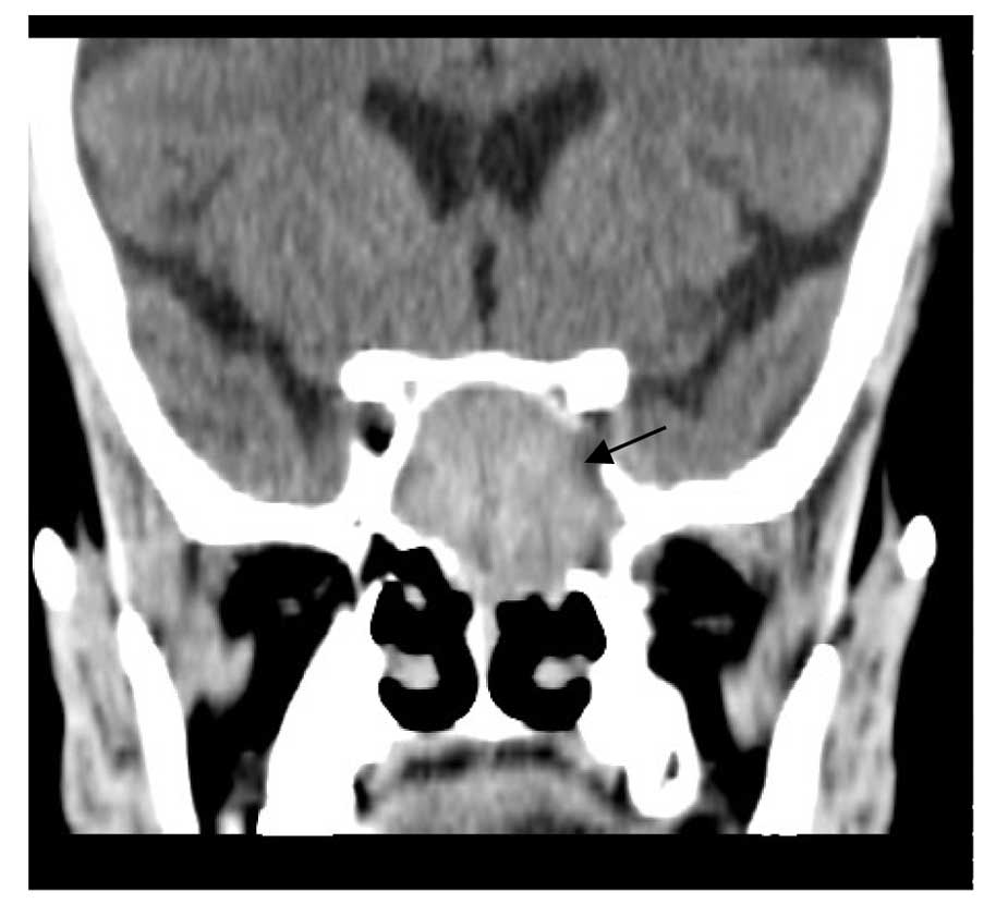
loss. Computed tomography (CT; SOMATOM Definition AS; Siemens
Healthcare GmbH, Forchheim, Germany) and magnetic resonance imaging
(MRI) scans (MAGNETOM Symphony 1.5T System; Siemens Healthcare
GmbH) demonstrated a homogenous soft-tissue lesion occupying the
left sphenoid sinus and invading the left cavernous sinus (Fig. 1). The patient received minimally
invasive biopsy of tissue obtained from the left sphenoid sinus in
the Department of Otorhinolaryngology, Head and Neck Surgery of The
Affiliated Baiyun Hospital of Guizhou Medical University. The
sample tissue was described as a red, friable, vascular tumor.
Intraoperative frozen section diagnosis was suggestive of a small
round cell tumor. In the next 3 days, the patients headache
gradually improved, but the patient developed sudden and complete
visual loss in the morning of the fourth day. Despite conservative
therapy with a high intravenous dose of methylprednisolone (500 mg
intravenously; Pfizer, Inc., New York, NY, USA), vision was not
recovered.
The final histological diagnosis was diffuse large
B-cell type PNHL. The patient was referred to the Medical Oncology
Clinic of Guizhou Medical University (Guiyang, China) for a staging
work-up, and received chemotherapy [standard 6 cycles of
cyclophosphamide (750 mg/m2 intravenously),
hydroxydaunorubicin (50 mg/m2 intravenously), oncovin
(1.4 mg/m2 intravenously) and prednisone (100 mg/day
orally) chemotherapy with adjuvant rituximab (375 mg/m2
intravenously); Bristol-Myers Squibb, New York, NY, USA] combined
with 40 Gy radiotherapy. At the time of writing the present study,
the patient had been disease-free for 3 years and received monthly
follow-ups in the Medical Oncology Clinic of Guizhou Medical
University.
Case 2
A 72-year-old female patient was admitted to The
Affiliated Baiyun Hospital of Guizhou Medical University on
September 2013 presenting with headache and blurred vision on the
left eye for 1 month, in addition to ptosis. The patient had a
history of chronic lymphocytic leukemia (CLL), which had manifested
7 years prior to admission to hospital, and had been treated with 4
cycles of fludarabine (25 mg/m2
intravenously)/cyclophosphamide (250 mg/m2
intravenously). The best corrected visual acuity was determined by
counting fingers on the left eye with mydriasis. Bone marrow biopsy
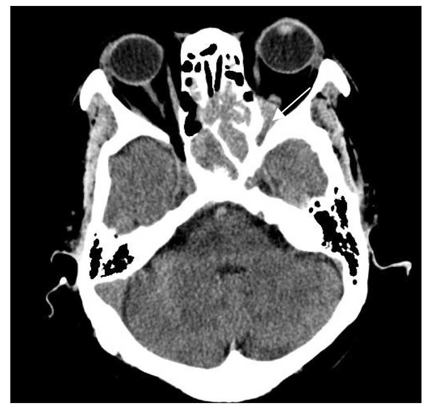
detected evidences of recurrence of leukemia. CT and MRI scans of
the brain and visual orbits demonstrated a mass occupying the left
sphenoid sinus with orbital apex involvement (Fig. 2); however, the clinician faced the
challenge of how to rescue the visual sight without the final
pathology result, which normally requires 4–5 days to be
identified. Based on the experience obtained from case 1, where the
suffering of the patient increased following endoscopic sinus
biopsy, it was considered that the possibility of sight loss would
be greater if the standard procedure was followed, that is, if
chemotherapy was started 3–4 days after the final pathological
diagnosis. Following a discussion with a hematologist in The
Affiliated Baiyun Hospital of Guizhou Medical University, it was
suggested that, due to the patients history of CLL, rituximab would
be administered following surgical biopsy to prevent sight
loss.
Following the consent of the patient, a single dose
of rituximab (375 mg/m2) was administered intravenously
following surgical biopsy. As a result of the treatment, the
mydriasis in the left eye was partly resolved on the morning of the
second day. No enhancing nerve lesion was observed during follow-up
examination, and her vision returned to baseline on the seventh
day. The final histological diagnosis was diffuse large B-cell type
PNHL, which was positive for cluster of differentiation (CD)20
(mouse monoclonal; dilution, 1:100; cat no., M0755; Dako, Glostrup,
Denmark) and negative for CD3 (mouse monoclonal; dilution, 1:80;
cat no., M7254; Dako). Immunohistochemistry was performed as
previously described (6). The patient
received standard cyclophosphamide, hydroxydaunorubicin, oncovin,
prednisone chemotherapy with adjuvant rituximab, as described for
case 1. At the time of writing the present study, the patient was
receiving monthly follow-ups and chemotherapy.
Discussion
Tumors of the sphenoid sinus are seldom associated
with symptoms such as nasal obstruction, epistaxis or nasal
discharge. By contrast, common symptoms include headache and
various other cranial neuropathies (7,8). The vital
anatomical regions in the vicinity of the sphenoid sinus are
commonly affected by this type of tumors (4).
The 2 cases described in the present study are
rather unique, since the patient from case 1, who did not present
with visual disturbance, lost her sight following surgical biopsy,
whereas the patient from case 2, who presented with visual
disturbance, did not experience the same outcome, possibly due to
the differences type of intervention. PNHL of the sphenoid sinus
poses a series of challenges to clinicians, due to its nonspecific
symptoms and unique anatomical sites of occurrence (9).
Previous studies have reported that T-cell and
natural killer/T-cell lymphomas mainly occur in the nasal cavity,
whereas B-cell lymphomas commonly occur in the sinus without nasal
disease (10,11). Clinically aggressive lymphomas,
including Burkitt's lymphoma and diffuse large B-cell lymphoma of
the sphenoid sinus, exhibit atypical characteristics, including
destruction of adjacent bone such as the skull base and
infiltration of the dura mater with intracranial invasion (12). To the best of our knowledge, only a
few cases of PNHL of the sphenoid sinus, including the present
ones, have been reported in the English literature to date
(13). Half of the reported patients
presented with visual disturbance. Thus, PNHL of the sphenoid sinus
should be suspected if a patient presents with a mass in the sinus
and visual disturbance.
The association between surgical biopsy and vision
loss has not yet been established (14). In cases of sudden vision loss,
paranasal lymphoma involving the optical nerves should be
considered, in addition to compression of the optical nerve, as a
possible cause (9). The conclusion
drawn from the outcomes of the present and previous studies is that
even minimally invasive biopsy may cause vision loss in patients
with PNHL of the sphenoid sinus. Furthermore, radiological evidence
of bone destruction in the lateral wall of the sphenoid sinus in
patients with no complaints of visual disturbance should be
concerning.
In the present study, case 1, who did not present
with visual disturbance, lost her sight following surgical biopsy,
whereas case 2, who presented with visual disturbance, did not
suffer the same result, possibly due to aggressive intervention. In
clinical practice, it is known that definite histological diagnosis
is required prior to the initiation of any treatment. The proximity
of the vital optical nerve to the sphenoid sinus is responsible for
the high incidence of visual disturbance in patients with lymphomas
of the sphenoid sinus (15). It
remains controversial whether or not patients with a very strong
clinical suspicion of lymphoma should receive aggressive lymphoma
treatment to prevent or recover certain complications (16). Therefore, the establishment of the
optimal treatment option for such patients is required.
References
|
1
|
Abbondanzo SL and Wenig BM: Non-Hodgkins
lymphoma of the sinonasal tract. A clinicopathologic and
immunophenotypic study of 120 cases. Cancer. 75:1281–1291. 1995.
View Article : Google Scholar : PubMed/NCBI
|
|
2
|
Yasumoto M, Taura S, Shibuya H and Honda
M: Primary malignant lymphoma of the maxillary sinus: CT and MRI.
Neuroradiology. 42:285–289. 2000. View Article : Google Scholar : PubMed/NCBI
|
|
3
|
Lee H, Choi KE, Park M, Lee SH and Baek S:
Primary diffuse large B-cell non-Hodgkin lymphoma of the paranasal
sinuses presenting as cavernous sinus syndrome. J Craniofac Surg.
25:e338–e339. 2014. View Article : Google Scholar : PubMed/NCBI
|
|
4
|
Peng KA, Kita AE, Suh JD, Bhuta SM and
Wang MB: Sinonasal lymphoma: Case series and review of the
literature. Int Forum Allergy Rhinol. 4:670–674. 2014. View Article : Google Scholar : PubMed/NCBI
|
|
5
|
Bhatia A, Saikia UN, Kumar Y and Dey P:
Fine needle aspiration cytology of spindle cell variant of diffuse
large B-cell lymphoma: A diagnostic dilemma. Cytopathology.
19:197–199. 2008. View Article : Google Scholar : PubMed/NCBI
|
|
6
|
Mubarak A, Wolters VM, Houwen RH and ten
Kate FJ: Immunohistochemical CD3 staining detects additional
patients with celiac disease. World J Gastroenterol. 21:7553–7557.
2015. View Article : Google Scholar : PubMed/NCBI
|
|
7
|
Alexander FW: Primary tumors of the
sphenoid sinus. Laryngoscope. 73:537–546. 1963. View Article : Google Scholar : PubMed/NCBI
|
|
8
|
Lawson W and Reino AJ: Isolated sphenoid
sinus disease: An analysis of 132 cases. Laryngoscope.
107:1590–1595. 1997. View Article : Google Scholar : PubMed/NCBI
|
|
9
|
Roth DB and Siatkowski RM: Bilateral
blindness as the initial presentation of lymphoma of the sphenoid
sinus. Am J Ophthalmol. 129:256–258. 2000. View Article : Google Scholar : PubMed/NCBI
|
|
10
|
Kim GE, Koom WS, Yang WI, Lee SW, Keum KC,
Lee CG, Suh CO, Hahn JS, Roh JK and Kim JH: Clinical relevance of
three subtypes of primary sinonasal lymphoma characterized by
immunophenotypic analysis. Head Neck. 26:584–593. 2004. View Article : Google Scholar : PubMed/NCBI
|
|
11
|
Yen TT, Wang RC, Jiang RS, Chen SC, Wu SH
and Liang KL: The diagnosis of sinonasal lymphoma: A challenge for
rhinologists. Eur Arch Otorhinolaryngol. 269:1463–1469. 2012.
View Article : Google Scholar : PubMed/NCBI
|
|
12
|
Bisdas S, Fetscher S, Feller AC, Baghi M,
Knecht R, Gstoettner W, Vogl TJ and Balzer JO: Primary B cell
lymphoma of the sphenoid sinus: CT and MRI characteristics with
correlation to perfusion and spectroscopic imaging features. Eur
Arch Otorhinolaryngol. 264:1207–1213. 2007. View Article : Google Scholar : PubMed/NCBI
|
|
13
|
Park YM, Cho JH, Cho JY, Huh JS and Ahn
JY: Non-Hodgkins lymphoma of the sphenoid sinus presenting as
isolated oculomotor nerve palsy. World J Surg Oncol. 5:862007.
View Article : Google Scholar : PubMed/NCBI
|
|
14
|
Kitthaweesin K and Theerakul T:
Neuro-ophthalmic manifestations in sinusitis patients who underwent
endoscopic sinus surgery. J Med Assoc Thai. 95:1543–1547.
2012.PubMed/NCBI
|
|
15
|
Inaki S, Okamura H and Chikamori Y: Adult
T-cell leukemia/lymphoma originating in the paranasal sinus. Arch
Otolaryngol Head Neck Surg. 114:1471–1473. 1988. View Article : Google Scholar : PubMed/NCBI
|
|
16
|
Touitou V, LeHoang P and Bodaghi B:
Primary CNS lymphoma. Curr Opin Ophthalmol. 26:526–533. 2015.
View Article : Google Scholar : PubMed/NCBI
|