Introduction
Ovarian cancer (OC) is the leading cause of
mortality resulting from gynecological cancers, and worldwide,
~114,000 patients succumb to OC annually (1). Although OC patients exhibit a 5-year
survival rate of 90% when treated at an early stage, 80% of
patients cannot be diagnosed until the advanced stages; therefore,
the 5-year survival rate is 30% (2).
OC recurrence occurs in 70% of patients following surgery and
platinum-based chemotherapy combined treatments, which causes
patients to become insensitive to chemotherapy (3). Consequently, tumor recurrence and drug
resistance are two common set-backs in the treatment of OC
(4,5).
Cancer stem cells (CSCs) are a type of tumor cell that possess an
unlimited potential for self-renewal and may differentiate into
multiple tumor cell types (6).
Numerous studies have demonstrated that CSCs possess various
characteristics, including a silent phenotype, enhanced DNA repair
capacity, expression of ATP-binding cassette (ABC) transporters and
anti-apoptotic proteins, and resistance to conventional
chemotherapy and radiotherapy (6–14).
Conventional chemotherapy may eliminate the majority of tumor
cells, but it has little impact on rare stem cell-like cells; and
remaining CSCs cause tumor recurrence and metastasis. Therefore,
studies concerning cancer should consider CSCs as a vital target to
achieve the complete ablation of tumors, and should not solely
focus on temporarily shrinking the tumor mass.
Goodell et al (15) reported that a small cell population
isolated from murine bone marrow demonstrated distinct
fluorescence-activated cell sorting (FACS) results compared with
the main cell population, termed the side population (SP) cells.
Numerous studies have demonstrated that SP cells, isolated from
numerous tumors, richly contain tumor-initiating cells that possess
stem cell characteristics (16–20). A
low-fluorescence staining phenotype is mediated by ABC transporters
(21), which provide a functional
method for isolating SP cells.
Although SP cells have been successfully isolated
from certain human and mouse ovarian cell lines (22,23), the
present study established an immortalized OC cell line from primary
cells in ascites and identified SP cells from this cell line.
Additionally, the present study investigated the biological
characteristics of the SP cells, including differentiation and
tumorsphere and colony formation, in addition to xenografted tumor
formation and ascites, metastasis and drug resistance of the
xenograft tumors.
Materials and methods
Establishment of an ovarian cancer
cell line
Primary cells were isolated from ascites of an
ovarian serous cystadenocarcinoma patient. Briefly, primary cells
were harvested by centrifugation at 300 × g for 5 min and red blood
cells were removed by 1X BD lysis buffer (BD Biosciences, Franklin
Lakes, NJ, USA) on ice for 1 min, followed by centrifugation at 300
× g for 3 min. Primary cells were cultured for 3 weeks in
Dulbecco's modified Eagle's medium (DMEM), supplemented with 10%
fetal bovine serum (FBS) (Gibco®; Thermo Fisher
Scientific, Inc., Waltham, MA, USA). Floating cells were collected
and re-cultured. Subsequent to subculturing for 15 passages,
primary cells were identified by a tumor xenograft model; the tumor
tissues were examined with hematoxylin and eosin staining and CA125
immunostaining.
Isolation of side population
cells
The cells were trypsinized, resuspended at
1.0×10⁶ cells/ml in pre-warmed DMEM containing 2% flow
cytometry staining buffer (CycleTEST™ PLUS DNA Reagent kit; BD
Biosciences) and incubated at 37°C for 10 min. The cells were
labeled with 5 µg/ml Invitrogen™ Hoechst 33342 dye (Thermo Fisher
Scientific, Inc.) at 37°C for 80 min, alone or combined with 50 mM
verapamil (Sigma-Aldrich, St. Louis, MO, USA), an inhibitor of ABC
transporters. The cells were counterstained with 1 µg/ml propidium
iodide. In total, 100,000 cells were analyzed on a BD Influx cell
sorter (BD Biosciences) and data were processed by BD FACSDiva
version 6.1.1 software (BD Biosciences).
Tumorsphere formation assay
A total of 500 SP and non-SP (NSP) cells were plated
onto a 24-well ultra-low attachment plate, and cultured in a
DMEM/F12 serum-free medium (Gibco®; Thermo Fisher
Scientific, Inc.) supplemented with 4 µg/ml insulin
(Sigma-Aldrich), 10% human leukocyte antigen B27
(Gibco®; Thermo Fisher Scientific, Inc.), 20 ng/ml
epidermal growth factor (EGF; Sigma-Aldrich), and 20 ng/ml basic
fibroblast growth factor (bFGF; Sigma-Aldrich), for 10 days.
Tumorspheres >50 mm in diameter were counted under a
phase-contrast microscope (IX50; Olympus Corporation, Tokyo,
Japan).
Soft agar colony formation assay
A total of 200 SP and NSP cells were resuspended in
a 0.8 ml growth medium (DMEM with EGF, bFGF and B27) containing
0.3% low-melting agarose (Sigma-Aldrich) and plated 3 times onto a
24-well plate pre-coated with a base layer of 0.8 ml growth medium
containing 0.6% low-melting agarose. The plates were incubated for
14–15 days until the size of colonies was large enough to count.
Colonies >75 µm in diameter or colonies that possessed >50
cells were counted as positive colonies.
Xenograft tumor assay
In total, 45 female 5-week old non-obese
diabetic-severe combined immune deficiency (NOD/SCID) mice weighing
16–20 g were purchased from Vital River Laboratories, Co., Ltd.
(Beijing, China). The mice were housed in a sterilized room with 12
h light/dark cycle, at a temperature of 22°C with 40–60% humidity.
Food and water were provided ad libitum. Animal experiment
protocols were approved by the Ethical Committee of the First
Affiliated Hospital of Jilin University (Changchun, China).
Subcutaneously, 1.0×10⁶ OC cells were injected into the
dorsal flank of 5-week-old female NOD/SCID mice, and tumor
formation and growth were observed after 6 weeks. SP and NSP cells
were sorted by FACS and resuspended in serum-free DMEM. In 0.1 ml
DMEM, 3.0×10³ or 3.0×10⁴ NSP and SP cells
were subcutaneously injected into the dorsal flank of 5-week-old
female NOD/SCID mice, or intraperitoneally injected into 5-week-old
female NOD/SCID mice (n=5). Formation of tumor and ascites were
examined subsequent to 3 weeks.
Drug resistance assay
SP, NSP and unsorted cells were seeded in 96-well
plates at 3,000 cells/well. Subsequent to 24-h culture, cells were
treated with 0.25, 0.50, 1.00, 2.00, 4.00, 8.00 and 16.00 µg/ml
cisplatin (Sigma-Aldrich) for 72 h. All treatments were run in
triplicates. Cells without cisplatin treatment were used as
negative controls. Following treatment, all cells were treated with
10 ml cell counting kit-8 (CCK-8) reagent (Dojindo Molecular
Technologies, Kumamoto, Japan) in 100 ml complete high-glucose DMEM
(Gibco®; Thermo Fisher Scientific, Inc.) and cultured in
normal culture conditions (DMEM with 10% FBS, 5% CO2 at
37°C) for 2.5 h. In total, 3 wells without cells were used as blank
controls. Absorbance at 490 nm was examined by a Model 550
microplate reader (Bio-Rad Laboratories, Inc., Hercules, CA, USA).
Cell viability was determined by the following formula: Cell
viability (%) = [optical density (OD) values of drug-treated cells
– OD values of blank controls] / (OD values of negative cells – OD
values of blank control) × 100. Cisplatin doses of 50% growth
inhibition (IC50) in SP, NSP and unsorted cells were
calculated using Prism software, version 4.0 (GraphPad Software,
Inc., La Jolla, CA, USA).
To additionally verify SP cell drug resistance,
unsorted cells were treated with the IC50 dose of
cisplatin for 3 h and cultured in a drug-free complete medium for
72 h. The SP cell ratio was examined by FACS. To investigate the
association between drug resistance and ABC transporters, SP cells
were treated with IC50 dose of cisplatin or cisplatin
with verapamil for 3 h, and were then cultured in a drug-free
complete medium for 72 h. Cell viability was examined using the
CCK-8 kit.
Statistical analysis
Statistical significance of differences between two
groups was analyzed using two-tailed unpaired Student's t-test.
Statistical analysis was performed using GraphPad Prism 5 software
(GraphPad Software, Inc., La Jolla, CA, USA). Data are presented as
the mean ± standard error of the mean. P<0.05 was considered to
indicate a statistically significantly difference.
Results
Establishment of human ovarian cancer
cell line from ascites
Primary OC cells were purified from ascites of an
ovarian serous cystadenocarcinoma patient (stage IIIc; grade 3) and
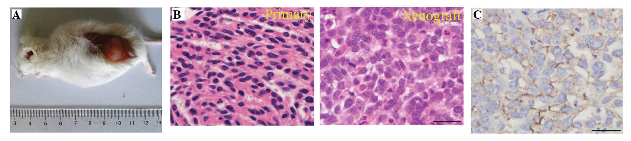
cultured for >50 passages over 2 years. Subcutaneously,
1.0×10⁶ OC cells were injected into the dorsal thigh of
female NOD/SCID mice and tumor formation was examined subsequent to
8 weeks (Fig. 1A). No histological or
cytological differences were observed between primary and
xenografted tumors; the two tumors demonstrated poorly
differentiated serous cystadenocarcinoma (Fig. 1B). Furthermore, the expression of
ovarian serous cystadenocarcinoma marker CA125 was observed in the
xenografted tumors (Fig. 1C)
(24).
Isolation of SP cells from established
OC cell line
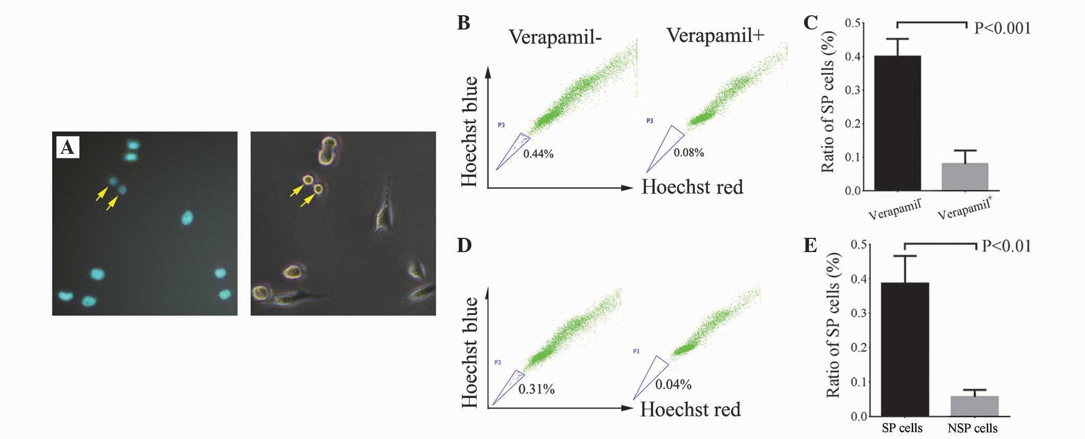
SP cells possess low-fluorescence staining
properties (21). FACS was used in
the present study to isolate SP cells using Hoechst 33342 staining.
Verapamil is an ABC transporter protein inhibitor that may
effectively reduce the SP cell ratio (25). The present study observed weak Hoechst
staining in certain cells, indicating that these are SP cells
(Fig. 2A). The SP cell ratio in the
OC cell line was 0.38% (n=5), while the SP cell ratio was
significantly reduced in the presence of verapamil (P=0.001;
Fig. 2B and C).
Biological characteristics of SP
cells: Differentiation potential of SP and NSP cells
To evaluate the differentiation potential of
isolated SP cells, 1×10³ SP and NSP cells were cultured
for 3 weeks and the SP cell ratios were compared between the two
groups. The average SP ratio was 0.39% following SP cell culture
for 3 weeks in vitro, which was a similar ratio to unsorted
OC cells. The average SP ratio was 0.06% in NSP cells subsequent to
3 weeks of culture, which was significantly lower compared with SP
and unsorted OC cells (P=0.004; Fig. 2D
and E). These results indicate that SP cells exhibited
competent self-renewal and differentiation capacities in
vitro, but NSP cells did not.
In vitro tumorsphere formation of SP
and NSP cells
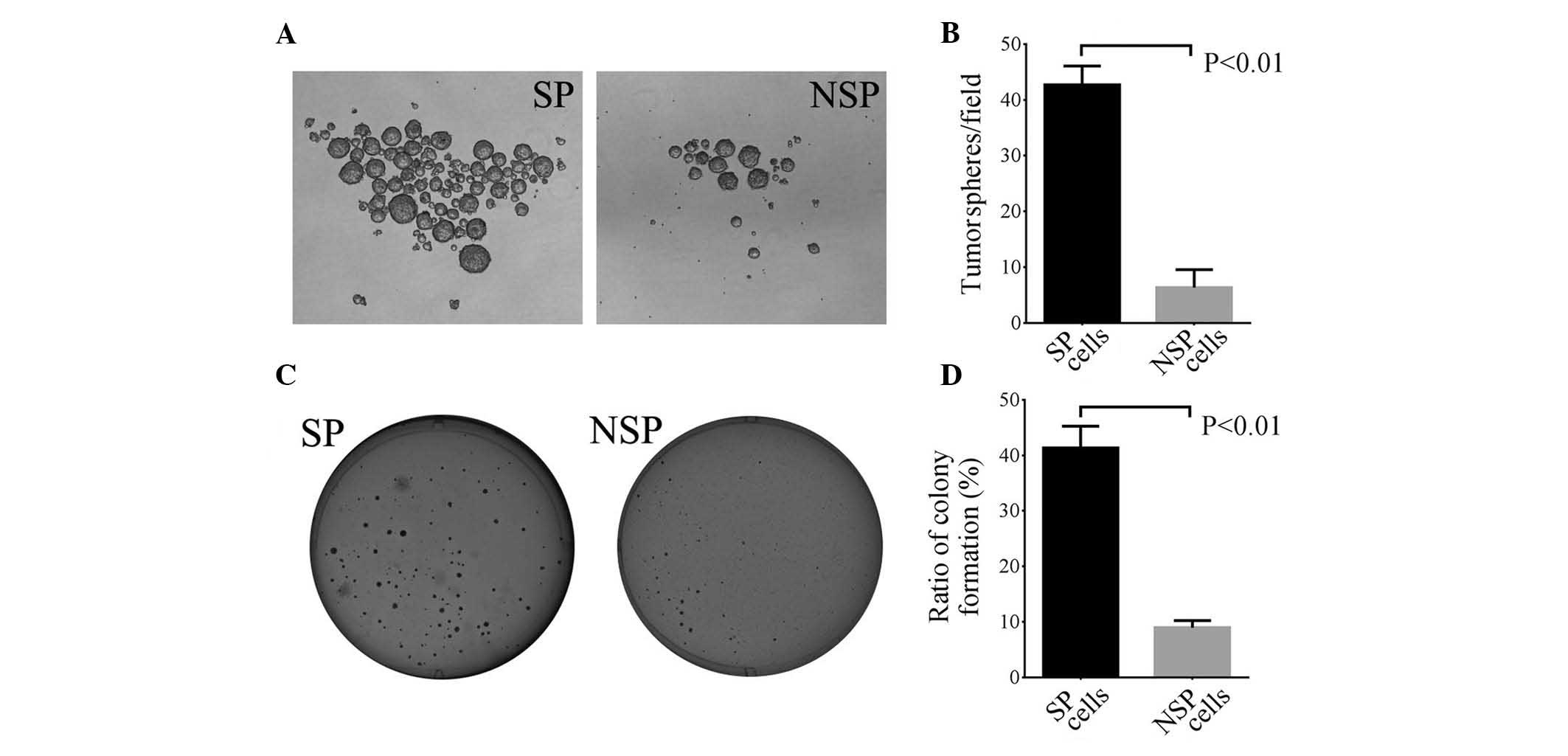
In total, 500 single SP and NSP cells were cultured
in a serum-free medium supplemented with growth factor for 10 days,
and tumorsphere formation was calculated. The present study
demonstrated that the average number of tumorspheres formed in SP
and NSP cells were 42.7 and 6.3, respectively (P=0.009; Fig. 3A and B). These data indicate that SP
cells possess improved self-renewal capacities compared with NSP
cells.
SP and NSP cell colony formation
To further verify the present findings, 500 SP and
NSP cells were seeded in soft agar medium and the cells were
cultured for 14–15 days to observe the colony formation ratio. The
present study observed that the colony formation ratio in SP cells
(41.33%) was significantly higher compared with NSP cells (8.93%)
(P=0.008; Fig. 3C and D). These data
suggest that SP cells have greater colony formation capacities than
NSP cells.
Tumorigenesis potential of SP and NSP
cells
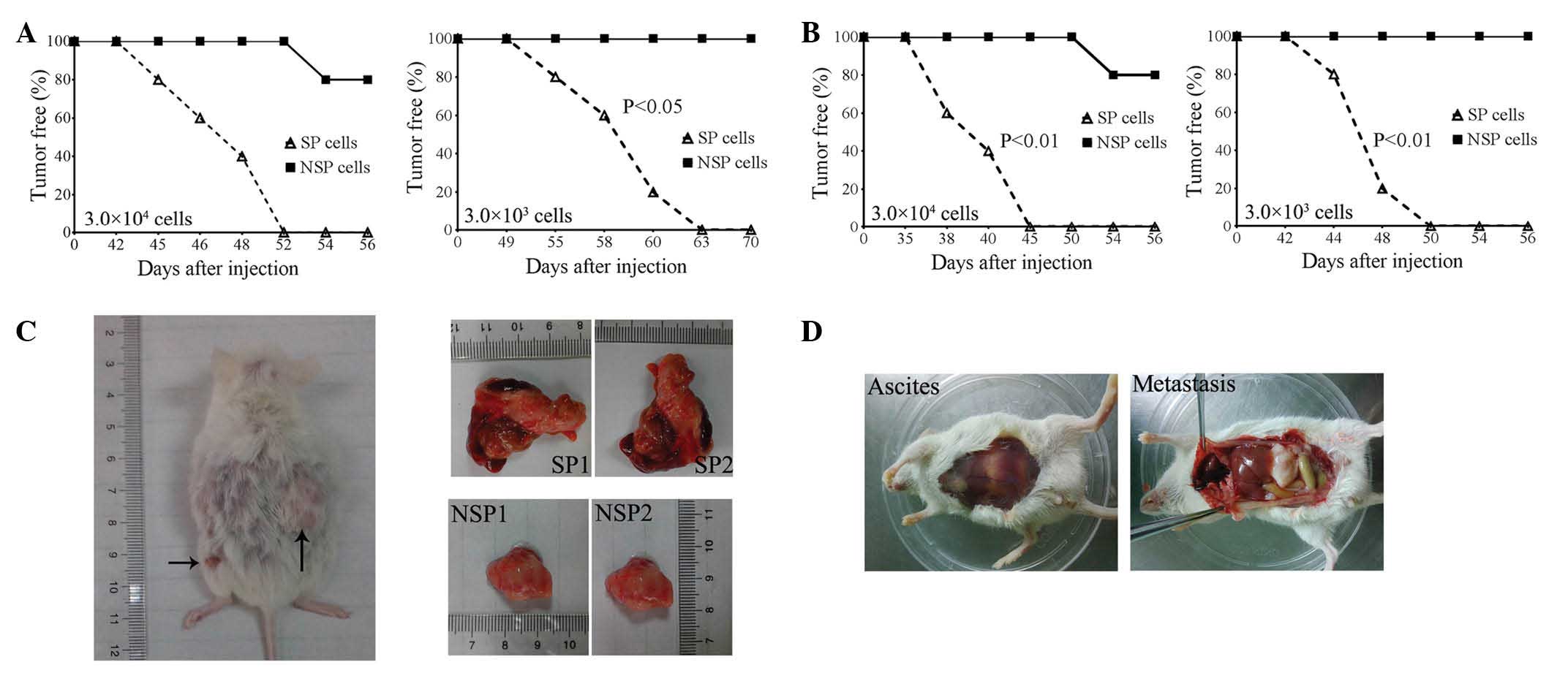
To evaluate the tumorigenesis potential of SP and
NSP cells, 3.0×10⁴ and 3.0×10³ SP and NSP
cells were subcutaneously injected into the dorsal flank of the
NOD/SCID mice. Xenografted tumors were observed in all 5 mice at 52
(3.0×10⁴ cells) and 63 (3.0×10³ cells) days
subsequent to the injection of SP cells. None of the mice formed
tumors 70 days following the injection of 3.0×10³ NSP
cells, and only 1 xenografted tumor was observed in 1 mouse at 54
days subsequent to injection of 3.0×10⁴ NSP cells
(Fig. 4A). Furthermore, the present
study also evaluated the intraperitoneal tumorigenesis of SP and
NSP cells. Following intraperitoneal injection of
3.0×10⁴ cells into NOD/SCID mice, tumors were observed
in the abdominal cavities of all 5 mice by 45 days post-injection,
however, only 1 mouse demonstrated ascites and metastasis after 56
days (Fig. 4B and C). At 56 days post
subcutaneous injection of 3.0×10⁴ cells, the xenografted
tumor size from SP cells was clearly larger than the size of the
tumor from NSP cells (Fig. 4C).
Similarly, the present study observed blood ascites and clear
metastasis in the abdominal cavity of all 5 mice following the
intraperitoneal injection of 3.0×10³ SP cells for 50
days, but this was not observed in any of the mice following the
injection of the same amount of NSP cells for 56 days (Fig. 4B and D). These data indicate that the
tumorigenesis potential of SP cells was significantly greater
compared with NSP cells.
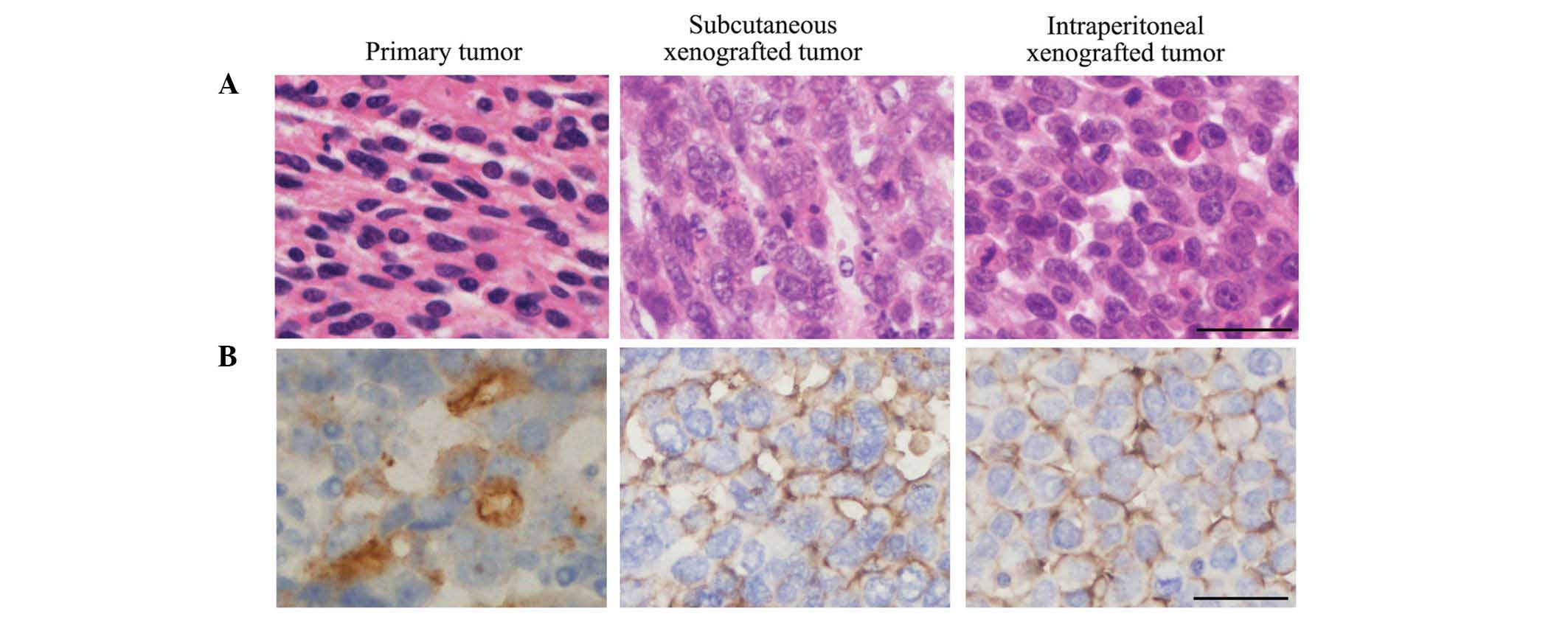
Histological and cytological
characteristics
In addition, the present study investigated the
histological and cytological differences between the primary and
xenografted tumors. Similar to the primary tumors, xenografted
tumors exhibited poorly differentiated serous cystadenocarcinoma
histological and cytological features following subcutaneous and
intraperitoneal injection (Fig. 5A).
Furthermore, CA125 expression was observed in two types of
xenografted tumors (Fig. 5B). These
results verify that SP cells possess competent self-renewal and
differentiation capacities in vivo.
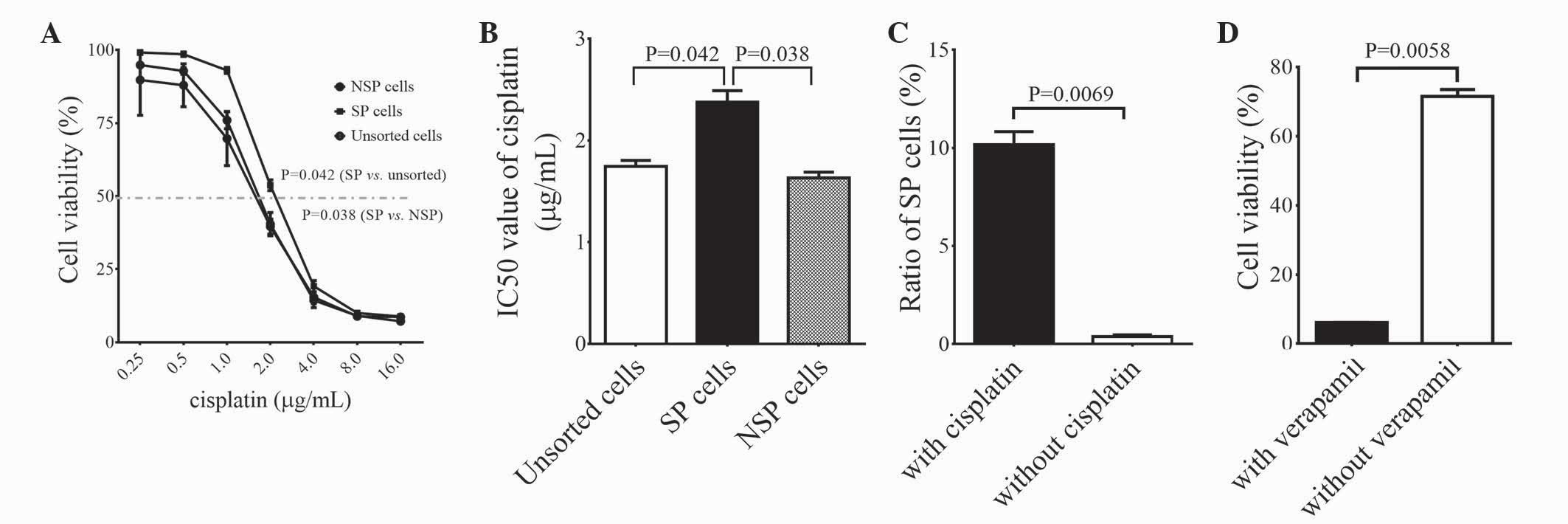
Drug resistance of SP cells
To investigate the drug resistance properties of SP
cells, the present study treated SP, NSP and unsorted cells with
0.25, 0.50, 1.00, 2.00, 4.00, 8.00 and 16.00 g/ml of cisplatin for
72 h and examined the cell viability using a CCK-8 kit. It was
observed that the cell viability of SP cells was significantly
higher compared with NSP and unsorted cells when treated with
1.0–4.0 g/ml of cisplatin (P=0.038 vs. NSP cells; P=0.042 vs.
unsorted cells; Fig. 6A). However,
there was no marked difference in cell viability following
cisplatin treatment between NSP and unsorted cells (Fig. 6A). IC50 values for
unsorted, SP and NSP cells were 1.75, 2.38 and 1.63 and µg/ml,
respectively, and the IC50 value of SP cells was
significantly higher than those of the NSP and unsorted cells
(P=0.038 vs. NSP cells; P=0.042 vs. unsorted cells; Fig. 6B). These results indicate that SP
cells exhibit greater drug resistance capacities compared with NSP
and unsorted cells. Furthermore, the SP cell ratio to unsorted
cells was significantly elevated following cisplatin treatment,
from 0.38 to 10.18% (P=0.007; Fig.
6C). These data support the theory that SP cells are more
resistant to cisplatin treatment compared with NSP and unsorted
cells.
To clarify the association between drug resistance
of SP cells and ABC transporters, the SP cells were treated with a
IC50 dose of cisplatin or cisplatin + verapamil. The
results demonstrated that cell viability was 71.41% following
cisplatin treatment, while it was reduced to 6.00% following
treatment with cisplatin + verapamil (P=0.006; Fig. 6D). These data indicate that verapamil
increases the sensitivity of SP cells to cisplatin treatment,
suggesting that the drug resistance of SP cells to cisplatin
depends on the drug efflux function of ABC transporters.
Discussion
Cancer cell lines are widely used in research to
study the biology of cancer and examine cancer treatments. These
cell lines can be cultured for long periods of time and share the
tumor-associated biological characteristics of the original tumor
(26). The present study successfully
established an OC cell line from primary cells in ascites of an OC
patient. This cell line had been passaged for 50 generations over 2
years and exhibited immortalized characteristics. Injection of this
cell line into NOD/SCID mice formed xenografted tumors that
revealed histological and cytological features similar to primary
tumors. Therefore, this cell line would be a valuable tool for
future functional studies on OC cancer.
Bonnet and Dick (27)
first identified leukemia stem cells in 1997, which are tumor stem
cells that have been isolated from various solid tumors, including
breast, cerebral, colon, prostate, cervix and ovarian tumors
(28–33). Numerous studies have demonstrated that
certain molecular markers, including cluster of differentiation
(CD)133, CD44 and CD117, may be used to isolate tumor stem cells
(24,34,35).
However, certain cancer stem cells may lack these specific cell
surface markers, or one type of stem-cell marker may only be
specific to a particular tumor (36).
SP cells within tumors are a small proportion of cancer cells with
stem-like properties, which can be isolated by FACS due to their
increased ability to export certain compounds, including Hoechst
33342 and chemotherapeutic agents. SP sorting is a type of
functional sorting that has general applicability, compared with
sorting stem cells by cell surface markers. Previous studies have
demonstrated that SP cells partly overlap with corresponding stem
cells, but SP cells in certain tumors, even the same type of tumor
from various individuals, do not have stem cell properties
(37–40). Therefore, the present study sought to
investigate important characteristics, including self-renewal,
differentiation ability and high tumorigenicity of SP cells from an
established OC cell line.
The present study verified the average population of
SP cells in our established cell line, which was 0.38%, and the
present results are similar to a previous study (0.01–5.00%)
(23). Stem cells have strong
self-renewal capacities and form floating spheroids in a serum-free
medium supplemented with growth factor, while differentiated tumor
cells become apoptotic (6,24). The present study additionally
demonstrated that isolated SP cells exhibited significantly
increased capacities in cell differentiation, colony formation and
in vivo tumorigenesis compared with NSP cells. Furthermore,
xenografted tumors from subcutaneous and intraperitoneal injections
of SP cells exhibited histological and cytological features similar
to primary tumors. These data indicate that isolated SP cells
possess a number of important similar characteristics to OC stem
cells, such as self-renewal, differentiation and high
tumorigenicity. In addition, the present study identified that
1.0×10⁴ NSP cells was able to form tumors in a small
proportion of mice, which contradicts previous studies (39,41). It is
possible that NSP cells may become contaminated with SP cells that
induce tumor formation during the long culture period of ~8
weeks.
It is well established that OC stem-like SP cells
are tumorigenic and chemoresistant (41,42). The
present study observed that isolated SP cells were more resistant
to the first-line OC chemotherapy drug cisplatin. In addition,
cisplatin treatment significantly increased the SP cell ratio. This
observation is consistent with a recent study, in which SP cell
ratio in ascites of patients with OC following chemotherapy was
significantly higher compared with ascites of those that did not
undergo chemotherapy (41). In OC
clinical treatments, the majority of patients would gradually
develop drug resistance following chemotherapy, which may be caused
by SP cell accumulation. Furthermore, it has been demonstrated that
ABC transporters are important in cisplatin-induced drug resistance
in OC (43,44). The present study identified an ABC
transporter inhibitor (verapamil) that potently abrogated SP cell
resistance to cisplatin, which is consistent with previous
observations in OC cell lines (12).
These data indicate that SP cell drug resistance to cisplatin
depends on the efflux function of ABC transporters, suggesting a
promising therapy for drug-resistant OC. In addition, previous
studies demonstrated that increased chemoresistance of SP cells is
associated with diverse mechanisms, including alterations in
signaling pathways and enzymes, reduced apoptosis and increased DNA
repair (19,45,46).
However, additional study is required to verify whether these
mechanisms exist in SP cells from OC.
In summary, the present study successfully isolated
and established an OC cell line. Isolated SP cells from the
established OC cell line possessed similar biological
characteristics as cancer stem cells, including self-renewal,
differentiation, proliferation, tumorigenesis and drug resistance.
This cell line and SP cells provide valuable models for studying OC
tumorigenesis and drug resistance mechanisms that may aid in
developing specific therapies for targeting OC SP or stem
cells.
Acknowledgements
The present study was supported by the Jilin
University Bethune plan B (grant no. B2012227) and Jilin Province
Science and Technology Development Program (grant no.
20140520032JH).
References
|
1
|
Pecorelli S, Favalli G, Zigliani L and
Odicino F: Cancer in women. Int J Gynaecol Obstet. 82:369–379.
2003. View Article : Google Scholar : PubMed/NCBI
|
|
2
|
Ozols RF: Update on the management of
ovarian cancer. Cancer J. 8(Suppl 1): S22–S30. 2002.PubMed/NCBI
|
|
3
|
Ozols RF: Treatment goals in ovarian
cancer. Int J Gynecol Cancer. 15(Suppl 1): 3–11. 2005. View Article : Google Scholar : PubMed/NCBI
|
|
4
|
Cannistra SA: Cancer of the ovary. N Engl
J Med. 351:2519–2529. 2004. View Article : Google Scholar : PubMed/NCBI
|
|
5
|
Ozols RF, Bookman MA, Connolly DC, Daly
MB, Godwin AK, Schilder RJ, Xu X and Hamilton TC: Focus on
epithelial ovarian cancer. Cancer Cell. 5:19–24. 2004. View Article : Google Scholar : PubMed/NCBI
|
|
6
|
Reya T, Morrison SJ, Clarke MF and
Weissman IL: Stem cells, cancer, and cancer stem cells. Nature.
414:105–111. 2001. View
Article : Google Scholar : PubMed/NCBI
|
|
7
|
Zhou BB, Zhang H, Damelin M, Geles KG,
Grindley JC and Dirks PB: Tumour-initiating cells: Challenges and
opportunities for anticancer drug discovery. Nat Rev Drug Discov.
8:806–823. 2009. View
Article : Google Scholar : PubMed/NCBI
|
|
8
|
Gottesman MM: Mechanisms of cancer drug
resistance. Annu Rev Med. 53:615–627. 2002. View Article : Google Scholar : PubMed/NCBI
|
|
9
|
Schatton T, Murphy GF, Frank NY, Yamaura
K, Waaga-Gasser AM, Gasser M, Zhan Q, Jordan S, Duncan LM,
Weishaupt C, et al: Identification of cells initiating human
melanomas. Nature. 451:345–349. 2008. View Article : Google Scholar : PubMed/NCBI
|
|
10
|
Yu F, Yao H, Zhu P, Zhang X, Pan Q, Gong
C, Huang Y, Hu X, Su F, Lieberman J and Song E: let-7 regulates
self renewal and tumorigenicity of breast cancer cells. Cell.
131:1109–1123. 2007. View Article : Google Scholar : PubMed/NCBI
|
|
11
|
Zhou S, Schuetz JD, Bunting KD, Colapietro
AM, Sampath J, Morris JJ, Lagutina I, Grosveld GC, Osawa M,
Nakauchi H and Sorrentino BP: The ABC transporter Bcrp1/ABCG2 is
expressed in a wide variety of stem cells and is amolecular
determinant of the side-population phenotype. Nature Med.
7:1028–1034. 2001. View Article : Google Scholar : PubMed/NCBI
|
|
12
|
Ito K, Hirao A, Arai F, Matsuoka S, Takubo
K, Hamaguchi I, Nomiyama K, Hosokawa K, Sakurada K and Nakagata N:
Regulation of oxidative stress by ATM isrequired for self-renewal
of haematopoietic stem cells. Nature. 31:997–1002. 2004. View Article : Google Scholar
|
|
13
|
Bao S, Wu Q, McLendon RE, Hao Y, Shi Q,
Hjelmeland AB, Dewhirst MW, Bigner DD and Rich JN: Glioma stem
cells promote radioresistance by preferential activation of the DNA
damage response. Nature. 444:756–760. 2006. View Article : Google Scholar : PubMed/NCBI
|
|
14
|
Diehn M, Cho RW, Lobo NA, Kalisky T, Dorie
MJ, Kulp AN, Qian D, Lam JS, Ailles LE, Wong M, et al: Association
of reactive oxygen species levels and radioresistance in cancer
stem cells. Nature. 458:780–783. 2009. View Article : Google Scholar : PubMed/NCBI
|
|
15
|
Goodell MA, Brose K, Paradis G, Conner AS
and Mulligan RC: Isolation and functional properties of murine
hematopoietic stem cells that are replicating in vivo. J Exp
Med. 183:1797–1806. 1996. View Article : Google Scholar : PubMed/NCBI
|
|
16
|
Meirelles K, Benedict LA, Dombkowski D,
Pepin D, Preffer FI, Teixeira J, Tanwar PS, Young RH, MacLaughlin
DT, Donahoe PK and Wei X: Human ovarian cancer stem/progenitor
cells are stimulated by doxorubicin but inhibited by Mullerian
inhibiting substance. Proc Natl Acad Sci USA. 109:2358–2363. 2012.
View Article : Google Scholar : PubMed/NCBI
|
|
17
|
Christgen M, Geffers R, Ballmaier M,
Christgen H, Poczkaj J, Krech T, Kreipe H and Lehmann U:
Down-regulation of the fetal stem cell factor SOX17 by H33342: A
mechanism responsible for differential gene expression in breast
cancer side population cells. J Biol Chem. 285:6412–6418. 2010.
View Article : Google Scholar : PubMed/NCBI
|
|
18
|
Oates JE, Grey BR, Addla SK, Samuel JD,
Hart CA, Ramani VA, Brown MD and Clarke NW: Hoechst 33342 side
population identification is a conserved and unified mechanism in
urological cancers. Stem Cells Dev. 18:1515–1522. 2009. View Article : Google Scholar : PubMed/NCBI
|
|
19
|
Luo Y, Ellis LZ, Dallaglio K, Takeda M,
Robinson WA, Robinson SE, Liu W, Lewis KD, McCarter MD, Gonzalez R,
et al: Side population cells from human melanoma tumors reveal
diverse mechanisms for chemoresistance. J Invest Dermatol.
132:2440–2450. 2012. View Article : Google Scholar : PubMed/NCBI
|
|
20
|
Mas A, Cervelló I, Gil-Sanchis C, Faus A,
Ferro J, Pellicer A and Simón C: Identification and
characterization of the human leiomyoma side population as putative
tumor-initiating cells. Fertil Steril. 98:741–751, e6. 2012.
View Article : Google Scholar : PubMed/NCBI
|
|
21
|
Gottesman MM, Fojo T and Bates SE:
Multidrug resistance in cancer: Role of ATP-dependent transporters.
Nat Rev Cancer. 2:48–58. 2002. View
Article : Google Scholar : PubMed/NCBI
|
|
22
|
Dou J, Jiang C, Wang J, Zhang X, Zhao F,
Hu W, He X, Li X, Zou D and Gu N: Using ABCG2-molecule-expressing
side population cells to identify cancer stem-like cells in a human
ovarian cell line. Cell Biol Int. 35:227–234. 2011. View Article : Google Scholar : PubMed/NCBI
|
|
23
|
Szotek PP, Pieretti-Vanmarcke R, Masiakos
PT, Dinulescu DM, Connolly D, Foster R, Dombkowski D, Preffer F,
Maclaughlin DT and Donahoe PK: Ovarian cancer side population
defines cells with stem cell-like characteristics and Mullerian
Inhibiting Substance responsiveness. Proc Natl Acad Sci USA.
103:11154–11159. 2006. View Article : Google Scholar : PubMed/NCBI
|
|
24
|
Zhang S, Balch C, Chan MW, Lai HC, Matei
D, Schilder JM, Yan PS, Huang TH and Nephew KP: Identification and
characterization of ovarian cancer-initiating cells from primary
human tumors. Cancer Res. 68:4311–4320. 2008. View Article : Google Scholar : PubMed/NCBI
|
|
25
|
Jakubikova J, Adamia S, Kost-Alimova M,
Klippel S, Cervi D, Daley JF, Cholujova D, Kong SY, Leiba M, Blotta
S, et al: Lenalidomide targets clonogenic side population in
multiple myeloma: Pathophysiologic and clinical implications.
Blood. 117:4409–4419. 2011. View Article : Google Scholar : PubMed/NCBI
|
|
26
|
Douglas EJ, Fiegler H, Rowan A, Halford S,
Bicknell DC, Bodmer W, Tomlinson IP and Carter NP: Array
comparative genomic hybridization analysis of colorectal cancer
cell lines and primary carcinomas. Cancer Res. 64:4817–4825. 2004.
View Article : Google Scholar : PubMed/NCBI
|
|
27
|
Bonnet D and Dick JE: Human acute myeloid
leukemia is organized as a hierarchy that originates from a
primitive hematopoietic cell. Nat Med. 3:730–737. 1997. View Article : Google Scholar : PubMed/NCBI
|
|
28
|
Al-Hajj M, Wicha MS, Benito-Hernandez A,
Morrison SJ and Clarke MF: Prospective identification of
tumorigenic breast cancer cells. Proc Natl Acad Sci USA.
100:3983–3988. 2003. View Article : Google Scholar : PubMed/NCBI
|
|
29
|
Collins AT, Berry PA, Hyde C, Stower MJ
and Maitland NJ: Prospective identification of tumorigenic prostate
cancer stem cells. Cancer Res. 65:10946–10951. 2005. View Article : Google Scholar : PubMed/NCBI
|
|
30
|
O'Brien CA, Pollett A, Gallinger S and
Dick JE: A human colon cancer cell capable of initiating tumour
growth in immunodeficient mice. Nature. 445:106–110. 2007.
View Article : Google Scholar : PubMed/NCBI
|
|
31
|
Singh SK, Hawkins C, Clarke ID, Squire JA,
Bayani J, Hide T, Henkelman RM, Cusimano MD and Dirks PB:
Identification of human brain tumour initiating cells. Nature.
432:396–401. 2004. View Article : Google Scholar : PubMed/NCBI
|
|
32
|
Zhang SL, Wang YS, Zhou T, Yu XW, Wei ZT
and Li YL: Isolation and characterization of cancer stem cells from
cervical cancer HeLa cells. Cytotechnology. 64:477–484. 2012.
View Article : Google Scholar : PubMed/NCBI
|
|
33
|
Bapat SA, Mali AM, Koppikar CB and Kurrey
NK: Stem and progenitor-like cells contribute to the aggressive
behavior of human epithelial ovarian cancer. Cancer Res.
65:3025–3029. 2005.PubMed/NCBI
|
|
34
|
Baba T, Convery PA, Matsumura N, Whitaker
RS, Kondoh E, Perry T, Huang Z, Bentley RC, Mori S, Fujii S, et al:
Epigenetic regulation of CD133 and tumorigenicity of
CD133+ ovarian cancer cells. Oncogene. 28:209–218. 2009.
View Article : Google Scholar : PubMed/NCBI
|
|
35
|
Clarke MF, Dick JE, Dirks PB, Eaves CJ,
Jamieson CH, Jones DL, Visvader J, Weissman IL and Wahl GM: Cancer
stem cells - perspectives on current status and future directions:
AACR Workshop on cancer stem cells. Cancer Res. 66:9339–9344. 2006.
View Article : Google Scholar : PubMed/NCBI
|
|
36
|
Burkert J, Otto WR and Wright NA: Side
populations of gastrointestinal cancers are not enriched in stem
cells. J Pathol. 214:564–573. 2008. View Article : Google Scholar : PubMed/NCBI
|
|
37
|
Kai K, D'Costa S, Yoon BI, Brody AR, Sills
RC and Kim Y: Characterization of side population cells in human
malignant mesothelioma cell lines. Lung Cancer. 70:146–151. 2010.
View Article : Google Scholar : PubMed/NCBI
|
|
38
|
Mitsutake N, Iwao A, Nagai K, Namba H,
Ohtsuru A, Saenko V and Yamashita S: Characterization of side
population in thyroid cancer cell lines: Cancer stem-like cells are
enriched partly but not exclusively. Endocrinology. 148:1797–1803.
2007. View Article : Google Scholar : PubMed/NCBI
|
|
39
|
Lichtenauer UD, Shapiro I, Geiger K,
Quinkler M, Fassnacht M, Nitschke R, Rückauer KD and Beuschlein F:
Side population does not define stem cell-like cancer cells in the
adrenocortical carcinoma cell line NCI h295R. Endocrinology.
149:1314–1322. 2008. View Article : Google Scholar : PubMed/NCBI
|
|
40
|
Wu C, Wei Q, Utomo V, Nadesan P, Whetstone
H, Kandel R, Wunder JS and Alman BA: Side population cells isolated
from mesenchymal neoplasms have tumor initiating potential. Cancer
Res. 67:8216–8222. 2007. View Article : Google Scholar : PubMed/NCBI
|
|
41
|
Rizzo S, Hersey JM, Mellor P, Dai W,
Santos-Silva A, Liber D, Luk L, Titley I, Carden CP, Box G, et al:
Ovarian cancer stem cell-like side populations are enriched
following chemotherapy and overexpress EZH2. Mol Cancer Ther.
10:325–335. 2011. View Article : Google Scholar : PubMed/NCBI
|
|
42
|
Hu L, McArthur C and Jaffe RB: Ovarian
cancer stem-like side-population cells are tumourigenic and
chemoresistant. Br J Cancer. 102:1276–1283. 2010. View Article : Google Scholar : PubMed/NCBI
|
|
43
|
Moreno-Smith M, Halder JB, Meltzer PS,
Gonda TA, Mangala LS, Rupaimoole R, Lu C, Nagaraja AS, Gharpure KM,
Kang Y, et al: ATP11B mediates platinum resistance in ovarian
cancer. J Clin Invest. 123:2119–2130. 2013. View Article : Google Scholar : PubMed/NCBI
|
|
44
|
Januchowski R, Zawierucha P, Andrzejewska
M, Ruciński M and Zabel M: Microarray-based detection and
expression analysis of ABC and SLC transporters in drug-resistant
ovarian cancer cell lines. Biomed Pharmacother. 67:240–245. 2013.
View Article : Google Scholar : PubMed/NCBI
|
|
45
|
Chen Y, Li D, Wang D, Liu X, Yin N, Song
Y, Lu SH, Ju Z and Zhan Q: Quiescence and attenuated DNA damage
response promote survival of esophageal cancer stem cells. J Cell
Biochem. 113:3643–3652. 2012. View Article : Google Scholar : PubMed/NCBI
|
|
46
|
Li XX, Dong Y, Wang W, Wang HL, Chen YY,
Shi GY, Yi J and Wang J: Emodin as an effective agent in targeting
cancer stem-like side population cells of gallbladder carcinoma.
Stem Cells Dev. 22:554–566. 2013. View Article : Google Scholar : PubMed/NCBI
|