Introduction
Hematopoietic tissue malignancies are a known late
side effect of previous treatments in patients that have received
chemotherapy or radiotherapy for a neoplastic or other
non-malignant condition (1–3). In 2008, the World Health Organization
(WHO) published a review for the classification of acute leukemia
(4–6)
that included a novel category for neoplasms that may emerge
following a previous treatment. In addition to improving treatment
outcomes, the evolution of treatment options over the past decade
has resulted in an increased risk of secondary malignancy (3,7). The
treatment options include the use of numerous novel drugs as
molecular targeted agents, novel and more intense protocols that
use increased maintenance regimens and neoadjuvant therapies
(8–10).
The diagnosis of therapy-related acute myeloid
leukemia (t-AML) is based on the previous exposure of a patient to
cytotoxic agents (4,6). In addition to this causal effect, the
mechanism remains obscure; however, the mechanism appears to result
from the direct mutational effect induced by prior therapy in the
cells. Each cytotoxic agent may damage DNA, which may lead to
various cytogenetic abnormalities and contribute to various
biological characteristics in t-AML (11–14). The
time between the primary malignancy diagnosis and AML development,
which is termed the latency time, varies with the treatment and
cumulative dose administered, and the dose intensification of the
previous cytotoxic treatment (15–18).
Morphological and cytogenetic findings are also affected by
previous treatments (3,7,15,19,20).
There are two distinct groups in t-AML, according to
the previous treatment administered, consisting of the group of
patients that were treated with alkylating agents or radiotherapy,
and the group that was treated with topoisomerase II inhibitors
(3,21). In the first group of patients, bone
marrow myelodysplastic changes usually precede t-AML, which allows
a longer latency period of 5–7 years (21,22).
Poorer cytogenetic findings are also a hallmark of the group of
patients treated with alkylating agents or radiotherapy, and are
the cytogenetic findings are often accompanied by a complex
karyotype with loss of cytogenetic material (21–24).
Patients previously treated with topoisomerase II inhibitors
usually present with balanced translocations. A shorter latency
period, which can be ~12 months in certain cases, exhibits a
rapidly progressive leukemia (21–25). The
common usage of a combination of topoisomerase II inhibitors and
alkylating agents led to the disappearance of these two groups
(8–10).
t-AML is a rare and frequently fatal disease
(2,19,23,26–28).
The poor disease outcome may be explained by numerous potential
factors, which include the prognosis of the primary malignancy and
the toxic products of previous cytotoxic treatments that may
compromise chemotherapy for AML (2,19,23,26–28). In
addition to these facts, the frequent association between t-AML and
poor cytogenetics appear to result in chemotherapy resistance
(21,23,29).
Patients and methods
Patients
In the last 10 years, 231 patients were diagnosed
with acute leukemia in the Onco-Hematology Department of the
Portuguese Institute of Oncology of Porto (Porto, Norte, Portugal),
the oncological referral hospital in North Portugal. Of these
patients, 38 were diagnosed with t-AML. The medical records of the
selected patients were reviewed in the present study. Data
regarding the patient demographics, primary diagnosis and
treatment, age at therapy-related myeloid neoplasm (t-MN) onset,
latency time, cytogenetic characteristics, AML therapy and outcome
were collected. The diagnosis of t-MN was based on the WHO 2008
classification, which concerns the previous medical history of the
patient and the presence of acute leukemia laboratory findings.
Bone marrow consisting of >20% myeloblasts conferred an AML
diagnosis.
The ethics committee of Instituto Português de
Oncologia do Porto (Porto, Portugal) approved the present study.
Written informed consent was obtained from the patients for
participation in the study.
Cytogenetics
G-banding was performed on bone marrow samples
obtained from the patients using standard techniques (30). The cytogenetic results were presented
according to the International System for Human Cytogenetic
Nomenclature (31). Patients were
then analyzed according to cytogenetic risk subgroups, as defined
by the Southwest Oncology Group (SWOG) (32).
Statistical analyses
An analysis of the data was performed SPSS software
for Windows (version 17.0; SPSS, Inc., Chicago, IL, USA).
Differences in proportions were evaluated using the χ2
test. The probability of survival was calculated. The means and
life tables were computed using the product limit estimate of
Kaplan-Meier and analyzed by the Breslow (generalized Wilcoxon)
test, which is a statistical test for equality of survival
distributions. P<0.05 was considered to indicate a statistically
significant difference. The hazard ratio (HR) was also assessed
using multivariate Cox regression analyses of the 1-, 3- and 5-year
overall survival (OS) rates. Using the HR value, the probability
(P) of survival was calculated as previously described in the
literature: HR = odds = P / (1 - P); P = HR / (1 + HR) (33).
The survival duration was defined as the time
between diagnosis and either mortality or time of the last clinical
evaluation of the patient. Only patients achieving complete
remission were evaluated for disease-free survival, which was
defined as the time between diagnosis and relapse. The latency
interval was defined as the time period between first cytotoxic
therapy and t-MN diagnosis.
Results
Patients
In total, 231 adult patients with AML were
identified. The median age at diagnosis was 51 years (range, 15–78)
and 58% (n=134) were females. t-AML was identified in 38 patients
and the remaining 193 were diagnosed with de novo AML.
Patients diagnosed with de novo AML were younger compared
with t-AML patients (median age, 50 vs. 56 years; P<0.000).
De novo AML and t-AML were more prevalent in females than in
males (55.4 vs. 71.1%), but this was not a statistically
significant difference (Table I).
 | Table I.Characteristics of patients with
de novo AML and t-AML. |
Table I.
Characteristics of patients with
de novo AML and t-AML.
| Characteristic | De novo AML,
n (%) | t-AML, n (%) | P-value |
|---|
| Total | 193 (83.5) | 38 (16.5) |
|
| Gender |
|
|
|
|
Male | 86 | 11 | 0.08* |
|
Female | 107 | 27 |
|
| Age, years
(range) | 50 (15–78) | 56 (17–78) |
<0.000+ |
| Cytogenetics |
|
|
|
|
Abnormal | 74
(46.6) | 27 (94.7) | 0.009* |
|
Normal | 77
(39.9) | 9
(23.7) |
|
|
Missing | 26
(13.5) | 2 (5.3) |
|
|
CBF | 53
(27.5) | 17 (44.7) |
<0.000* |
| 5,7 and
8 alterations | 13 (6.7) | 3 (7.9) | 0.912* |
|
t(9,11) | 8
(4.1) | 2 (5.3) | 0.845* |
|
Complex | 16 (8.3) | 5
(13.2) | 0.441* |
| SWOG risk
score |
|
|
|
|
Favorable | 56
(30.3) | 9 (23.7) | 0.656* |
|
Intermediate | 97
(52.4) | 14 (36.8) | 0.228* |
|
Unfavorable | 32
(17.3) | 11 (28.9) | 0.042* |
| Complete
response | 150 (84.3) | 26 (68.4) | 0.872* |
Cytogenetics
An abnormal karyotype is the most frequent finding
in t-AML patients when compared with AML patients (94.7 vs. 46.6%;
P<0.000; Table I). Although there
were no significant differences between the prevalence of
cytogenetic abnormalities when stratifying patients using the SWOG
score, t-AML patients were more frequently placed in the
unfavorable risk score group than AML patients (28.9 vs. 17.3%;
P=0.042). Between the favorable and intermediate risk groups, no
differences were found (Table I).
When analyzing the cytogenetic alterations in t-AML
by comparing the solid tumors and the hematological malignancy
groups, there were no significant differences. The loss of
cytogenetic material was more frequent among t-AML patients
following hematological malignancies. Balanced translocations,
including core binding factor (CBF) abnormalities, were more
frequent subsequent to diagnosis with solid tumors (Table II).
| Table II.Cytogenetic abnormalities, according
to the type of tumor. |
Table II.
Cytogenetic abnormalities, according
to the type of tumor.
| Karyotype | Solid tumor, n
(%) | Hematological
malignancy, n (%) | P-value |
|---|
| Total | 28
(100.0) | 8 (80) |
|
| Normal | 6
(21.4) | 3 (30) | 0.355 |
| CBF and balanced
translocations | 17 (60.7) | 2 (20) | 0.743 |
| Alterations on
chromosomes 5,7 and 8 | 2 (7.1) | 1 (10) | 0.629 |
| Complex | 3
(10.7) | 2 (20) | 0.303 |
Previous disease and treatment
The majority of patients with t-AML were previously
diagnosed with a solid tumor (n=28; 74%). The most common solid
tumor was breast cancer (n=15; 39.5%), followed by uterine cancer
(n=4; 10.5%) and prostate cancer (n=2; 5.3%). In total, 7 cases
(18.4%) were associated with several neoplastic conditions, each
unique to a single case. In the hematological malignancies group, a
previous lymphoproliferative disorder was identified in all
patients, with non-Hodgkin's lymphoma observed in 8 patients
(21.1%) and Hodgkin's lymphoma in 2 cases (5.3%).
Chemotherapy alone was performed on 15 patients
(39.5%), 13 patients (34.2%) were treated only with radiotherapy
and the remaining patients had received previous chemo- and
radiotherapy. The majority of patients treated with chemotherapy
alone were treated with a combination of alkylating agents and
topoisomerase II inhibitors (n=12; 80%). Only 3 patients (20%) were
treated with alkylating agents. The median time between the first
antineoplastic treatment and t-AML was 3 years (range, 1–19
years).
According to the previous treatment received, a
shorter latency time was observed in the group treated with the
combination of chemo- and radiotherapy, which had a median latency
time of 3.5 years (range, 2–13). However, the differences between
the patients treated with chemotherapy alone, radiotherapy alone or
a combination of the two lacked significance (P=0.619). The
complete response to induction treatment was similar in de
novo and therapy related AML groups (P=0.872).
Survival
The median follow-up time of all patients was 23
months (range, 1–141 months). The median OS time in the entire
cohort (n=231) was 39 months [95% confidence interval (CI),
10.458–67.542] and the OS rate in the subsequent 5 years was
45.9%.
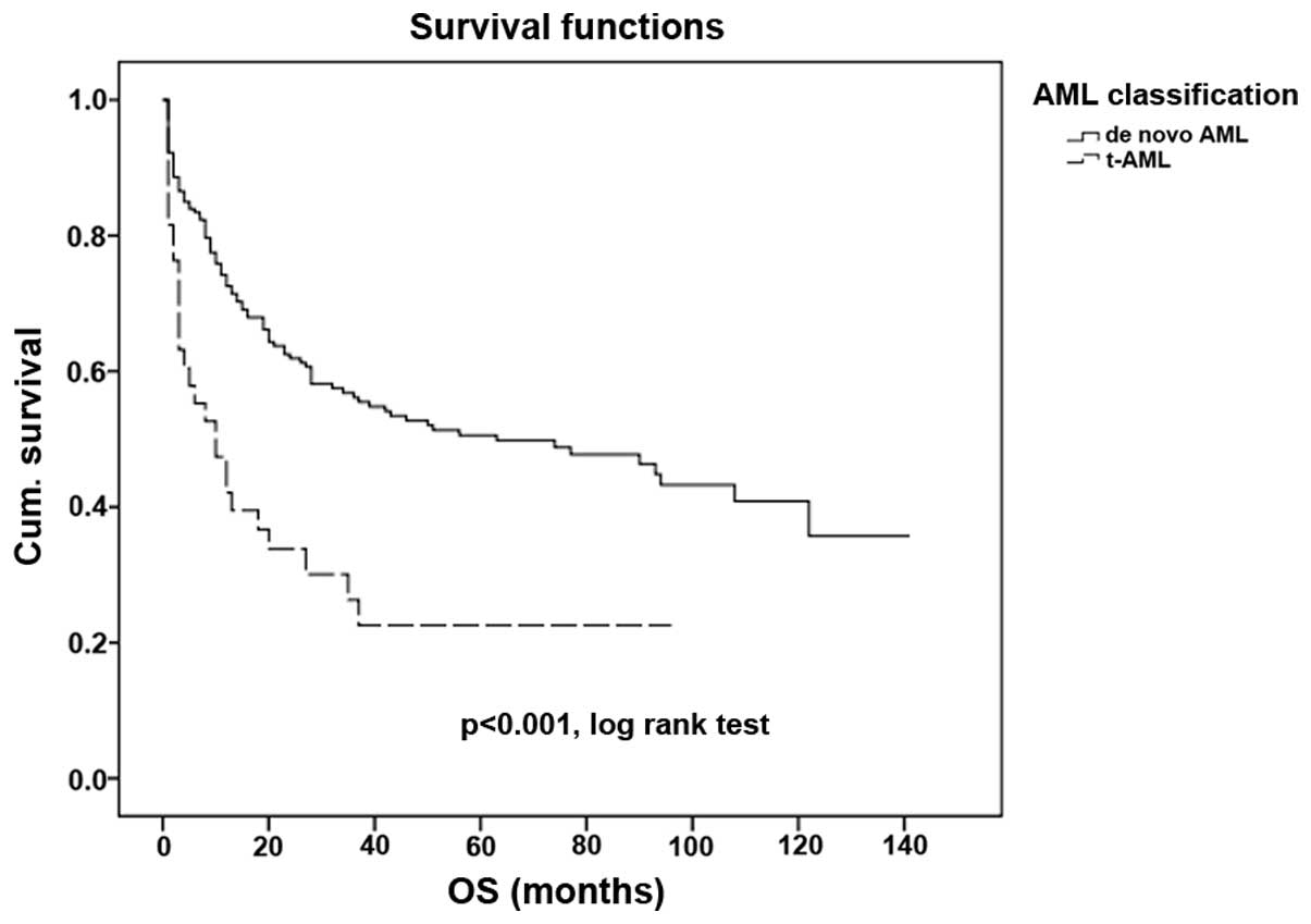
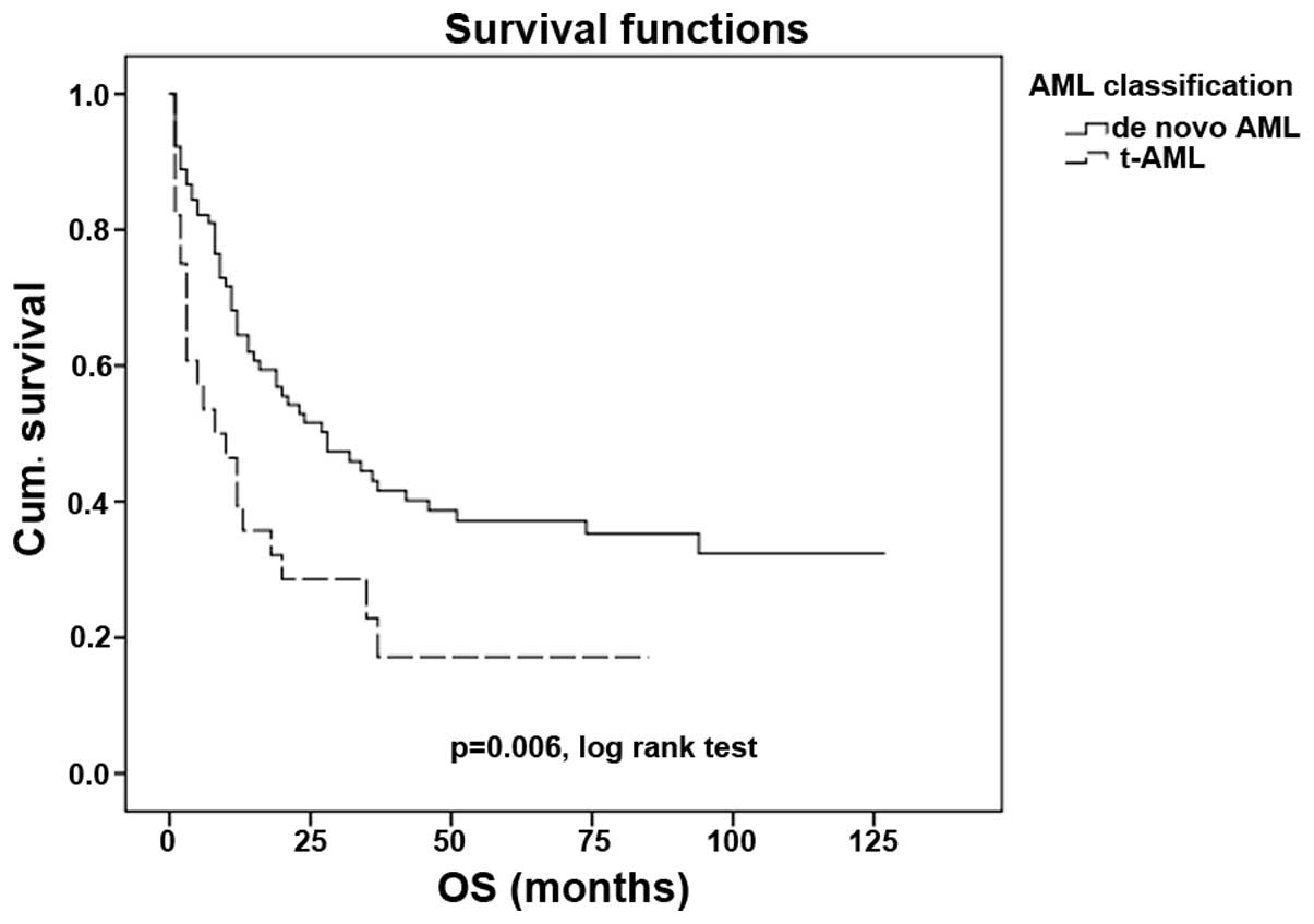
The outcome of patients with t-AML is significantly
poorer when compared with the outcome of patients with de
novo AML (P<0.001; Fig. 1),
with median OS times of 10 and 64 months, respectively. The OS
rates at 5 years were 22.6 and 50.6% in patients with t-AML and
de novo AML, respectively.
Cytogenetics and age are the strongest prognostic
markers for the outcome in de novo AML. In the present
study, cytogenetics (P=0.009; 95% CI, 1.042–1.336) and age
(P=0.008; 95% CI, 1.153–2.616) were of independent prognostic value
in all patients. However, in the t-AML cohort, cytogenetics
(P=0.392; 95% CI, 0.862–1.459) and age (P=0.517; 95% CI,
0.539–3.423) lacked prognostic value.
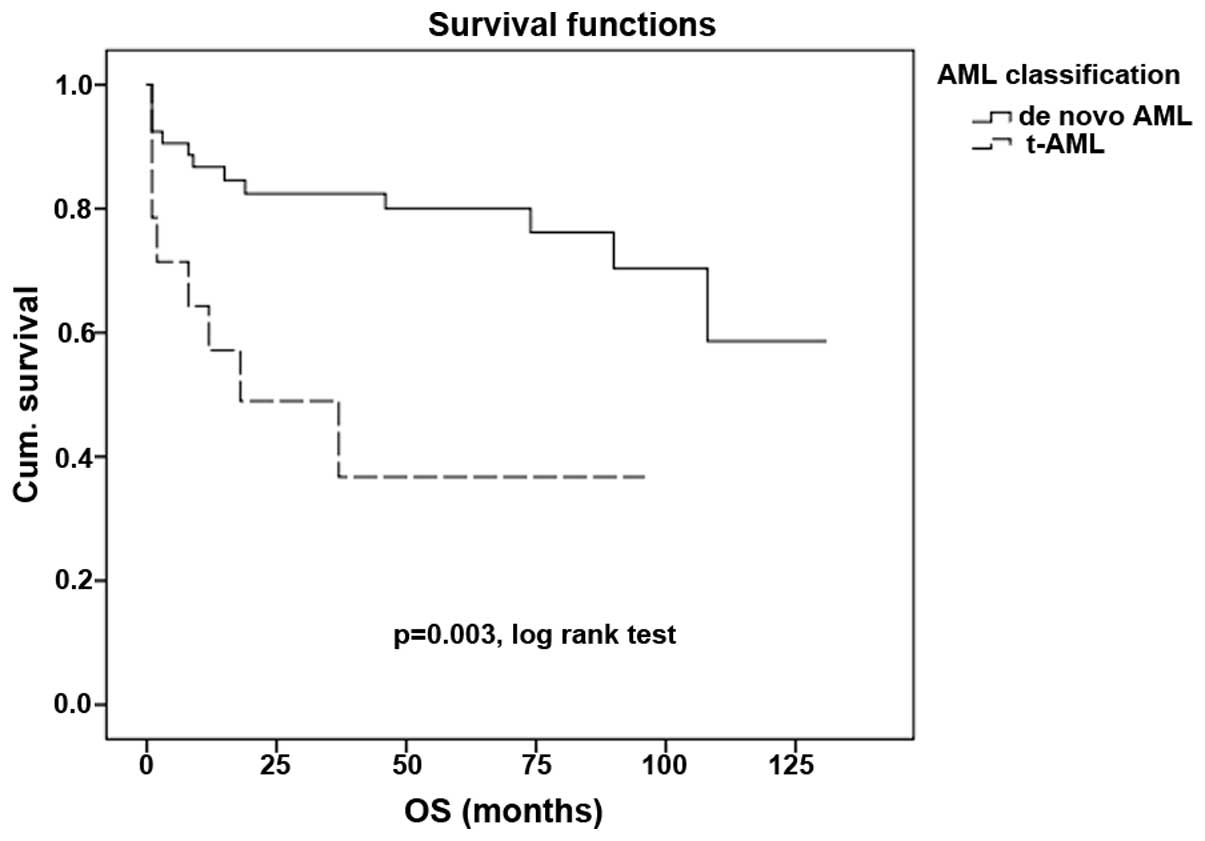
Other core binding factor (CBF) abnormalities that
are often associated with a good prognosis often lose this
association in patients with secondary leukemia (P<0.001;
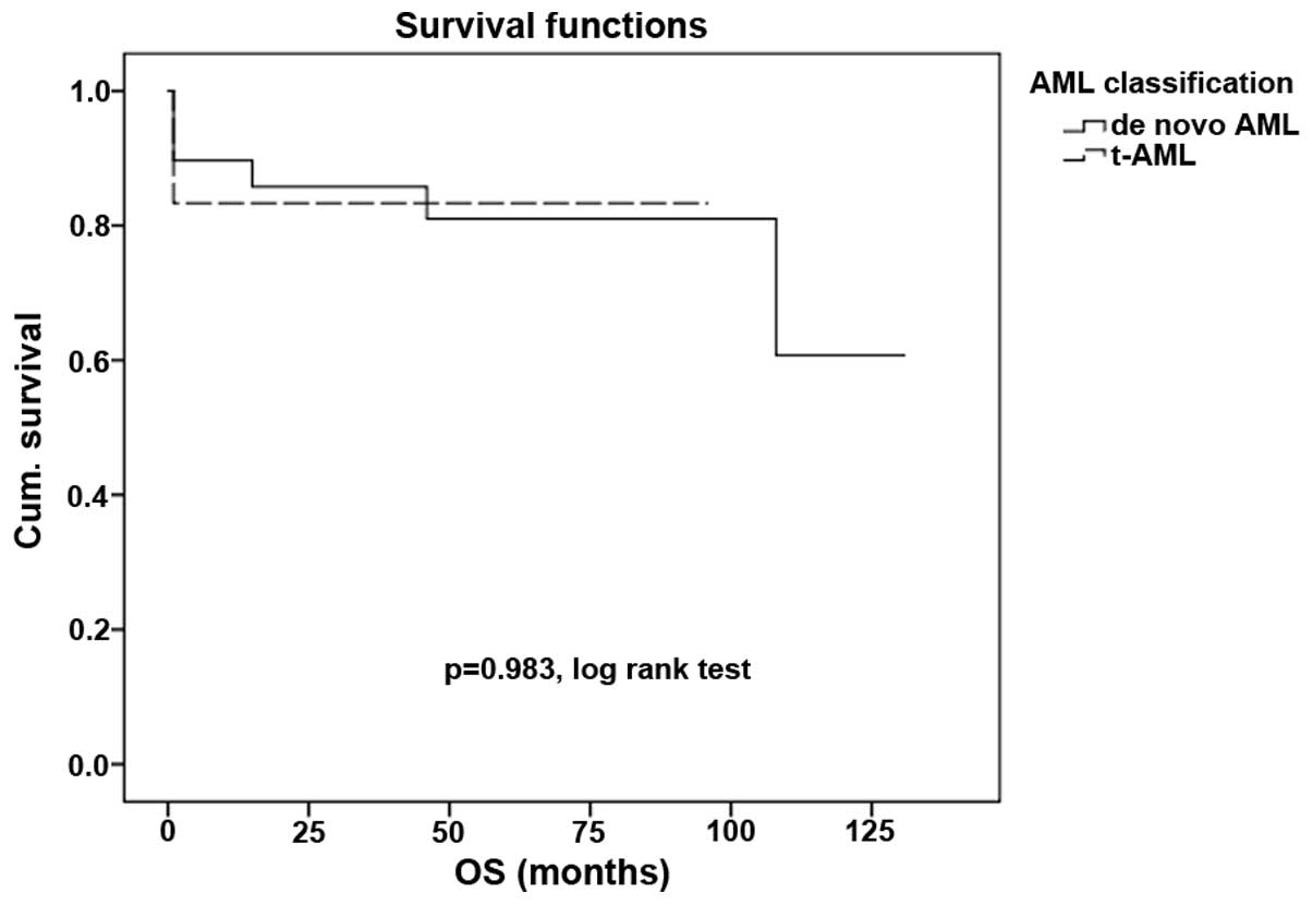
Fig. 2). However, in the t(15,17) group
of patients, even those with t-AML demonstrated the preservation of
a good prognosis, with no statistically significant difference in
survival compared with the de novo AML subgroup (P=0.983;
Fig. 3).
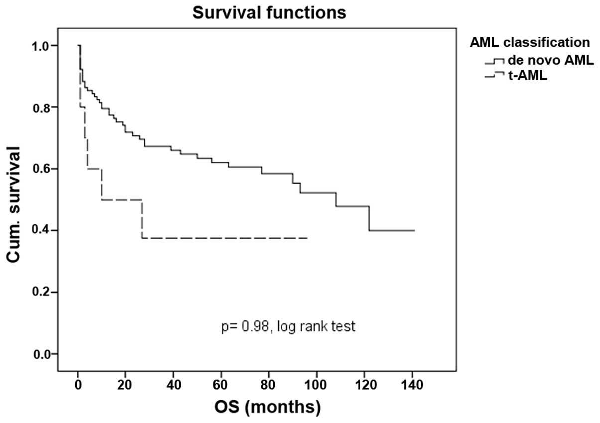
An older age is associated with a poorer outcome in
patients with t-AML. By analyzing the study population in two
subgroups, one containing patients of ≤50 years and another
containing patients of >50 years, the outcome of t-AML patients
in the younger subgroup was similar in t-AML and de novo AML
patients (P=0.98; Fig. 4). Older
patients with t-AML are more likely to experience a poor prognosis
when compared with their de novo counterpart (P=0.006;
Fig. 5).
Multivariable Cox-regression analyses demonstrated a
significant adverse impact on the 5-year OS rate of t-AML patients
(P<0.001; HR, 3.363; 95% CI, 1.951–5.796), a significant HR of
complete response following induction treatment (P<0.001; HR,
5.376; 95% CI, 3.303–8.750) and a significant occurrence of relapse
(P<0.001; HR, 2.827; 95% CI, 1.790–4.466; Table III).
| Table III.Multivariate analyses of 5-year
overall survival. |
Table III.
Multivariate analyses of 5-year
overall survival.
|
|
| 5-year overall
survival |
|---|
|
|
|
|
|---|
| Factors | P-value | HR | 95% CI |
|---|
| t-AML | <0.001 | 3.363 | 1.951–5.796 |
| Age |
0.065 | 1.523 | 0.974–2.382 |
| Gender |
0.736 | 0.928 | 0.601–1.433 |
| SWOG risk
score |
0.864 | 1.048 | 0.614–1.787 |
| Complete
response | <0.001 | 5.376 | 3.303–8.750 |
| Relapse | <0.001 | 2.827 | 1.790–4.466 |
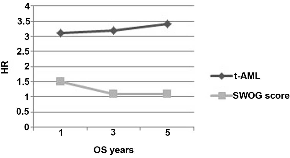
By independently analyzing AML and the SWOG risk
score as prognostic factors, and comparing the HR for OS rate at 1,
3 and 5 years, a constant and gradual increased HR was observed for
the t-AML group (Fig. 6). This trend
was not observed in the same analysis for the SWOG risk, which
demonstrated a decreasing HR over the years. The 5-year OS rates
demonstrate a separation in the two curves, which becomes even
clearer over time. The mortality risk in the t-AML group at 1 year
is 75.6%, and this increased to 76 and 77% at 3 and 5 years,
respectively. In the SWOG group, the mortality risk decreased
between 60% at 1 year and 52% at 3 and 5 years (32).
Discussion
Long-term side effects of previous cytotoxic
treatments are one of the most challenging problems faced by
oncologists (2,3,7). The
improved survival rates associated with the most efficient novel
drugs allow patients to survive the primary cancer and develop a
secondary malignancy induced by the cell toxicity of the primary
treatment. Numerous studies have now been conducted to characterize
this particular type of acute leukemia (2,8,19,28,34);
however, to the best of our knowledge, the present study is the
first to analyze therapy-induced secondary malignancy in Portuguese
patients.
The frequency of t-AML in the present cohort was
16.5% (8,27), which is increased compared with
previously reported data from other populations. This may be
explained by the focused activity of the present hospital, an
oncology referral center, which may lead to a clustering of all
oncological conditions.
The response to the induction regimen was not
significantly different between patients with t-AML and those with
de novo AML. This suggests that t-AML patients do not
demonstrate a worse response to chemotherapy compared with de
novo AML patients, and also suggests that the comparably poor
outcome of patients with t-AML is likely to be a result of toxicity
or relapse from acute leukemia or the initial tumor (35).
Cytogenetics acts as a good prognostic marker in
patients with AML (8,23,24,27,36–38).
Patients with t-AML are more prone to a poor prognosis and
cytogenetic findings. Core binding factor abnormalities are
associated with an improved outcome in patients with AML (39–41).
However, in the present cohort, t(15,17)
appeared to have a good effect on the outcome of disease, even in
patients with t-AML (42–45).
t-AML patients are generally older than patients
with de novo AML. Older age has previously been linked with
a poorer prognosis (42).
Comorbidities and primary resistance disease have been associated
with a poorer outcome (43–45). By analyzing the present data, younger
t-AML patients were concluded to behave equally to de novo
AML patients of the same age. However, older t-AML patients
demonstrated a significantly poorer outcome compared with de
novo AML patients of the same age.
In the present study, t-AML was a solid independent
prognostic marker, persisting over 5 years, and was associated with
a trend of increased HR. By contrast, the SWOG risk score behaved
differently, losing relevance as a prognostic marker over time. A
possible explanation for this observation may be that the
prognostic relevance of the SWOG risk is scored only at diagnosis
and not following the onset of relapse, when other cytogenetic
abnormalities may occur, de novo or in association with the
initial anomaly. From the present study, t-AML may be concluded to
be a serious late side effect of a previous oncological treatment.
The incidence of t-AML may be increasing due to the use of more
effective therapeutic strategies, which may lead to an increased
life expectancy, therefore making it possible for late side effects
to appear.
Despite the poor prognosis of t-AML, certain groups
of patients may be identified that have a similar outcome to de
novo AML patients (46–48). Promyelocytic leukemia continues to
demonstrate a good prognosis, even when appearing as a secondary
event following a previous malignancy (49,50); this
is perhaps due to the therapy used in this particular leukemia
type, including specific target agents in addition to
chemotherapy.
Improvements to the outcome of t-AML depend on a
deeper understanding of the disease etiology, and also depend on
the use of novel drugs with increased efficacy and limited
toxicity, and a preventive strategy (27). Additional studies are required,
particularly to better characterize this subset of patients.
Clinical trials that use novel drugs often exclude t-AML patients
by omitting secondary leukemia from novel drug approval.
References
|
1
|
Larson RA: Therapy-related myeloid
neoplasms. Haematologica. 94:454–459. 2009. View Article : Google Scholar : PubMed/NCBI
|
|
2
|
Morton LM, Dores GM, Tucker MA, Kim CJ,
Onel K, Gilbert ES, Fraumeni JF and Curtis RE: Evolving risk of
therapy-related acute myeloid leukemia following cancer
chemotherapy among adults in the United States, 1975–2008. Blood.
121:2996–3004. 2013. View Article : Google Scholar : PubMed/NCBI
|
|
3
|
Leone G, Fianchi L and Voso MT:
Therapy-related myeloid neoplasms. Curr Opin Oncol. 23:672–680.
2011. View Article : Google Scholar : PubMed/NCBI
|
|
4
|
Vardiman JW, Thiele J, Arber DA, Brunning
RD, Borowitz MJ, Porwit A, Harris NL, Le Beau MM,
Hellström-Lindberg E, Tefferi A and Bloomfield CD: The 2008
revision of the World Health Organization (WHO) classification of
myeloid neoplasms and acute leukemia: Rationale and important
changes. Blood. 114:937–951. 2009. View Article : Google Scholar : PubMed/NCBI
|
|
5
|
Vardiman JW, Harris NL and Brunning RD:
The World Health Organization (WHO) classification of the myeloid
neoplasms. Blood. 100:2292–2302. 2002. View Article : Google Scholar : PubMed/NCBI
|
|
6
|
Vardiman JW: The World Health Organization
(WHO) classification of tumors of the hematopoietic and lymphoid
tissues: An overview with emphasis on the myeloid neoplasms. Chem
Biol Interact. 184:16–20. 2010. View Article : Google Scholar : PubMed/NCBI
|
|
7
|
Leone G, Fianchi L, Pagano L and Voso MT:
Incidence and susceptibility to therapy-related myeloid neoplasms.
Chem Biol Int. 184:39–45. 2010. View Article : Google Scholar
|
|
8
|
Kayser S, Döhner K, Krauter J, Köhne CH,
Horst HA, Held G, von Lilienfeld-Toal M, Wilhelm S, Kündgen A,
Götze K, et al: German-Austrian AMLSG: The impact of
therapy-related acute myeloid leukemia (AML) on outcome in 2853
adult patients with newly diagnosed AML. Blood. 117:2137–2145.
2011. View Article : Google Scholar : PubMed/NCBI
|
|
9
|
Siegel R, Ma J, Zou Z and Jemal A: Cancer
statistics, 2014. CA Cncer J Clin. 64:9–29. 2014. View Article : Google Scholar
|
|
10
|
Siegel R, DeSantis C, Virgo K, Stein K,
Mariotto A, Smith T, Cooper D, Gansler T, Lerro C, Fedewa S, et al:
Cancer treatment and survivorship statistics, 2012. CA Cancer J
Clin. 62:220–241. 2012. View Article : Google Scholar : PubMed/NCBI
|
|
11
|
Wong TN, Ramsingh G, Young AL, Miller CA,
Touma W, Welch JS, Lamprecht TL, Shen D, Hundal J, Fulton RS, et
al: Role of TP53 mutations in the origin and evolution of
therapy-related acute myeloid leukaemia. Nature. 518:552–555. 2015.
View Article : Google Scholar : PubMed/NCBI
|
|
12
|
Li L, Li M, Sun C, Francisco L,
Chakraborty S, Sabado M, McDonald T, Gyorffy J, Chang K, Wang S, et
al: Altered hematopoietic cell gene expression precedes development
of therapy-related myelodysplasia/acute myeloid leukemia and
identifies patients at risk. Cancer cell. 20:591–605. 2011.
View Article : Google Scholar : PubMed/NCBI
|
|
13
|
Pedersen-Bjergaard J, Andersen MK,
Andersen MT and Christiansen DH: Genetics of therapy-related
myelodysplasia and acute myeloid leukemia. Leukemia. 22:240–248.
2008. View Article : Google Scholar : PubMed/NCBI
|
|
14
|
Pedersen-Bjergaard J, Pedersen M, Roulston
D and Philip P: Different genetic pathways in leukemogenesis for
patients presenting with therapy-related myelodysplasia and
therapy-related acute myeloid leukemia. Blood. 86:3542–3552.
1995.PubMed/NCBI
|
|
15
|
Brusamolino E, Gotti M and Fiaccadori V:
The risk of therapy-related myelodysplasia/acute myeloid leukemia
in Hodgkin lymphoma has substantially decreased in the ABVD era
abolishing mechlorethamine and procarbazine and limiting volumes
and doses of radiotherapy. Mediterr J Hematol Infect Dis.
4:e20120222012. View Article : Google Scholar : PubMed/NCBI
|
|
16
|
Brusamolino E, Baio A, Orlandi E, Arcaini
L, Passamonti F, Griva V, Casagrande W, Pascutto C, Franchini P and
Lazzarino M: Long-term events in adult patients with clinical stage
IA- IIA nonbulky Hodgkin's lymphoma treated with four cycles of
doxorubicin, bleomycin, vinblastine, and dacarbazine and adjuvant
radiotherapy: A single-institution 15-year follow-up. Clin Cancer
Res. 12:6487–6493. 2006. View Article : Google Scholar : PubMed/NCBI
|
|
17
|
Brusamolino E, Anselmo AP, Klersy C,
Santoro M, Orlandi E, Pagnucco G, Lunghi F, Maurizi-Enrici R,
Baroni CD, Lazzarino M, et al: The risk of acute leukemia in
patients treated for Hodgkin's disease is significantly higher aft
[see bined modality programs than after chemotherapy alone and is
correlated with the extent of radiotherapy and type and duration of
chemotherapy: A case-control study. Haematologica. 83:812–823.
1998.PubMed/NCBI
|
|
18
|
Delwail V, Jais JP, Colonna P and Andrieu
JM: Fifteen-year secondary leukaemia risk observed in 761 patients
with Hodgkin's disease prospectively treated by MOPP or ABVD
chemotherapy plus high-dose irradiation. Br J Haematol.
118:189–194. 2002. View Article : Google Scholar : PubMed/NCBI
|
|
19
|
Koontz MZ, Horning SJ, Balise R, Greenberg
PL, Rosenberg SA, Hoppe RT and Advani RH: Risk of therapy-related
secondary leukemia in Hodgkin lymphoma: The Stanford University
experience over three generations of clinical trials. J Clin Oncol.
31:592–598. 2013. View Article : Google Scholar : PubMed/NCBI
|
|
20
|
Eichenauer DA and Engert A:
Therapy-related myeloid neoplasms in patients treated for Hodgkin
lymphoma. Mediterr J Hematol Infect Dis. 3:e20110462011. View Article : Google Scholar : PubMed/NCBI
|
|
21
|
Godley LA and Larson RA: Therapy-related
myeloid leukemia. Semin Oncol. 35:418–429. 2008. View Article : Google Scholar : PubMed/NCBI
|
|
22
|
Rund D, Krichevsky S, Bar-Cohen S,
Goldschmidt N, Kedmi M, Malik E, Gural A, Shafran-Tikva S,
Ben-Neriah S and Ben-Yehuda D: Therapy-related leukemia: Clinical
characteristics and analysis of new molecular risk factors in 96
adult patients. Leukemia. 19:1919–1928. 2005. View Article : Google Scholar : PubMed/NCBI
|
|
23
|
Chen Y, Estrov Z, Pierce S, Qiao W,
Borthakur G, Ravandi F, Kadia T, Brandt M, O'Brien S, Jabbour E, et
al: Myeloid neoplasms after breast cancer: “Therapy-related” not an
independent poor prognostic factor. Leuk Lymphoma. 56:1012–1019.
2015. View Article : Google Scholar : PubMed/NCBI
|
|
24
|
Larson RA: Cytogenetics, not just previous
therapy, determines the course of therapy-related myeloid
neoplasms. J Clin Oncol. 30:2300–2302. 2012. View Article : Google Scholar : PubMed/NCBI
|
|
25
|
Olney HJ, Mitelman F, Johansson B, Mrózek
K, Berger R and Rowley JD: Unique balanced chromosome abnormalities
in treatment-related myelodysplastic syndromes and acute myeloid
leukemia: report from an international workshop. Genes Chromosomes
Cancer. 33:413–423. 2002. View Article : Google Scholar : PubMed/NCBI
|
|
26
|
Paschka P, Du J, Schlenk RF, Gaidzik VI,
Bullinger L, Corbacioglu A, Späth D, Kayser S, Schlegelberger B,
Krauter J, et al: Secondary genetic lesions in acute myeloid
leukemia with inv(16) or t(16;16): A study of the German-Austrian
AML Study Group (AMLSG). Blood. 121:170–177. 2013. View Article : Google Scholar : PubMed/NCBI
|
|
27
|
Churpek JE and Larson RA: The evolving
challenge of therapy-related myeloid neoplasms. Best Pract Res Clin
Haematol. 26:309–317. 2013. View Article : Google Scholar : PubMed/NCBI
|
|
28
|
Huh HJ, Lee SH, Yoo KH, Sung KW, Koo HH,
Kim K, Jang JH, Jung C, Kim SH and Kim HJ: Therapy-related myeloid
neoplasms in 39 Korean patients: A single institution experience.
Ann Lab Med. 33:97–104. 2013. View Article : Google Scholar : PubMed/NCBI
|
|
29
|
Schoch C, Kern W, Schnittger S, Hiddemann
W and Haferlach T: Karyotype is an independent prognostic parameter
in therapy-related acute myeloid leukemia (t-AML): An analysis of
93 patients with t-AML in comparison to 1091 patients with de
novo AML. Leukemia. 18:120–125. 2004. View Article : Google Scholar : PubMed/NCBI
|
|
30
|
Wang HC and Fedoroff S: Banding in human
chromosomes treated with trypsin. Nat New Biol. 235:52–54. 1972.
View Article : Google Scholar : PubMed/NCBI
|
|
31
|
Gonzalez Garcia JR and Meza-Espinoza JP:
Use of the International System for Human Cytogenetic Nomenclature
(ISCN). Blood. 108:3952–3953. 2006. View Article : Google Scholar : PubMed/NCBI
|
|
32
|
Slovak ML, Kopecky KJ, Cassileth PA,
Harrington DH, Theil KS, Mohamed A, Paietta E, Willman CL, Head DR,
Rowe JM, et al: Karyotypic analysis predicts outcome of
preremission and postremission therapy in adult acute myeloid
leukemia: A Southwest Oncology Group/Eastern Cooperative Oncology
Group Study. Blood. 96:4075–4083. 2000.PubMed/NCBI
|
|
33
|
Spruance SL, Reid JE, Grace M and Samore
M: Hazard ratio in clinical trials. Antimicrob Agents Chemother.
48:2787–2792. 2004. View Article : Google Scholar : PubMed/NCBI
|
|
34
|
Suvajdžić N, Cvetković Z, Dorđević V,
Kraguljac-Kurtović N, Stanisavljević D, Bogdanović A, Djunić I,
Colović N, Vidović A, Elezović I and Tomin D: Prognostic factors
for therapy-related acute myeloid leukaemia (t-AML) - a single
centre experience. Biomed Pharmacother. 66:285–292. 2012.
View Article : Google Scholar : PubMed/NCBI
|
|
35
|
Quesnel B, Kantarjian H, Bjergaard JP,
Brault P, Estey E, Lai JL, Tilly H, Stoppa AM, Archimbaud E,
Harousseau JL, et al: Therapy-related acute myeloid leukemia with
t(8;21), inv(16), and t(8;16): A report on 25 cases and review of
the literature. J Clin Oncol. 11:2370–2379. 1993.PubMed/NCBI
|
|
36
|
Larson RA and Le Beau MM: Prognosis and
therapy when acute promyelocytic leukemia and other “good risk”
acute myeloid leukemias occur as a therapy-related myeloid
neoplasm. Mediterr J Hematol Infect Dis. 3:e20110322011. View Article : Google Scholar : PubMed/NCBI
|
|
37
|
Feldman EJ: Does therapy-related AML have
a poor prognosis, independent of the cytogenetic/molecular
determinants? Best Pract Res Clin Haematol. 24:523–526. 2011.
View Article : Google Scholar : PubMed/NCBI
|
|
38
|
Grimwade D, Hills RK, Moorman AV, Walker
H, Chatters S, Goldstone AH, Wheatley K, Harrison CJ and Burnett
AK: National Cancer Research Institute Adult Leukaemia Working
Group: Refinement of cytogenetic classification in acute myeloid
leukemia: determination of prognostic significance of rare
recurring chromosomal abnormalities among 5876 younger adult
patients treated in the United Kingdom Medical Research Council
trials. Blood. 116:354–365. 2010. View Article : Google Scholar : PubMed/NCBI
|
|
39
|
Hospital MA, Prebet T, Bertoli S, Thomas
X, Tavernier E, Braun T, Pautas C, Perrot A, Lioure B, Rousselot P,
et al: Core-binding factor acute myeloid leukemia in first relapse:
A retrospective study from the French AML Intergroup. Blood.
124:1312–1319. 2014. View Article : Google Scholar : PubMed/NCBI
|
|
40
|
Solh M, Yohe S, Weisdorf D and Ustun C:
Core-binding factor acute myeloid leukemia: Heterogeneity,
monitoring, and therapy. Am J Hematol. 89:1121–1131. 2014.
View Article : Google Scholar : PubMed/NCBI
|
|
41
|
Duployez N, Willekens C, Marceau-Renaut A,
Boudry-Labis E and Preudhomme C: Prognosis and monitoring of
core-binding factor acute myeloid leukemia: Current and emerging
factors. Expert Rev Hematol. 8:43–56. 2015. View Article : Google Scholar : PubMed/NCBI
|
|
42
|
Foran JM: Frontline therapy of AML: Should
the older patient be treated differently? Curr Hematol Malig Rep.
9:100–108. 2014. View Article : Google Scholar : PubMed/NCBI
|
|
43
|
van der Holt B, Van den Heuvel-Eibrink MM,
Van Schaik RHN, van der Heiden IP, Wiemer EAC, Vossebeld PJM,
Löwenberg B and Sonneveld P: ABCB1 gene polymorphisms are not
associated with treatment outcome in elderly acute myeloid leukemia
patients. Clin Pharmacol Ther. 80:427–439. 2006. View Article : Google Scholar : PubMed/NCBI
|
|
44
|
Leith CP, Kopecky KJ, Chen IM, Eijdems L,
Slovak ML, McConnell TS, Head DR, Weick J, Grever MR, Appelbaum FR
and Willman CL: Frequency and clinical significance of the
expression of the multidrug resistance proteinsMDR1/P-glycoprotein,
MRP1, and LRP in acute myeloid leukemia: A Southwest Oncology Group
Study. Blood. 94:1086–1099. 1999.PubMed/NCBI
|
|
45
|
Benderra Z, Faussat AM, Sayada L, Perrot
JY, Tang R, Chaoui D, Morjani H, Marzac C, Marie JP and Legrand O:
MRP3, BCRP, and P-glycoprotein activities are prognostic factors in
adult acute myeloid leukemia. Clin Cancer Res. 11:7764–7772. 2005.
View Article : Google Scholar : PubMed/NCBI
|
|
46
|
Appelbaum FR, Kopecky KJ, Tallman MS,
Slovak ML, Gundacker HM, Kim HT, Dewald GW, Kantarjian HM, Pierce
SR and Estey EH: The clinical spectrum of adult acute myeloid
leukaemia associated with core binding factor translocations. Br J
Haematol. 135:165–173. 2006. View Article : Google Scholar : PubMed/NCBI
|
|
47
|
Schlenk RF, Benner A, Krauter J, Buchner
T, Sauerland C, Ehninger G, Schaich M, Mohr B, Niederwieser D,
Krahl R, Pasold R, et al: Individual patient data-based
meta-analysis of patients aged 16 to 60 years with core binding
factor acute myeloid leukemia: A survey of the German Acute Myeloid
Leukemia Intergroup. J Clin Oncol. 22:3741–3750. 2004. View Article : Google Scholar : PubMed/NCBI
|
|
48
|
Aldoss I and Pullarkat V: Therapy-related
acute myeloid leukemia with favorable cytogenetics: still
favorable? Leuk Res. 36:1547–1551. 2012. View Article : Google Scholar : PubMed/NCBI
|
|
49
|
Andersen MK and Pedersen-Bjergaard J:
Therapy-related MDS and AML in acute promyelocytic leukemia. Blood.
100:1928–1929. 2002. View Article : Google Scholar : PubMed/NCBI
|
|
50
|
Beaumont M, Sanz M, Carli PM, Maloisel F,
Thomas X, Detourmignies L, Guerci A, Gratecos N, Rayon C, San
Miguel J, et al: Therapy-related acute promyelocytic leukemia. J
Clin Oncol. 21:2123–2137. 2003. View Article : Google Scholar : PubMed/NCBI
|