Introduction
Hepatocellular carcinoma (HCC) is a prevalent type
of cancer worldwide, with >600,000 individuals succumbing to the
disease each year (1). In China, HCC
exhibits an incidence of 30.3 cases per 100,000 individuals
(2). Similar to other solid tumors,
the main curative therapy for HCC is surgery, which generally is
only successful if the cancer is diagnosed at an early stage
(3). The conventional chemotherapies
and radiotherapies used to treat advanced or late-stage HCC tumors,
despite being reasonably effective, have also demonstrated various
side effects, including hepatotoxicity (4) and hematotoxicity (5), and only a small percentage of patients
may have the chance to undergo surgery for radical therapy, which
complicates the safe administration of systemic therapy.
Numerous types of cancer cells and microorganisms
have relatively more anionic phospholipids in the outer layer of
their external membrane compared with normal eukaryotic cells.
Several studies have demonstrated that certain antimicrobial
peptides (AMPs) are more cytotoxic against transformed cells than
against non-transformed cells (6,7). In
addition, certain AMPs, when administered locally to solid tumors,
exhibit anticancer activity (8). The
cecropins, which were first isolated by Boman et al
(9) from Hyalophora cecropia
pupae (9,10), are a family of AMPs. To date, >20
types of cecropins have been identified, some of which have been
reported to possess antitumor activities against various cancer
cells, including bladder cancer (11), HCC (12), gastric carcinoma (13), fibrosarcoma (14) and leukemia cells (15). The mechanisms of action of cecropins
against prokaryotic cells have been widely investigated (16). Cecropins exert their cytolytic
activity by folding into an amphipathic helix, following selective
binding and insertion into the target membrane, leading to the
breakdown of the membrane structure, thus causing leakage of the
cell contents and resulting in cell death (17). In eukaryotic cells, cecropins target
non-polar lipid cell membranes, whereby they form transmembrane
channels, which leads to irreversible cytolysis and eventually cell
death (18). In addition, certain
cecropins translocate spontaneously across eukaryotic membranes
into the cytoplasm, whereby they depolarize inner mitochondrial
membranes, thus causing disruption of the mitochondrial membrane
potential, release of mitochondrial cytochrome c (cyt
c) and induction of apoptosis (19).
CecropinXJ, a polypeptide composed of 33 amino acid
residues, is a cationic AMP isolated from the larvae of Bombyx
mori (20). CecropinXJ exhibits
98% homology with cecropin B (20).
In the present study, the cecropinXJ gene was cloned into the
pYES2/CT/α-Factor expression vector, and expressed in
Saccharomyces cerevisiae (21).
CecropinXJ was previously demonstrated to exhibit
various antibacterial activities (22). In addition, previous studies have
reported that cecropinXJ is able to inhibit the proliferation and
induce the apoptosis of tumor cells (23), although its antitumor mechanism
remains unclear. In the present study, the cytotoxicity and
mechanism of cecropinXJ against the human HCC cell line Huh-7 was
investigated. The results revealed that cecropinXJ suppressed the
proliferation and induced the apoptosis of Huh-7 cells in
vitro through mitochondrial apoptosis pathways, suggesting that
cecropinXJ is a potential anticancer drug.
Materials and methods
Preparation of the AMP cecropinXJ and
reagents
CecropinXJ of B. mori was prepared using the S.
cerevisiae eukaryotic pYES2/CT/α-Factor expression system
(Invitrogen; Thermo Fisher Scientific, Inc., Waltham, MA, USA), and
purified with Ni-nitrilotriacetic acid agarose, as previously
reported (21). The concentration of
purified recombinant cecropinXJ protein was determined with a
Bradford protein assay kit (BioTek China, Beijing, China). Prior to
use, the peptide was dissolved in Dulbecco's modified Eagle's
medium (DMEM) (HyClone; GE Healthcare Life Sciences, Logan, UT,
USA) at a concentration of 50 mmol/l, and sterilized by filtration
through a 0.22-µm filter.
Fetal bovine serum (FBS) was obtained from Gibco
(Thermo Fisher Scientific, Inc., Waltham, MA, USA), while dimethyl
sulfoxide (DMSO) was purchased from Beijing Solarbio Science &
Technology Co., Ltd. (Beijing, China),
3-(4,5-dimethylthiazol-2-yl)-2,5-diphenyltetrazolium bromide (MTT)
was acquired from, 3,3′-dihexyloxacarbocyanine iodide
[DiOC6(3)] was obtained
from Sigma-Aldrich (St. Louis, MO, USA), cell cycle staining
solution was purchased from Multi Sciences (Lianke) Biotech Co.,
Ltd. (Hangzhou, China) and Annexin V-fluorescein isothiocyanate
(FITC)/propidium iodide (PI) staining apoptosis detection kit was
acquired from BestBio (Shanghai, China). Monoclonal rabbit
anti-caspase 3 (#9664), monoclonal rabbit anti-poly(ADP-ribose)
polymerase (PARP; #9532), monoclonal rabbit anti-cytochrome c
(#11940), monoclonal mouse anti-B-cell lymphoma 2 (Bcl-2; #15071),
monoclonal mouse anti-Bcl-2-associated death promoter (Bad; #9296),
monoclonal rabbit anti-Bcl-2-associated X protein (Bax; #14796),
monoclonal rabbit anti-glyceraldehyde 3-phosphate dehydrogenase
(GAPDH; #2118) (dilution, 1:1,000) were all obtained from Cell
Signaling Technology, Inc., Danvers, MA, USA. CytoBuster™ protein
extraction reagent was purchased from Novagen (Merck Millipore,
Darmstadt, Germany), bicinchoninic acid (BCA) assay kit was
acquired from Beyotime Institute of Biotechnology (Haimen, China)
and enhanced chemiluminescence detection kit was obtained from
CWbio. Co. Ltd. (Beijing, China).
Cell culture
The human HCC cell line Huh-7 was purchased from the
Chinese Academy of Medical Sciences (Beijing, China). Cells were
cultured in DMEM supplemented with 10% FBS, 100 µg/ml streptomycin
and 100 U/ml penicillin in a humidified atmosphere of 5%
CO2 in air at 37°C.
Cell viability assay
To evaluate the effects of cecropinXJ on the
proliferation of Huh-7 cells, cell viability was measured by MTT
assay. Huh-7 cells in the logarithmic phase of growth were
collected, seeded in 96-well plates at a density of
2×10³ cells/well and cultured overnight. Following 24 h,
Huh-7 cells were treated with or without cecropinXJ at various
concentrations (1, 5, 10 and 50 µmol/l) for 0, 24, 48, 72, 96 and
120 h. Bovine serum albumin (10 µmol/l; Beijing Solarbio Science
& Technology Co., Ltd.) served as a negative control, while 10
µmol/l cisplatin (Beijing Solarbio Science & Technology Co.,
Ltd.) served as a positive control. Upon incubation, the culture
medium was removed, and 100 µl MTT solution (5 mg/ml) was added to
each well, followed by incubation at 37°C for 4 h. Then, 150 µl
DMSO was added to each well, and the plates were incubated at 37°C
for additional 10 min. Absorbance was measured at 540 and 655 nm
using a 96-well microplate reader (Bio-Rad Laboratories, Inc.,
Hercules, CA, USA), and the ratio of optical density
(OD)540/655 was determined. Cell viability (%) was
calculated as (ODtreatment-ODblank)/(ODcontrol-ODblank)
× 100.
Cell cycle analysis of cecropinXJ
effects on Huh-7 cells by flow cytometry
Huh-7 cells at 5×10⁵ cells/ml were
inoculated into 100-mm dishes and incubated at 37°C for 24 h. Once
the medium had been removed, cells were treated for 24 h with
cecropinXJ at a final concentration of 5, 10 and 50 µmol/l.
Untreated cells served as the control. Upon culture, both floating
and adherent cells were collected, washed with cold
phosphate-buffered saline (PBS; Beijing Solarbio Science &
Technology Co., Ltd.) (pH 7.4) and fixed with 75% ethanol overnight
at −20°C. Cells were then treated with DNA staining solution at
37°C in the darkness for 30 min. Samples were analyzed by
fluorescence-activated cell sorting (FACS) with a flow cytometer
(BD Biosciences, Franklin Lakes, NJ, USA). Cell cycle analysis was
performed using the ModFit LT™ 3.0 DNA analysis software (Verity
Software House, Inc., Topsham, ME, USA).
Apoptosis rate determined by flow
cytometry
Huh-7 cells at 5×10⁵ cells/ml were
inoculated into 60-mm dishes and incubated at 37°C for 24 h. Upon
removal of the medium, 2 ml complete DMEM with cecropinXJ at a
final concentration of 5, 10 and 50 µmol/l was added to each dish,
and cells were incubated for 12, 24 and 48 h. Untreated cells
served as the control. Subsequently, cells were collected following
digestion with 0.25% trypsin, washed with PBS (two times) and
suspended in 400 µl binding buffer. Cell suspensions were stained
with 5 µl Annexin V-FITC and 10 µl PI, according to the
manufacturer's protocol, prior to be subjected to flow cytometry
analysis in a (BD Biosciences). The results are presented as the
percentage of Annexin V+ cells (mean ± standard error).
Measurement of the mitochondrial
membrane potential (Δψm)
Changes in the Δψm during apoptosis were measured
using DiOC6(3), which is a
lipophilic cationic dye. Huh-7 cells (5×10⁵ cells/ml)
were cultured in 100-mm dishes in the absence or presence of
cecropinXJ for 24 h at a concentration of 0, 5, 10 and 50 µmol/l.
Subsequently, cells were incubated with 40 nmol/l
DiOC6(3) for 15 min at
37°C. Cells were then washed twice in PBS and fluorescence was
measured using a fluorescence spectrophotometer with an excitation
wavelength of 482 nm and an emission wavelength of 504 nm.
Western blotting
Huh-7 cells were incubated with 0, 1, 5, 10 and 50
µmol/l cecropinXJ for 24 h, followed by two washes with ice-cold
PBS. The adherent and floating cells were next harvested and lysed
in 100 µl CytoBuster™ protein extraction reagent on ice for 15 min.
Following centrifugation at 12,000 rpm 4°C for 10 min, the protein
concentration of the supernatant was determined by BCA assay. The
protein lysates (40 µg/lane) were separated by 12% sodium dodecyl
sulfate-polyacrylamide gel electrophoresis, and transferred onto
nitrocellulose membranes. Upon being washed with Tris-buffered
saline and Tween 20 (TBS-T) buffer (20 mM Tris-HCl, 150 mM NaCl and
0.05% Tween 20), the membranes were blocked with 5% skimmed milk at
room temperature for 1 h, and then incubated with the
aforementioned primary antibodies (1:2,000) overnight at 4°C.
Subsequently, membranes were washed with TBS-T and incubated with
the corresponding HRP-conjugated secondary antibodies for 2 h at
room temperature. Upon washing with TBS-T, the membranes were
exposed using an ECL detection kit (CWbio. Co. Ltd.). Proteins were
visualized using the Odyssey® CLx Imaging system
(Li-Cor, Lincoln, NE, USA).
Statistical analysis
All results were confirmed in ≥3 independent
experiments. Data are expressed as the mean ± standard deviation.
Differences between two sample means were assessed by Student's t
test. P<0.05 was considered to indicate a statistically
significant difference.
Results
CecropinXJ suppressed Huh-7 cell
proliferation and decreased cell viability
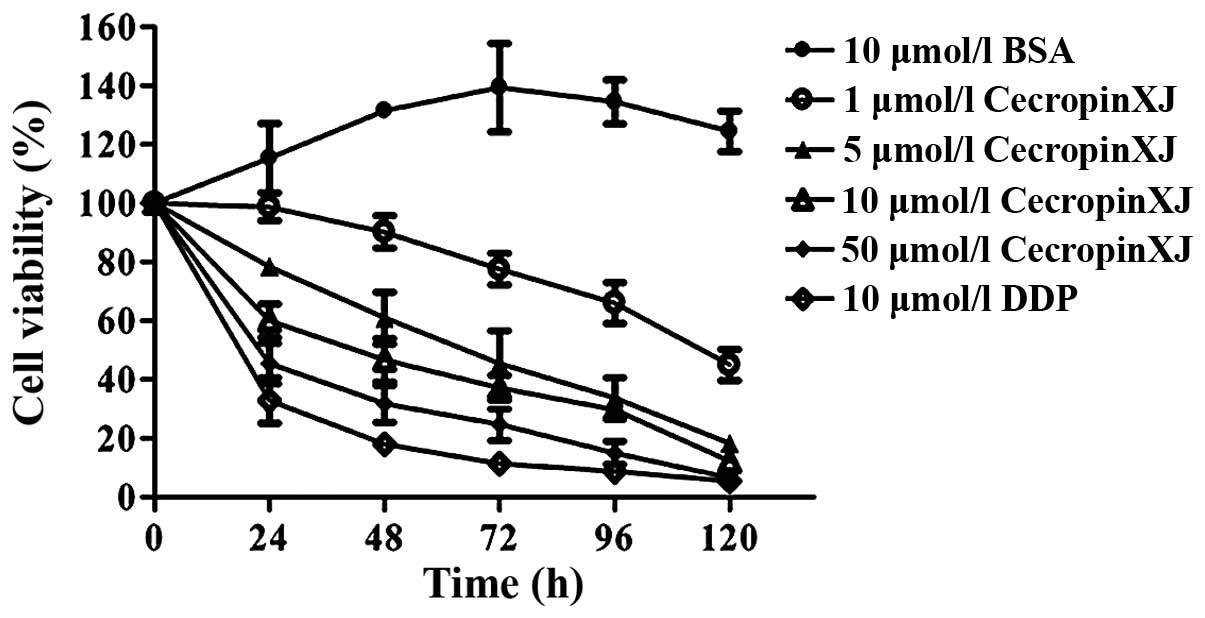
The results of MTT assay revealed that B. mori
cecropinXJ inhibited the proliferation of Huh-7 cells in a dose-
and time-dependent manner. CecropinXJ treatment for 24 h suppressed
the growth of Huh-7 cells, and this inhibitory effect was enhanced
by increasing concentrations of cecropinXJ. CecropinXJ at a
concentration of 50 µmol/l significantly inhibited the
proliferation of Huh-7 cells, with an inhibitory rate of ≤53%
(P<0.05). Furthermore, the inhibition effects of 50 µmol/l
cecropinXJ on Huh-7 cells was similar to those caused by 10 µmol/l
cisplatin (Fig. 1).
Detection of tumor cell apoptosis rate
by flow cytometry
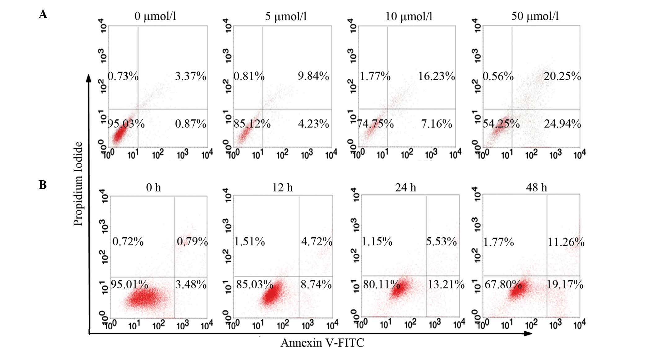
Since cell viability was significantly inhibited by
cecropinXJ, it was critical to determine which type of cell death
was induced by this AMP in Huh-7 cells (P<0.05). For that
purpose, an Annexin V/PI assay. Flow cytometry assay with Annexin
V/PI double staining revealed that cecropinXJ induced Huh-7 cell
apoptosis. The apoptosis rate increased, whereas the number of
necrotic cells did not significantly increase with cecropinXJ
concentration or time. CecropinXJ treatment (5–50 µmol/l) for 24 h
increased Huh7 cell apoptosis at both early and late stages, in a
dose-dependent manner. The percentage of total apoptotic cells
significantly increased from 3.76±0.53% (untreated) to 15.24±0.31%
at 5 µmol/l cecropinXJ, 23.13±0.26% at 10 µmol/l cecropinXJ and
44.77±0.42% at 50 µmol/l cecropinXJ (Fig.
2). Furthermore, the number of apoptotic cells increased with
10 µmol/l cecropinXJ treatment for 12 h, and was significantly
elevated at 24 h (P<0.05) (Fig.
2). This result indicated that cecropinXJ induced apoptotic
cell death in Huh-7 cells.
Cell cycle analysis of cecropinXJ on
Huh-7 cells by flow cytometry
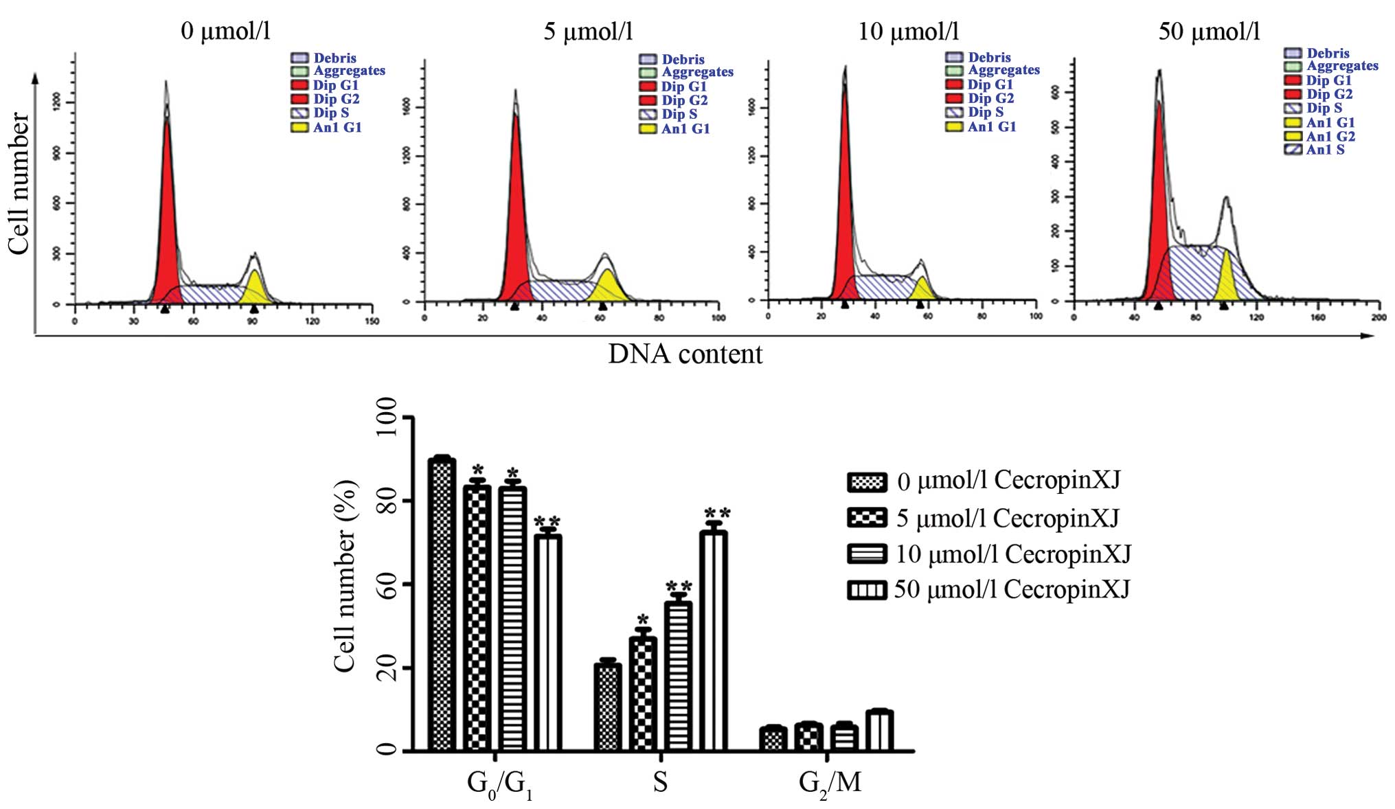
Flow cytometry was used to determine the effects of
cecropinXJ on cell cycle distribution (Fig. 3). Upon treatment with cecropinXJ for
24 h, the percentage of S-phase cells was significantly higher in
the treated group than in the control group (P<0.05). These
results suggested that cecropinXJ arrested the cell cycle at the S
phase in vitro.
CecropinXJ caused the loss of the
Δψm and the release of cyt c in Huh-7 cells
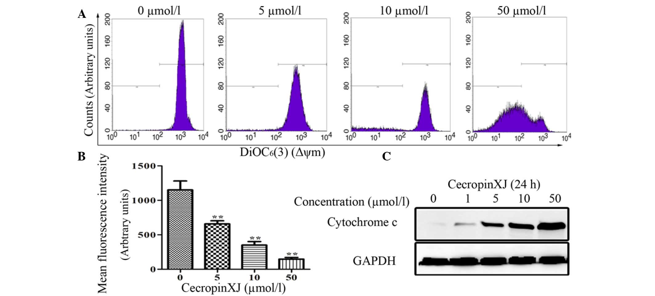
Since the loss of the Δψm acts as a key regulator in
the intrinsic apoptosis pathway (24), it was next examined whether the Δψm
was affected by cecropinXJ (Fig. 4).
The mean fluorescence intensity of cells treated with 5, 10 and 50
µmol/l cecropinXJ for 24 h decreased to 658.17±44.57, 350.07±52.23
and 147.27±23.99, respectively, compared with the untreated control
(1,196±76.12) (Fig. 4B). CecropinXJ
also induced the release of cyt c into the cytosol (Fig. 4C) and triggered the intrinsic
apoptosis pathway. This suggests that cecropinXJ may initiate
apoptosis through depolarization of the Δψm.
CecropinXJ induced caspase-dependent
apoptosis and regulated the expression of Bcl-2-family proteins in
Huh-7 cells
To further investigate whether cecropinXJ induced
apoptosis in Huh-7 cells through the caspase-dependent pathway, the
present study investigated whether caspase-3 and PARP were cleaved
in cecropinXJ-treated cells. According to the results of western
blot analysis, the above enzymes were cleaved in a dose-dependent
manner (Fig. 5), indicating that
cecropinXJ induced caspase-dependent apoptosis in the Huh-7 HCC
cell line. Bcl-2-family members are important in the mitochondrial
pathway of apoptosis, and are divided into pro- and anti-apoptotic
family members, according to whether they promote or inhibit
apoptosis (25). As indicated in
Fig. 5, the pro-apoptotic proteins
Bad and Bax were upregulated, whereas the anti-apoptotic protein
Bcl-2 was downregulated, following treatment with cecropinXJ.
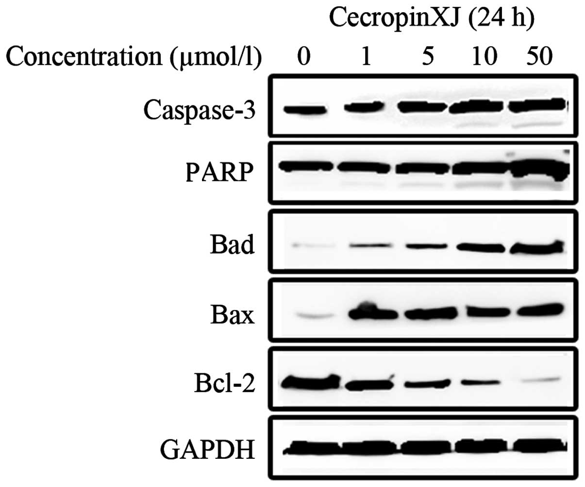 | Figure 5.Western blot analysis of the
activation of apoptosis-related enzymes and expression of proteins
of the Bcl-2 family. Huh-7 cells were treated with 0, 1, 5, 10 and
50 µmol/l cecropinXJ for 24 h, and their cellular extracts were
analyzed by western blotting for detection of the cleaved forms of
caspase-3 and poly(ADP-ribose) polymerase, and the expression
levels of Bcl-2, Bcl-2-associated death promoter and
Bcl-2-associated X protein, in order to evaluate whether cecropinXJ
induced cell apoptosis through the caspase-dependent pathway.
Glyceraldehyde 3-phosphate dehydrogenase was used as an internal
control. GAPDH, glyceraldehyde 3-phosphate dehydrogenase; PARP,
poly(ADP-ribose) polymerase; Bcl-2, B-cell lymphoma 2; Bad,
Bcl-2-associated death promoter; Bax, Bcl-2-associated X
protein. |
Discussion
AMPs are a kind of small peptides that specifically
interact with membranes and affect the proliferation and apoptosis
of various tumor cells (26,27). AMPs have gained great attention due to
their antitumor effects (28). Since
Moore et al (29) first
discovered the antitumor activity of cecropin B against mammalian
cancer cells, other studies have reported the cyototoxicity of
cecropin-family members on gastric, bladder, liver and other types
of cancer cells, but without causing damage to human normal cells
(30). Thus, cecropins may be good
candidates for the development of antitumor agents. In recent
years, a number of studies have reported that AMPs could inhibit
the growth of HCC, including Musca domestica cecropin
(12), melittin (31) and PR-39 (32). In the present study, cecropinXJ
inhibited the proliferation of Huh-7 cells, and the inhibitory rate
of 50 µmol/l cecropin on HCC was 36.6±0.1%, which was higher than
that of M. domestica cecropin (12).
Previous studies have demonstrated that antitumor
drugs generally inhibit tumor proliferation through the induction
of apoptosis in sensitive tumor cells, and their antitumor effects
are associated with the drug-induced activation of apoptosis in the
tumor cells. Therefore, the induction of apoptosis to treat tumors
has become a novel target for the development of antitumor drugs,
and constitutes a novel direction in tumor pharmacology research.
In the present study, flow cytometry revealed that cecropinXJ
induced apoptosis in Huh-7 HCC cells.
The mechanism of cecropins-induced apoptosis in
vitro has been previously reported (11). Cecropin A, a 37-residue linear AMP
produced by the cecropia moth, is able to induce apoptosis in HL-60
cells through a signaling mechanism that is mediated by the
mitochondria but is independent of caspase activation (19). In the present study, the
Δψm decreased following cecropinXJ treatment, which also
resulted in the release of cyt c into the cytoplasm of Huh-7
cells, suggesting that cecropinXJ-mediated apoptosis may be
associated with mitochondrial dysfunction. To further understand
the mechanism of cell apoptosis mediated by cecropinXJ, its effects
on the expression of caspase-family and Bcl-2-family proteins were
examined. The results indicated that caspase-3 and PARP were
cleaved, while Bad and Bax were upregulated and Bcl-2 was
downregulated in a time-dependent manner, indicating that
cecropinXJ induced caspase-dependent apoptosis in the Huh-7 HCC
cell line. These findings suggested the existence of a common
pathway that involves the activation of the family of proteolytic
enzymes known as caspases (33). One
of the pathways that lead to caspase activation is triggered by cyt
c, following its release from the mitochondria into the
cytoplasm, which is called the ‘intrinsic’ pathway of apoptosis
(34). Bcl-2 blocks the release of
cyt c from the mitochondria (35), suggesting that the mitochondria are
the principal sites for apoptotic regulation by the Bcl-2
family.
In conclusion, the present study has demonstrated
that cecropinXJ possesses antitumor activity against human HCC
Huh-7 cells in vitro. The mechanism of cecropinXJ-induced
apoptosis involves the loss of the Δψm, the release of
cyt c from the mitochondria and the activation of caspase-3
and PARP, which suggests that cecropinXJ induced cell apoptosis
possibly via the mitochondrial pathway. Therefore cecropinXJ may be
a potential candidate for the treatment of HCC.
Acknowledgements
The present study was supported by grants from the
National Natural Science Foundation of China (no. 31500752), the
Doctoral Start-up Fund of Xinjiang University (no. BS150241) and
the High-Tech Research and Development Program of Xinjiang (no.
201110101).
References
|
1
|
Somboon K, Siramolpiwat S and Vilaichone
RK: Epidemiology and survival of hepatocellular carcinoma in the
central region of Thailand. Asian Pac J Cancer Prev. 15:3567–3570.
2014. View Article : Google Scholar : PubMed/NCBI
|
|
2
|
Chen W, Zheng R, Zhang S, Zhao P, Li G, Wu
L and He J: The incidences and mortalities of major cancers in
China, 2009. Chin J Cancer. 32:106–112. 2013. View Article : Google Scholar : PubMed/NCBI
|
|
3
|
Llovet JM, Burroughs A and Bruix J:
Hepatocellular carcinoma. Lancet. 362:1907–1917. 2003. View Article : Google Scholar : PubMed/NCBI
|
|
4
|
Zolfagharzadeh F and Roshan VD:
Pretreatment hepatoprotective effect of regular aerobic training
against hepatic toxicity induced by doxorubicin in rats. Asian Pac
J Cancer Prev. 14:2931–2936. 2013. View Article : Google Scholar : PubMed/NCBI
|
|
5
|
Sostelly A, Henin E, Chauvenet L,
Hardy-Bessard AC, Jestin-Le Tallec V, Kirsher S, Leyronnas C,
Ligeza-Poisson C, Ramdane S, Salavt J, et al: Can we predict
chemo-induced hematotoxicity in elderly patients treated with
pegylated liposomal doxorubicin? Results of a population-based
model derived from the DOGMES phase II trial of the GINECO. J
Geriatr Oncol. 4:48–57. 2013. View Article : Google Scholar : PubMed/NCBI
|
|
6
|
Cruciani RA, Barker JL, Zasloff M, Chen HC
and Colamonici O: Antibiotic magainins exert cytolytic activity
against transformed cell lines through channel formation. Proc Natl
Acad Sci USA. 88:3792–3796. 1991. View Article : Google Scholar : PubMed/NCBI
|
|
7
|
Jin XB, Li XB, Zhu JY, Lu XM, Shen J, Chu
FJ and Mei HF: The target of Musca domestica cecropin on
human hepatocellular carcinoma BEL-7402 cells. Zhongguo Ji Sheng
Chong Xue Yu Ji Sheng Chong Bing Za Zhi. 29:271–273. 2011.(In
Chinese). PubMed/NCBI
|
|
8
|
Jin XB, Wang YJ, Liang LL, Pu QH, Shen J,
Lu XM, Chu FJ and Zhu JY: Cecropin suppresses human hepatocellular
carcinoma BEL-7402 cell growth and survival in vivo without
side-toxicity. Asian Pac J Cancer Prev. 15:5433–5436. 2014.
View Article : Google Scholar : PubMed/NCBI
|
|
9
|
Boman HG, Nilsson-Faye I, Paul K and
Rasmuson T: Insect immunity. I. Characteristics of an inducible
cell-free antibacterial reaction in hemolymph of Samia
cynthia pupae. Infect Immun. 10:136–145. 1974.PubMed/NCBI
|
|
10
|
Steiner H, Hultmark D, Engström A, Bennich
H and Boman HG: Sequence and specificity of two antibacterial
proteins involved in insect immunity. Nature. 292:246–248. 1981.
View Article : Google Scholar : PubMed/NCBI
|
|
11
|
Suttmann H, Retz M, Paulsen F, Harder J,
Zwergel U, Kamradt J, Wullich B, Unteregger G, Stöckle M and
Lehmann J: Antimicrobial peptides of the Cecropin-family show
potent antitumor activity against bladder cancer cells. BMC Urol.
8:52008. View Article : Google Scholar : PubMed/NCBI
|
|
12
|
Jin X, Mei H, Li X, Ma Y, Zeng AH, Wang Y,
Lu X, Chu F, Wu Q and Zhu J: Apoptosis-inducing activity of the
antimicrobial peptide cecropin of Musca domestica in human
hepatocellular carcinoma cell line BEL-7402 and the possible
mechanism. Acta Biochim Biophys Sin (Shanghai). 42:259–265. 2010.
View Article : Google Scholar : PubMed/NCBI
|
|
13
|
Pan WR, Chen PW, Chen YL, Hsu HC, Lin CC
and Chen WJ: Bovine lactoferricin B induces apoptosis of human
gastric cancer cell line AGS by inhibition of autophagy at a late
stage. J Dairy Sci. 96:7511–7520. 2013. View Article : Google Scholar : PubMed/NCBI
|
|
14
|
Lin WJ, Chien YL, Pan CY, Lin TL, Chen JY,
Chiu SJ and Hui CF: Epinecidin-1, an antimicrobial peptide from
fish (Epinephelus coioides) which has an antitumor effect
like lytic peptides in human fibrosarcoma cells. Peptides.
30:283–290. 2009. View Article : Google Scholar : PubMed/NCBI
|
|
15
|
Hui L, Leung K and Chen HM: The combined
effects of antibacterial peptide cecropin A and anti-cancer agents
on leukemia cells. Anticancer Res. 22:2811–2816. 2002.PubMed/NCBI
|
|
16
|
Yeaman MR and Yount NY: Mechanisms of
antimicrobial peptide action and resistance. Pharmacol Rev.
55:27–55. 2003. View Article : Google Scholar : PubMed/NCBI
|
|
17
|
Chou HT, Wen HW, Kuo TY, Lin CC and Chen
WJ: Interaction of cationic antimicrobial peptides with
phospholipid vesicles and their antibacterial activity. Peptides.
31:1811–1820. 2010. View Article : Google Scholar : PubMed/NCBI
|
|
18
|
Chen HM, Wang W, Smith D and Chan SC:
Effects of the anti-bacterial peptide cecropin B and its analogs,
cecropins B1 and B2, on liposomes, bacteria, and cancer cells.
Biochim Biophys Acta. 1336:171–179. 1997. View Article : Google Scholar : PubMed/NCBI
|
|
19
|
Cerón JM, Contreras-Moreno J, Puertollano
E, de Cienfuegos GÁ, Puertollano MA and de Pablo MA: The
antimicrobial peptide cecropin A induces caspase-independent cell
death in human promyelocytic leukemia cells. Peptides.
31:1494–1503. 2010. View Article : Google Scholar : PubMed/NCBI
|
|
20
|
Li JY, Zhang FC and Ma ZH: Prokaryotic
expression of cecropin gene isolated from the silkworm Bombyx
mori Xinjiang race and antibacterial activity of fusion
cecropin. Acta Entomologica Sinica. 47:407–411. 2004.(In
Chinese).
|
|
21
|
Xia L, Liu Z, Ma J, Sun S, Yang J and
Zhang F: Expression, purification and characterization of cecropin
antibacterial peptide from Bombyx mori in Saccharomyces
cerevisiae. Protein Expr Purif. 90:47–54. 2013. View Article : Google Scholar : PubMed/NCBI
|
|
22
|
Xia L, Zhang F, Liu Z, Ma J and Yang J:
Expression and characterization of cecropinXJ, a bioactive
antimicrobial peptide from Bombyx mori (Bombycidae, Lepidoptera) in
Escherichia coli. Exp Ther Med. 5:1745–1751. 2013.PubMed/NCBI
|
|
23
|
Xia L, Wu Y, Kang S, Ma J, Yang J and
Zhang F: CecropinXJ, a silkworm antimicrobial peptide, induces
cytoskeleton disruption in esophageal carcinoma cells. Acta Biochim
Biophys Sin (Shanghai). 46:867–876. 2014. View Article : Google Scholar : PubMed/NCBI
|
|
24
|
Cerón JM, Contreras-Moreno J, Puertollano
E, de Cienfuegos GÁ, Puertollano MA and de Pablo MA: The
antimicrobial peptide cecropin A induces caspase-independent cell
death in human promyelocytic leukemia cells. Peptides.
31:1494–1503. 2010. View Article : Google Scholar : PubMed/NCBI
|
|
25
|
Kuwana T and Newmeyer DD: Bcl-2-family
proteins and the role of mitochondria in apoptosis. Curr Opin Cell
Biol. 15:691–699. 2003. View Article : Google Scholar : PubMed/NCBI
|
|
26
|
Chen YL, Li JH, Yu CY, Lin CJ, Chiu PH,
Chen PW, Lin CC and Chen WJ: Novel cationic antimicrobial peptide
GW-H1 induced caspase-dependent apoptosis of hepatocellular
carcinoma cell lines. Peptides. 36:257–265. 2012. View Article : Google Scholar : PubMed/NCBI
|
|
27
|
Huh JE, Kang JW, Nam D, Baek YH, Choi DY,
Park DS and Lee JD: Melittin suppresses VEGF-A-induced tumor growth
by blocking VEGFR-2 and the COX-2-mediated MAPK signaling pathway.
J Nat Prod. 75:1922–1929. 2012. View Article : Google Scholar : PubMed/NCBI
|
|
28
|
Papo N and Shai Y: Host defense peptides
as new weapons in cancer treatment. Cell Mol Life Sci. 62:784–790.
2005. View Article : Google Scholar : PubMed/NCBI
|
|
29
|
Moore AJ, Devine DA and Bibby MC:
Preliminary experimental anticancer activity of cecropins. Pept
Res. 7:265–269. 1994.PubMed/NCBI
|
|
30
|
Chernysh S, Kim SI, Bekker G, Pleskach VA,
Filatova NA, Anikin VB, Platonov VG and Bulet P: Antiviral and
antitumor peptides from insects. Proc Natl Acad Sci USA.
99:12628–12632. 2002. View Article : Google Scholar : PubMed/NCBI
|
|
31
|
Li B, Gu W, Zhang C, Huang XQ, Han KQ and
Ling CQ: Growth arrest and apoptosis of the human hepatocellular
carcinoma cell line BEL-7402 induced by melittin. Onkologie.
29:367–371. 2006.PubMed/NCBI
|
|
32
|
Ohtake T, Fujimoto Y, Ikuta K, Saito H,
Ohhira M, Ono M and Kohgo Y: Proline-rich antimicrobial peptide,
PR-39 gene transduction altered invasive activity and actin
structure in human hepatocellular carcinoma cells. Br J Cancer.
81:393–403. 1999. View Article : Google Scholar : PubMed/NCBI
|
|
33
|
Thornberry NA and Lazebnik Y: Caspases:
Enemies within. Science. 281:1312–1316. 1998. View Article : Google Scholar : PubMed/NCBI
|
|
34
|
Liu X, Kim CN, Yang J, Jemmerson R and
Wang X: Induction of apoptotic program in cell-free extracts:
Requirement for dATP and cytochrome c. Cell. 86:147–157.
1996. View Article : Google Scholar : PubMed/NCBI
|
|
35
|
Yang J, Liu X, Bhalla K, Kim CN, Ibrado
AM, Cai J, Peng TI, Jones DP and Wang X: Prevention of apoptosis by
Bcl-2: Release of cytochrome c from mitochondria blocked.
Science. 275:1129–1132. 1997. View Article : Google Scholar : PubMed/NCBI
|