Introduction
Hepatocellular carcinoma (HCC), the most common of
the hepatobiliary malignancies, ranks as the fifth most common type
and the third primary cause associated with cancer mortality
(1). Recent data indicates that the
incidence of HCC appears to be increasing rapidly all over the
world (2). Although the majority of
cases are observed in developing nations, there is also evidence of
a rising incidence of HCC in both North America and Europe
(3,4).
Despite aggressive research, the prognosis for HCC patients remains
relatively poor, and a definitive cure remains elusive,
particularly in advanced cases.
At present, partial hepatectomy remains a potential
curative treatment for most early-stage HCC patients (solitary
tumor ≤5 cm in size, or ≤3 tumors each ≤3 cm in size and no
evidence of gross vascular invasion) (5–7). Although
partial liver resection may achieve a 5-year survival rate of
~50–70%, HCC recurrence rates at 5 years have been reported to
exceed 70% following such procedures (8–10). In
addition, because of the limitations of preserved liver function,
considerations of patient performance, and factors such as tumor
size and position, <30% patients are deemed suitable candidates
for partial resection surgery (11).
Liver transplantation is another potentially curative therapeutic
option for patients with early-stage HCC. However, the shortage of
donor organs and the numerous factors that complicate finding,
acquiring, and successfully transplanting a well-matched liver
severely restrict the use of transplantation as a treatment
alternative (5,12). In recent years, the techniques of
local-regional ablation and embolization have provided additional
local-regional treatment options for patients who are not suitable
surgical candidates (13,14). However these therapies are not
adequate for patients with advanced HCC.
Sorafenib, an oral multi-kinase inhibitor that
suppresses tumor cell proliferation and angiogenesis, has been
shown to effectively improve outcomes in cases of advanced or
metastatic HCC. However, the impact of this drug is quite limited,
as the mOS of advanced or metastatic HCC patients on sorafenib is
only 6.5–10.7 months (15,16). Recently, the development of novel
chemotherapy drugs such as Oxaliplatin, Capecitabine, and
Gemcitabine among others has facilitated the development of
systemic chemotherapy regimens for the treatment of HCC. However,
this approach has so far only demonstrated modest improvements in
outcomes, including an mOS of 6.47–12.2 months in the treatment of
advanced HCC (17–19).
Because of the limitations of surgical and
chemotherapeutic options, immunotherapy has gained steadily
increasing attention as a treatment option for advanced HCC. In
1991, Schmidt-Wolf, IG et al identified a type of anti-tumor
effector cells, known as cytokine-induced killer cells (CIK), that
proliferate rapidly in vitro, possess strong antitumor
activity against a broad spectrum of solid tumors, and cause
minimal adverse effects when active in the host's body (20). As expected, the anti-tumor effect of
CIK depends on the number of active CIK cells and on the overall
functional quality of the cells, a factor that is particularly
important when considering the techniques employed to culture CIK
cells for therapeutic delivery. Recent studies have reported that
RetroNectin can improve conglutination, extension, differentiation
and proliferation of cultured cells, all of which contribute to
more efficient stimulation of T cells (21–24). Our
previous study also confirmed this (25). In the present study, the clinical
effects of R-CIK therapy were compared with standard chemotherapy
and the predictive factors governing the efficacy of R-CIK
treatment were investigated in patients with advanced HCC.
Materials and methods
Patients
Between January 2010 and October 2013, 74 patients
with an initial diagnosis of advanced hepatocelluar carcinoma (AJCC
criteria) were enrolled in this study. Patients were assigned to
either arm 1 (n=37) to receive CIK treatment as a first line first
line therapy, or arm 2 (n=37) to receive chemotherapy [Oxaliplatin
(130 mg/m2) combined with Capecitabine (1,000
mg/m2)] as a first line treatment. The present study was
approved by the Ethics Committee at The Affiliated Cancer Hospital
of Zhengzhou University (Zhengzhou, China) and an approved consent
form was signed by all the patients. For purposes of analysis, two
subsets of HCC patients were identified. One subset consists of
patients who had previously received local-regional treatments
[e.g. transcatheter arterial chemoembolization (TACE),
radiofrequencey ablation (RFA), surgery] for early HCC, but who had
subsequently developed distant metastasis and/or multiple
intra-hepatic metastases (>3 nodules) that could not be
effectively treated with local-regional treatments and where the
patient could not reasonably be expected to pay the fee of
sorafenib therapy. The other subset of patients identified had
received no prior therapy and presented initially with distant
metastasis and/or multiple intra-hepatic metastases (>3 nodules)
that could be effectively with local-regional treatments and for
whom the fee of sorafenib therapy could not be paid. Both subsets
of patients were included in the group considered to have advanced
HCC and were enrolled in the study. The clinical characteristics of
the 74 patients are detailed in Table
I.
 | Table I.Patient characteristics. |
Table I.
Patient characteristics.
| Characteristic | Arm 1 | Arm 2 | P |
|---|
| Number of
patients | 37 | 37 |
|
| Age (years) |
|
| 0.227 |
|
<60 | 26 | 21 |
|
|
≥60 | 11 | 16 |
|
| Gender |
|
| 0.207 |
|
Male | 29 | 33 |
|
|
Female | 8 | 4 |
|
| Diameter of tumor
(cm) |
|
| 0.321 |
| ≥5,
<7 | 10 | 14 |
|
|
>7 | 27 | 23 |
|
| KPS (scores) |
|
| 0.227 |
|
>70 | 21 | 26 |
|
|
≤70 | 16 | 11 |
|
| Child-Pugh |
|
| 0.075 |
|
A+B | 27 | 33 |
|
| C | 10 | 4 |
|
| AFP (ng/ml) |
|
| 0.480 |
|
>400 | 20 | 23 |
|
|
≤400 | 17 | 14 |
|
| Hepatitis B |
|
| 0.553 |
|
Yes | 31 | 29 |
|
| No | 6 | 8 |
|
| Vascular
invasion |
|
| 0.566 |
|
Yes | 15 | 13 |
|
| No | 21 | 24 |
|
| Cirrhosis |
|
| 0.183 |
|
Yes | 30 | 25 |
|
| No | 7 | 12 |
|
| Extrahepatic
metastasis |
|
| 0.480 |
|
Yes | 23 | 20 |
|
| No | 14 | 17 |
|
| Ascites |
|
| 0.407 |
|
Yes | 10 | 7 |
|
| No | 27 | 30 |
|
| Once TACE |
|
| 0.344 |
|
Yes | 20 | 24 |
|
| No | 17 | 13 |
|
| Once RFA |
|
| 0.295 |
|
Yes | 8 | 12 |
|
| No | 29 | 25 |
|
| Once surgery |
|
| 0.626 |
|
Yes | 12 | 14 |
|
| No | 25 | 23 |
|
| Albumin (g/l) |
|
| 0.782 |
|
<35 | 8 | 9 |
|
|
≥35 | 29 | 28 |
|
| Bilirubin
(µmol/l) |
|
| 0.483 |
|
>ULN | 18 | 15 |
|
|
≤ULN | 19 | 22 |
|
| ALT (U/l) |
|
| 0.451 |
|
>ULN | 13 | 10 |
|
|
≤ULN | 24 | 27 |
|
| AST (U/l) |
|
| 0.639 |
|
>ULN | 17 | 15 |
|
|
≤ULN | 20 | 22 |
|
| γ-GGT (U/l) |
|
| 0.632 |
|
>ULN | 24 | 22 |
|
|
≤ULN | 13 | 15 |
|
| PT (s) |
|
| 0.812 |
|
>ULN | 15 | 14 |
|
|
≤ULN | 22 | 23 |
|
| PLT (/l) |
|
| 0.259 |
|
>LLN | 27 | 31 |
|
|
≤LLN | 10 | 6 |
|
| Median
R-CIK cycles | 5 (3–24) | 0 |
|
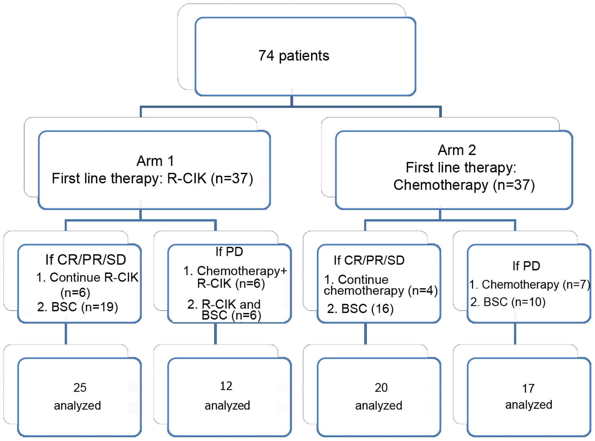
Study design and response
Patients were assigned to one of two treatment
groups. Patients in arm 1 received R-CIK as a first line therapy,
while patients in arm 2 received chemotherapies [Oxaliplatin (130
mg/m2) combined with Capecitabine (1,000
mg/m2)]. The primary endpoint of the study was mOS, and
secondary endpoints were mPFS and 1- and 2-year survival rates.
During treatment, patients received follow-up and reviews every 2
months to evaluate the changes in patients' conditions. The
follow-up deadline was December 8, 2014. Responses to therapy were
evaluated using RECIST 1.1 criteria (26). According to these criteria, clinical
effects were divided into complete response (CR), partial response
(PR), stable disease (SD) and progressive disease (PD). PFS was
calculated from the first treatment to the time at which patients
were found to meet the criteria for PD. OS was calculated from the
time of their first treatment to the time of death or to the time
the patient was re-censored at the last instance of contact with
the live patient. Adverse reactions of these regimes were evaluated
using the National Cancer Institute Common Toxicity Criteria 3.0
(NCI-CTC 3.0), accordingly, reactions were divided into grades 1–4
(27). In arm 1, when patients
emerged as meeting the criteria for PD, R-CIK combined with
chemotherapy (Oxaliplatin combined with Capecitabine) or R-CIK
and/or best supportive care (BSC) was be applied to these patients;
when patients were identified to meet the criteria for CR/PR/SD,
R-CIK or BSC would be applied to these patients. However, in arm 2,
when patients met the criteria for PD, other chemotherapy methods
and/or BSC would be applied to these patients; when patients in arm
2 were found to qualify as showing CR/PR/SD, chemotherapy or BSC
was continued. Details of the courses of therapy in the two
treatment arms are shown in Fig.
1.
Preparation of R-CIK
The method used for CIK cell preparation in the
present study was slightly different from methods detailed in
previous literature (25,28). Peripheral blood mononuclear cells
(PBMCs) were collected from 50 ml peripheral blood draws taken from
patients. These cells were coated with RetroNectin (10 µg/ml;
Takara Biomedical Technology Co., Ltd, Beijing, China) and anti-CD3
antibody (5 µg/ml; catalog no. 7381803; GE Healthcare Bio-Sciences,
Pittsburgh, PA, USA) for 24 h and then cultured in GT-T551 medium
(Takara Bio Inc., Otsu, Japan), which contained recombinant human
interleukin-2 (rhIL-2; 1,000 U/ml), interferon-γ (IFN-γ; 1,000
U/ml) and 5% inactive autogeneic plasma, at 37°C with 5%
CO2 for 4 days. Then the cells were cultured with fresh
IL-2 and 2% inactive autogeneic plasma-containing medium for 5
days. At day 15, R-CIKs were harvested and analyzed for phenotype.
All the products were free of bacterial, mycoplasma, and fungal
contamination. The endotoxin level was <5 EU in all samples.
Phenotypic assessment of R-CIK
R-CIKs (1×10⁶) and PBMCs
(1×10⁶) were harvested and were double stained with 10
µl fluorescein isothiocyanate (FITC) CD3 (catalog no., 555342) and
phycoerythrin (PE) CD4 (catalog no., 555347), CD8 (catalog no.,
555635), CD16 (catalog no., 555408) and CD56 (catalog no., 555517)
(BD Biosciences, San Jose, CA, USA), all at a 1:200 dilution.
Samples were incubated at 4°C for 30 min, then washed twice with
PBS and re-suspended in 500 ml PBS. Fluorescence was detected by
FACS Calibur flow cytometer (BD Biosciences) and data on 10,000
cells were acquired and analyzed. Propidium iodide and annexin
V-FITC (BD Biosciences) were used to measure viability and
apoptosis, according to the manufacturer's protocol.
Cytotoxicity assessment of R-CIK
At the end of cell expansion, R-CIKs' anti-tumor
activity was tested using an overnight cytotoxicity assay with
K-562 (chronic myeloid leukaemia) cell line cells used as targets.
The cytotoxic activity of cells was investigated in a lactate
dehydrogenase (LDH) release assay. This non-radioactive assay is a
colorimetric alternative to the 51Cr release assay and
quantitatively measures LDH that is released upon cell lysis in the
same way that 51Cr is released. Every experiment, at
each effector cell concentration, was performed in a triplicate set
of wells and, the mean value was calculated.
Statistical analysis
SPSS software, version 17.0(SPSS, Inc., Chicago, IL,
USA) was used to perform the statistical analysis. Data for
phenotypic analysis of R-CIK cells and cytotoxicity were analyzed
by independent sample t-tests. For survival time analysis, the
Kaplan-Meier method was used. Prognostic factors of survival time
were analyzed using log-rank test and multivariable analysis. For
all data, P<0.05 was considered to indicate a statistically
significant difference.
Results
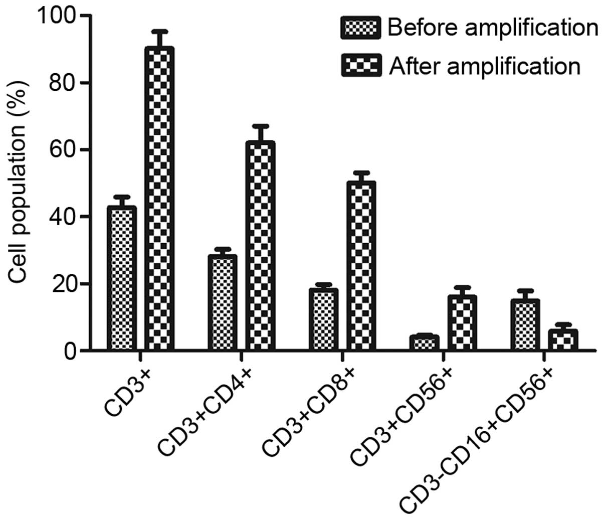
Phenotypic analysis of R-CIKs
Phenotypic analysis of R-CIKs in the patients prior
to culture and after 15 days of culture indicated that the
percentages of CD3+, CD3+/CD4+, CD3+/CD8+ and CD3+/CD56+, increased
from 42.67±3.21, 28.12±2.16, 18.13±1.66 and 4.12±0.56% to
90.21±5.02, 62.01±5.01, 50.13±3.03 and 24.03±2.81% respectively,
(all P<0.05). In contrast, the population of CD3-/CD16+/CD56+
cells reduced from 14.87±2.99 to 5.89±1.87% (P<0.05). The
details are presented in Fig. 2.
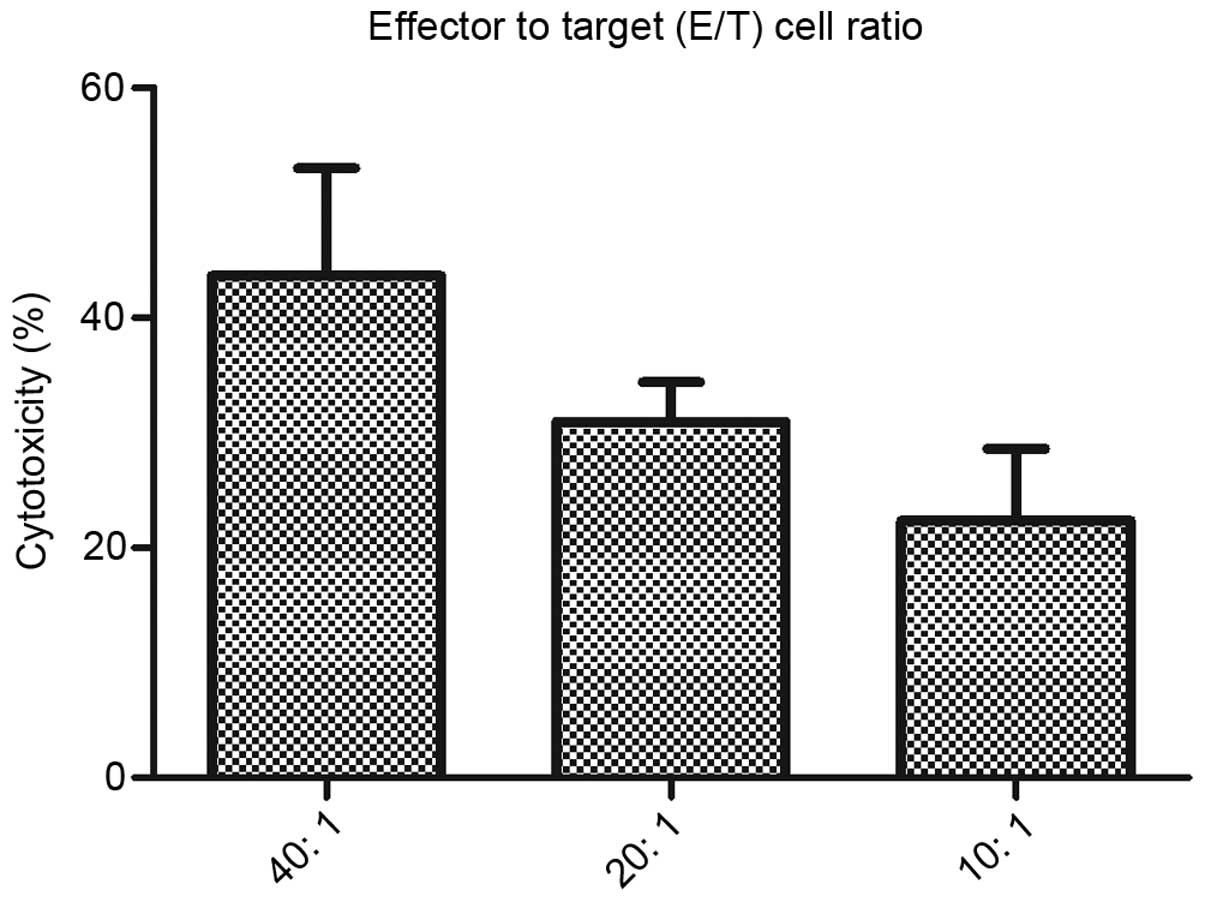
Cytotoxicity of R-CIK in vitro
A sample of each R-CIK expansion obtained from a
total of 37 patients was tested for cytotoxicity against K-562, an
NK-sensitive leukemia target cell line. At effector to target (E/T)
cell ratios of 40:1, 20:1 and 10:1 the median levels of
cytotoxicity were 43.66±9.36%, 30.95±3.49% and 22.39±6.24%,
respectively. The details are presented in Fig. 3.
Treatment outcomes
Follow-up with all patients occurred from January
2010 to October 2013, the outcomes observed in arm 1 patients were
significantly improved compared with the outcomes in arm 2
patients. Specially, the mOS of arm 1 vs. arm 2 was 14.03 vs. 9.46
months (χ2=4.406 P=0.036) (Fig. 4A). The 1-year survival rate was 42.47
vs. 24.89% (95% CI 24.91–59.01% vs. 12.10–40.02%, P=0.066), the
2-year survival rate was 21.24 vs. 5.53% (95% CI 4.60–45.86% vs.
0.46–21.06%, P=0.106), and the mPFS of arm 1 vs. arm 2 was 4.37 vs.
3.90(x2=0.182 P=0.670) (Fig
4B), indicating no significant differences in these measures.
Although the mPFS, 1-year survival rates, and 2-year survival rates
demonstrated no significant differences between the two treatment
arms, the mOS of arm 1 was significantly prolonged when compared
with arm 2. Therefore, patients with advanced HCC who could not
receive local-regional treatments benefited significantly from
R-CIK treatment.
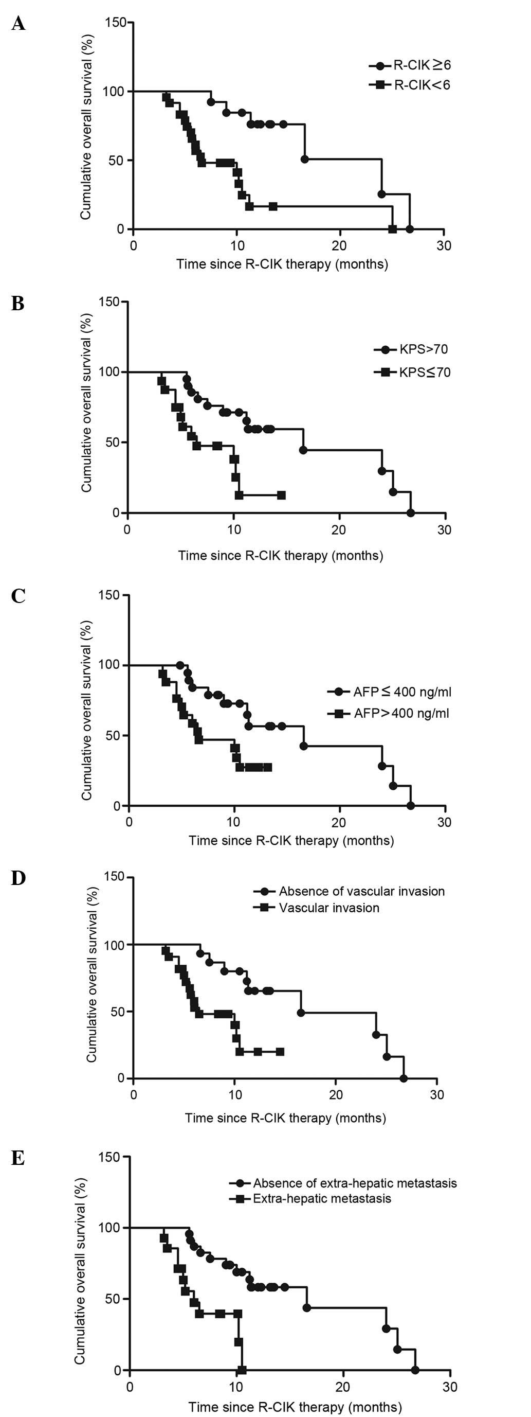
Prognosis factors of R-CIK treatment
based on mOS
Univariate analyses of arm 1 revealed that receiving
≥6 cycles of R-CIK (Fig. 5A), a KPS
>70 (Fig. 5B), AFP ≤400 ng/ml
(Fig. 5C), absence of vascular
invasion (Fig. 5D), and absence of
extra-hepatic metastasis (Fig. 5E)
were all potential predictive factors (P<0.05), (Table II). Multivariate analyses similarly
identified these factors as potentially predictive (P<0.05)
(Table III).
| Table II.Univariate analysis. |
Table II.
Univariate analysis.
| Variable | Median OS
(months) | P | Median PFS
(months) | P |
|---|
| Age (years) |
| 0.697 |
| 0.468 |
|
<60 | 13.674 |
| 4.546 |
|
≥60 | 10.269 |
| 3.955 |
|
| Gender |
| 0.605 |
| 0.517 |
|
Male | 14.440 |
| 4.507 |
|
|
Female | 9.390 |
| 3.875 |
|
| Diameter of tumor
(cm) |
| 0.406 |
| 0.508 |
| ≥5,
<7 | 14.086 |
| 4.760 |
|
|
>7 | 11.244 |
| 4.226 |
|
| KPS (scores) |
| 0.004 |
| 0.401 |
|
>70 | 17.014 |
| 4.638 |
|
|
≤70 | 7. 964 |
| 4.019 |
|
| Child-Pugh |
| 0.145 |
| 0.339 |
|
A+B | 15.057 |
| 4.578 |
|
| C | 8.151 |
| 3.810 |
|
| AFP (ng/ml) |
| 0.026 |
| 0.125 |
|
>400 | 8.270 |
| 3.763 |
|
|
≤400 | 16.768 |
| 4.833 |
|
| Hepatitis B |
| 0.991 |
| 0.961 |
|
Yes | 13.327 |
| 4.217 |
|
| No | 13.868 |
| 4.400 |
|
| Vascular
invasion |
| 0.004 |
| 0.304 |
|
Yes | 8.423 |
| 4.055 |
|
| No | 18.300 |
| 4.833 |
|
| Cirrhosis |
| 0.283 |
| 0.669 |
|
Yes | 8.762 |
| 3.943 |
|
| No | 14.781 |
| 4.470 |
|
| Extrahepatic
metastasis |
| 0.001 |
| 0.116 |
|
Yes | 7.030 |
| 3.657 |
|
| No | 16.928 |
| 4.804 |
|
| Ascites |
| 0.979 |
| 0.508 |
|
Yes | 9.878 |
| 4.226 |
|
| No | 14.064 |
| 4.760 |
|
| Once TACE |
| 0.731 |
| 0.263 |
|
Yes | 14.452 |
| 4.745 |
|
| No | 10.142 |
| 3.929 |
|
| Once RFA |
| 0.720 |
| 0.331 |
|
Yes | 13.543 |
| 5.100 |
|
| No | 11.943 |
| 4.169 |
|
| Once surgery |
| 0.258 |
| 0.227 |
|
Yes | 16.149 |
| 5.033 |
|
| No | 11.215 |
| 4.052 |
|
| Albumin (g/l) |
| 0.605 |
| 0.228 |
|
<35 | 9.391 |
| 4.131 |
|
|
≥35 | 14.444 |
| 5.238 |
|
| Bilirubin
(µmol/l) |
| 0.792 |
| 0.396 |
|
>ULN | 10.553 |
| 4.100 |
|
|
≤ULN | 13.571 |
| 4.656 |
|
| ALT (U/L) |
| 0.283 |
| 0.355 |
|
>ULN | 11.271 |
| 4.113 |
|
|
≤ULN | 16.081 |
| 4.846 |
|
| AST (U/L) |
| 0.625 |
| 0.310 |
|
>ULN | 10.681 |
| 4.070 |
|
|
≤ULN | 13.161 |
| 4.724 |
|
| γ-GGT (U/L) |
| 0.787 |
| 0.238 |
|
>ULN | 13.743 |
| 3.846 |
|
|
≤ULN | 10.104 |
| 4.654 |
|
| PT (s) |
| 0.620 |
| 0.404 |
|
>ULN | 10.636 |
| 4.136 |
|
|
≤ULN | 13.027 |
| 4.713 |
|
| PLT (/L) |
| 0.238 |
| 0.657 |
|
>LLN | 17.092 |
| 4.481 |
|
|
≤LLN | 12.913 |
| 4.070 |
|
| R-CIK cycles |
| 0.002 |
| 0.154 |
| ≥6 | 19.317 |
| 5.077 |
|
|
<6 | 10.173 |
| 3.988 |
|
| Table III.Multivariate analysis. |
Table III.
Multivariate analysis.
| Parameters | Hazard ratio | 95% confidence
interval | P-value |
|---|
| R-CIK (≥6 cycles
vs. 6 cycles) | 0.224 | 0.079–0.632 | 0.005 |
| AFP (ng/ml)
(>400 vs. ≤400) | 0.363 | 0.142–0.924 | 0.034 |
| KPS (>70 scores
vs. ≤70 scores) | 0.265 | 0.102–0.687 | 0.006 |
| Vascular invasion
(yes vs. no) | 0.227 | 0.078–0.661 | 0.007 |
| Extrahepatic
metastasis (yes vs. no) | 0.201 | 0.074–0.546 | 0.002 |
Adverse events
The incidences of adverse treatment effects are
described in Table IV. None of the
patients in arm 1 failed to complete immunotherapy or were ruled
out because of side effects. No severe adverse effects (grade 3 or
4) were associated with R-CIK therapy. Common side effects of
immunotherapy (grade 1 or 2) included fever and headache pain. None
of the patients in arm 2 failed to complete chemotherapy or were
ruled out because of side effects. Common side effects of
chemotherapy (grade 1 or 2) included nausea, leukopenia,
thrombocytopenia and liver dysfunction. There were 4 patients that
developed grade 3 or 4 side effects: Two patients developed fevers
and two were found to have leukopenia.
| Table IV.Adverse events distribution. |
Table IV.
Adverse events distribution.
|
| Arm 1 | Arm 2 |
|---|
|
|
|
|
|---|
| Side effects | Grade 1/2 | Grade 3/4 | Grade 1/2 | Grade 3/4 |
|---|
| Fever | 15 | 0 | 8 | 2 |
| Headache pain | 8 | 0 | 7 | 0 |
| Nausea | 5 | 0 | 15 | 0 |
| Leukopenia | 30 | 0 | 33 | 2 |
| Liver
dysfunction | 20 | 0 | 25 | 0 |
| Diarrhea | 2 | 0 | 4 | 0 |
|
Thrombocytopenia | 10 | 0 | 7 | 0 |
| Anemia | 5 | 0 | 7 | 0 |
Discussion
At present, advanced HCCs are not sufficiently
treated with traditional chemotherapy or radiotherapy. And while
local-regional treatments may be appropriate for cases of early
HCC, they are not suitable for patients with advanced HCC.
Fortunately, recent developments in immunology have given
researchers and clinical physicians the option of utilizing
immunotherapy as a significant component of cancer treatment
(29).
CIK cell therapy is emerging as an important form of
adoptive immunotherapy thanks to its high amplification efficiency
and strong anti-tumor activity. CIK cell therapy represents a
realistic novel option in the field of cancer therapy as it
consistently demonstrates strong anti-tumor activity and improves
the overall survival time of cancer patients when used alone, or
when combined with other conventional therapies. Since 1991, CIK
cell therapy has been evaluated as an adoptive immunotherapy for
cancer patients in a number of clinical trials, including in
patients with HCC (28,30–33). For
example, Pan et al (34)
reported that CIK cell treatment as an adjuvant therapy for
postoperative hepatocellular carcinoma patients could increase
overall survival rates compared with surgery alone. Similarly, Yu
et al (31) reported that
adding CIK treatment could prolong the overall survival time and
progression-free survival time of HCC patients found to be
unsuitable for surgical treatment in 2014. In 2015, Lee et
al reported (35) that adjuvant
immunotherapy with CIK cells increased recurrence-free and overall
survival combined with surgical resection, radiofrequency ablation,
and percutaneous ethanol injection in a multi-center, randomized,
open-label, phase 3 trial. These reports indicate that CIK cell
immunotherapy has significant potential benefits for cancer
patients. However, the specific question of whether CIK cell
therapy can improve survival time of patients with advanced HCC
remained unclear.
The effectiveness of CIK cell therapy depends on the
quality and quantity of CIK cells obtained for therapeutic
administration. In the CIK cell preparations in the present study,
the method of culture was slightly different from methods employed
in previous literature and it was possible to obtain more of the
CD3+/CD56+ cellular subset on day 15 than common CIK cells
preparation methods achieve (28).
RetroNectin (RN) was added to the culture media in our CIK cell
preparation (called R-CIK), which improves the conglutination,
extension, and differentiation of these cells (25). Our results indicate that the
percentages of CD3+, CD3+/CD4+, CD3+/CD8+ and CD3+/CD56+ cells were
significantly increased after stimulation and expansion in culture
(25). In addition, there was a
significant decrease in the CD3-/CD16+/CD56+ population. It is
widely accepted that the increase in CD4+ and CD8+ T cells during
CIK expansion is crucial to tumor immunity. CD3+/CD56+ cells, which
comprise a rare population of cells in normal peripheral blood, are
also significantly increased after stimulation and expansion. These
may be the most important cells for the anti-cancer effect of CIK
treatment (36) as they have shown
strong anti-cancer activity in a variety of malignant tumor cell
lines in vitro (20,25,37).
Therefore, we have good reason to hypothesize that our improved
methods have allowed us to obtain R-CIK cells with particularly
strong anti-tumor activity for transferring them to patients.
Although the present study failed to show a
significant difference in mPFS between the two treatment arms,
R-CIK treatment did significantly prolonged the mOS of advanced HCC
patients compared with the chemotherapy arm. In the calculations
from univariate analyses of arm 1, R-CIK cycles ≥6, KPS >70,
AFP≈≤400 ng/ml, the absence of vascular invasion and a lack of
extra-hepatic metastasis were all potential predictive factors
(P<0.05). In the calculations from multivariate analyses, these
factors were also found to have potential predictive value
(P<0.05). These results indicate that the likely effects of
R-CIK treatment can be reasonably well predicted based on
preliminary laboratory tests and imaging reports.
In conclusion, the present study indicated R-CIK
cell therapy can improve prognosis in advanced HCC and increasing
the number of cycles of R-CIK cell therapy is likely to result in
additional benefits. Discrepancies versus previous studies of CIK
therapy may be attributed to the differences in stimulation and
expansion methods, genetic and environmental backgrounds, staging
systems, and sample size. It will certainly be necessary to carry
out numerous additional studies in order to optimize techniques and
determine the full range of application for this type of therapy.
At the same time, the combination of R-CIK with conventional
chemotherapy, as well as R-CIK combined with Sorafenib also clearly
merit exploration.
R-CIK treatment can prolong mOS of advanced
hepatocellular carcinoma patients compared with conventional
chemotherapy alone. Patients who undergo >6 cycles of R-CIK,
have a KPS >70, an AFP ≤400 ng/ml, no vascular invasion and no
extra-hepatic metastases may have longer survival times in
comparison with other patients.
Acknowledgements
The authors are grateful for the collaboration
received from the participating college and its staff and Dr
Weiquan Lu and Dr Erjiang Zhao from the Department of Cancer
Prevention, Henan Cancer Hospital, China, for statistical analysis.
Professor Quanli Gao designed this study, Dr Wei Li and Dr Yaomei
Wang wrote the paper, Dr Lingdi Zhao and Dr Linping Xu did the
follow-up of all patients and Dr Daniel B. Kellner edited this
paper as a native English speaker.
References
|
1
|
Qiang L, Huikai L, Butt K, Wang PP and Hao
X: Factors associated with disease survival after surgical
resection in Chinese patients with hepatocellular carcinoma. World
J Surg. 30:439–445. 2006. View Article : Google Scholar : PubMed/NCBI
|
|
2
|
Bosch FX, Ribes J and Borràs J:
Epidemiology of primary liver cancer. Semin Liver Dis. 19:271–285.
1999. View Article : Google Scholar : PubMed/NCBI
|
|
3
|
El-Serag HB and Mason AC: Rising incidence
of hepatocellular carcinoma in the United States. N Engl J Med.
340:745–750. 1999. View Article : Google Scholar : PubMed/NCBI
|
|
4
|
Taylor-Robinson SD, Foster GR, Arora S,
Hargreaves S and Thomas HC: Increase in primary liver cancer in the
UK, 1979–94. Lancet. 350:1142–1143. 1997. View Article : Google Scholar : PubMed/NCBI
|
|
5
|
Jarnagin WR: Management of small
hepatocellular carcinoma: A review of transplantation, resection,
and ablation. Ann Surg Oncol. 17:1226–1233. 2010. View Article : Google Scholar : PubMed/NCBI
|
|
6
|
Page AJ, Cosgrove DC, Philosophe B and
Pawlik TM: Hepatocellular carcinoma: Diagnosis, management, and
prognosis. Surg Oncol Clin N Am. 23:289–311. 2014. View Article : Google Scholar : PubMed/NCBI
|
|
7
|
Truty MJ and Vauthey JN: Surgical
resection of high-risk hepatocellular carcinoma: Patient selection,
preoperative considerations, and operative technique. Ann Surg
Oncol. 17:1219–1225. 2010. View Article : Google Scholar : PubMed/NCBI
|
|
8
|
Asham EH, Kaseb A and Ghobrial RM:
Management of hepatocellular carcinoma. Surg Clin North Am.
93:1423–1450. 2013. View Article : Google Scholar : PubMed/NCBI
|
|
9
|
Chok KS, Ng KK, Poon RT, Lo CM and Fan ST:
Impact of postoperative complications on long-term outcome of
curative resection for hepatocellular carcinoma. Br J Surg.
96:81–87. 2009. View
Article : Google Scholar : PubMed/NCBI
|
|
10
|
Seo DD, Lee HC, Jang MK, Min HJ, Kim KM,
Lim YS, Chung YH, Lee YS, Suh DJ, Ko GY, et al: Preoperative portal
vein embolization and surgical resection in patients with
hepatocellular carcinoma and small future liver remnant volume:
Comparison with transarterial chemoembolization. Ann Surg Oncol.
14:3501–3509. 2007. View Article : Google Scholar : PubMed/NCBI
|
|
11
|
Ikai I, Itai Y, Okita K, Omata M, Kojiro
M, Kobayashi K, Nakanuma Y, Futagawa S, Makuuchi M and Yamaoka Y:
Report of the 15th follow-up survey of primary liver cancer.
Hepatol Res. 28:21–29. 2004. View Article : Google Scholar : PubMed/NCBI
|
|
12
|
Deans C and Leslie P: Hepatocellular
carcinoma. Lancet. 354:253–254. 1999. View Article : Google Scholar : PubMed/NCBI
|
|
13
|
Ribero D, Curley SA, Imamura H, Madoff DC,
Nagorney DM, Ng KK, Donadon M, Vilgrain V, Torzilli G, Roh M and
Vauthey JN: Selection for resection of hepatocellular carcinoma and
surgical strategy: Indications for resection, evaluation of liver
function, portal vein embolization, and resection. Ann Surg Oncol.
15:986–992. 2008. View Article : Google Scholar : PubMed/NCBI
|
|
14
|
Vivarelli M, Guglielmi A, Ruzzenente A,
Cucchetti A, Bellusci R, Cordiano C and Cavallari A: Surgical
resection versus percutaneous radiofrequency ablation in the
treatment of hepatocellular carcinoma on cirrhotic liver. Ann Surg.
240:102–107. 2004. View Article : Google Scholar : PubMed/NCBI
|
|
15
|
Bruix J, Raoul JL, Sherman M, Mazzaferro
V, Bolondi L, Craxi A, Galle PR, Santoro A, Beaugrand M,
Sangiovanni A, et al: Efficacy and safety of sorafenib in patients
with advanced hepatocellular carcinoma: Subanalyses of a phase III
trial. J Hepatol. 57:821–829. 2012. View Article : Google Scholar : PubMed/NCBI
|
|
16
|
Cheng AL, Kang YK, Chen Z, Tsao CJ, Qin S,
Kim JS, Luo R, Feng J, Ye S, Yang TS, et al: Efficacy and safety of
sorafenib in patients in the Asia-Pacific region with advanced
hepatocellular carcinoma: A phase III randomised, double-blind,
placebo-controlled trial. Lancet Oncol. 10:25–34. 2009. View Article : Google Scholar : PubMed/NCBI
|
|
17
|
Qin S, Bai Y, Lim HY, Thongprasert S, Chao
Y, Fan J, Yang TS, Bhudhisawasdi V, Kang WK, Zhou Y, et al:
Randomized, multicenter, open-label study of oxaliplatin plus
fluorouracil/leucovorin versus doxorubicin as palliative
chemotherapy in patients with advanced hepatocellular carcinoma
from Asia. J Clin Oncol. 31:3501–3508. 2013. View Article : Google Scholar : PubMed/NCBI
|
|
18
|
von Delius S, Lersch C, Mayr M, Stock K,
Schulte-Frohlinde E, Schmid RM and Eckel F: Capecitabine for
treatment of advanced hepatocellular carcinoma.
Hepatogastroenterology. 54:2310–2314. 2007.PubMed/NCBI
|
|
19
|
Zaanan A, Williet N, Hebbar M, Dabakuyo
TS, Fartoux L, Mansourbakht T, Dubreuil O, Rosmorduc O, Cattan S,
Bonnetain F, et al: Gemcitabine plus oxaliplatin in advanced
hepatocellular carcinoma: A large multicenter AGEO study. J
Hepatol. 58:81–88. 2013. View Article : Google Scholar : PubMed/NCBI
|
|
20
|
Schmidt-Wolf IG, Negrin RS, Kiem HP, Blume
KG and Weissman IL: Use of a SCID mouse/human lymphoma model to
evaluate cytokine-induced killer cells with potent antitumor cell
activity. J Exp Med. 174:139–149. 1991. View Article : Google Scholar : PubMed/NCBI
|
|
21
|
Ma H, Zhang Y, Wang Q, Li Y, He J, Wang H,
Sun J, Pan K, Chen M and Xia J: Therapeutic safety and effects of
adjuvant autologous RetroNectin activated killer cell immunotherapy
for patients with primary hepatocellular carcinoma after
radiofrequency ablation. Cancer Biol Ther. 9:903–907. 2010.
View Article : Google Scholar : PubMed/NCBI
|
|
22
|
Lee HJ, Lee YS, Kim HS, Kim YK, Kim JH,
Jeon SH, Lee HW, Kim S, Miyoshi H, Chung HM and Kim DK: Retronectin
enhances lentivirus-mediated gene delivery into hematopoietic
progenitor cells. Biologicals. 37:203–209. 2009. View Article : Google Scholar : PubMed/NCBI
|
|
23
|
Lamers CH, van Elzakker P, van Steenbergen
SC, Sleijfer S, Debets R and Gratama JW: Retronectin-assisted
retroviral transduction of primary human T lymphocytes under good
manufacturing practice conditions: Tissue culture bag critically
determines cell yield. Cytotherapy. 10:406–416. 2008. View Article : Google Scholar : PubMed/NCBI
|
|
24
|
Yu SS, Nukaya I, Enoki T, Chatani E, Kato
A, Goto Y, Dan K, Sasaki M, Tomita K, Tanabe M, et al: In vivo
persistence of genetically modified T cells generated ex vivo using
the fibronectin CH296 stimulation method. Cancer Gene Ther.
15:508–516. 2008. View Article : Google Scholar : PubMed/NCBI
|
|
25
|
Wang Z, Zhang Y, Liu Y, Wang L, Zhao L,
Yang T, He C, Song Y and Gao Q: Association of myeloid-derived
suppressor cells and efficacy of cytokine-induced killer cell
immunotherapy in metastatic renal cell carcinoma patients. J
Immunother. 37:43–50. 2014. View Article : Google Scholar : PubMed/NCBI
|
|
26
|
Watanabe H, Okada M, Kaji Y, Satouchi M,
Sato Y, Yamabe Y, Onaya H, Endo M, Sone M and Arai Y: New response
evaluation criteria in solid tumours-revised RECIST guideline
(version 1.1). Gan To Kagaku Ryoho. 36:2495–2501. 2009.(In
Japanese). PubMed/NCBI
|
|
27
|
Trotti A, Colevas AD, Setser A, Rusch V,
Jaques D, Budach V, Langer C, Murphy B, Cumberlin R, Coleman CN and
Rubin P: CTCAE v3.0: Development of a comprehensive grading system
for the adverse effects of cancer treatment. Semin Radiat Oncol.
13:176–181. 2003. View Article : Google Scholar : PubMed/NCBI
|
|
28
|
Liu L, Zhang W, Qi X, Li H, Yu J, Wei S,
Hao X and Ren X: Randomized study of autologous cytokine-induced
killer cell immunotherapy in metastatic renal carcinoma. Clin
Cancer Res. 18:1751–1759. 2012. View Article : Google Scholar : PubMed/NCBI
|
|
29
|
DeVita VJ Jr and Rosenberg SA: Two hundred
years of cancer research. N Engl J Med. 366:2207–2214. 2012.
View Article : Google Scholar : PubMed/NCBI
|
|
30
|
Hontscha C, Borck Y, Zhou H, Messmer D and
Schmidt-Wolf IG: Clinical trials on CIK cells: First report of the
international registry on CIK cells (IRCC). J Cancer Res Clin
Oncol. 137:305–310. 2011. View Article : Google Scholar : PubMed/NCBI
|
|
31
|
Yu X, Zhao H, Liu L, Cao S, Ren B, Zhang
N, An X, Yu J, Li H and Ren X: A randomized phase II study of
autologous cytokine-induced killer cells in treatment of
hepatocelluar carcinoma. J Clin Immunol. 34:194–203. 2014.
View Article : Google Scholar : PubMed/NCBI
|
|
32
|
Hui D, Qiang L, Jian W, Ti Z and Da-Lu K:
A randomized, controlled trial of postoperative adjuvant
cytokine-induced killer cells immunotherapy after radical resection
of hepatocellular carcinoma. Dig Liver Dis. 41:36–41. 2009.
View Article : Google Scholar : PubMed/NCBI
|
|
33
|
Shi M, Zhang B, Tang ZR, Lei ZY, Wang HF,
Feng YY, Fan ZP, Xu DP and Wang FS: Autologous cytokine-induced
killer cell therapy in clinical trial phase I is safe in patients
with primary hepatocellular carcinoma. World J Gastroenterol.
10:1146–1151. 2004.PubMed/NCBI
|
|
34
|
Pan K, Li YQ, Wang W, Xu L, Zhang YJ,
Zheng HX, Zhao JJ, Qiu HJ, Weng DS, Li JJ, et al: The efficacy of
cytokine-induced killer cell infusion as an adjuvant therapy for
postoperative hepatocellular carcinoma patients. Ann Surg Oncol.
20:4305–4311. 2013. View Article : Google Scholar : PubMed/NCBI
|
|
35
|
Lee JH, Lee JH, Lim YS, Yeon JE, Song TJ,
Yu SJ, Gwak GY, Kim KM, Kim YJ, Lee JW and Yoon JH: Adjuvant
immunotherapy with autologous cytokine-induced killer cells for
hepatocellular carcinoma. Gastroenterology. 148:1383–1391.e6. 2015.
View Article : Google Scholar : PubMed/NCBI
|
|
36
|
Lu PH and Negrin RS: A novel population of
expanded human CD3+CD56+ cells derived from T cells with potent in
vivo antitumor activity in mice with severe combined
immunodeficiency. J Immunol. 153:1687–1696. 1994.PubMed/NCBI
|
|
37
|
Shablak A, Hawkins RE, Rothwell DG and
Elkord E: T cell-based immunotherapy of metastatic renal cell
carcinoma: Modest success and future perspective. Clin Cancer Res.
15:6503–6510. 2009. View Article : Google Scholar : PubMed/NCBI
|