Introduction
Pulmonary carcinoids (PCs), which account for 2–5%
of all lung primary tumors (1) and
are comprised of the typical carcinoid (TC) and atypical carcinoid
(AC) types, are components of neuroendocrine neoplasms, a
heterogenous group of tumors whose prevalence is increasing
(2). TCs, which are up to four times
more frequent than ACs, are low-grade tumors characterized by
scarce mitotic figures, an absence of necrosis and a lower average
size in comparison with ACs. ACs are considered to be
intermediate-grade tumors with more mitotic figures than TCs, plus
areas of focal necrosis and a greater size than TCs (3). The mean 5-year survival rate for
patients with TCs is 88% and ranges from 25–56% for ACs (4–6). Surgical
resection of the tumor in the affected lung is the standard
treatment, but complete surgical excision can be difficult or
unattainable depending upon the location of the tumor in the chest
and whether it has metastasized. The outcome for patients with
widely metastatic disease is poor, and chemotherapy and radiation
therapy have limited success in treating PCs (7–9). Several
studies designed to identify molecular alterations associated with
the development and unpredictable progression of this type of tumor
have only been partially successful (10–12). A
better understanding of PC biology could lead to the development of
novel therapeutic strategies.
The human epidermal growth factor receptor (HER)
family, comprising the epidermal growth factor receptor (EGFR; also
known as HER1), HER2, HER3 and HER4, is not only essential for the
development and maintenance of normal tissue, but is also strongly
involved in the development of numerous tumor types (13,14). HER
dimerization is required for signal transduction to occur, through
formation of homodimeric or heterodimeric complexes. Dimer
formation leads to activation of the intrinsic tyrosine kinase
domain, followed by phosphorylation on specific tyrosine residues,
which serve as docking sites for downstream signaling proteins
(15). An increasing number of small
molecules and monoclonal antibodies are being recognized as
targeting the HER family (15–17).
In the present study, the expression pattern of
unphosphorylated (HER) and phosphorylated (p-HER) forms of HER
receptors were defined in archival tumor specimens of 37 patients
with PC tumors (29 with TC and 8 with AC), in order to assess the
expression of possible molecular targets of anti-HER therapy.
Materials and methods
Tissue samples
Archived formalin-fixed paraffin-embedded blocks
derived from resection material of 37 apparently sporadic PCs
consecutively diagnosed between January 2001 and December 2008 at
the Institute of Pathology, ‘S.S. Annunziata’ Hospital (Chieti,
Italy) were retrieved. All tumors were reviewed for diagnosis.
Cases were classified as TC (29 cases) or AC (8 cases) according to
the World Health Organization classification (18). For each sample, tumor and normal
tissues were available. The study was approved by the Ethical
Committee of the ‘S.S. Annunziata’ Hospital.
Tissue microarray (TMA) construction
and immunohistochemistry (IHC)
Duplicate TMAs were constructed by extracting cores
(2-mm in diameter) of histologically confirmed neoplastic areas, as
previously described (19). TMA
sections were stained using antibodies to HER1 (EGFR PharmDx kit;
Dako, Milano, Italy), pY1197-HER1 (rabbit polyclonal, 1:400
dilution; 30-min incubation; Thermo Fisher Scientific Inc.,
Waltham, MA, USA), HER2 (HercepTest kit; Dako), pY1248-HER2 (clone
PN2A; 1:25 dilution; overnight incubation; Dako), HER3 (clone RTJ1;
1:20 dilution; overnight incubation; Novocastra; Leica
Microsystems, Milano, Italy), pY1289-HER3 (clone 21D3; 1:50
dilution; overnight incubation; Cell Signaling Technology Inc.,
Danvers, MA, USA), HER4 (rabbit polyclonal; 1:50 dilution;
overnight incubation; Santa Cruz Technology Inc., Dallas, TX, USA)
and pY1162-HER4 (rabbit monoclonal; 1:100 dilution; overnight
incubation; Epitomics/Abcam, Cambridge, UK). Epitope retrieval was
performed in a heated citrate buffer (pH 6.0) for p-HER2, HER3,
HER4 and p-HER4, and in 1 mmol/l EDTA (pH 8.0) for p-HER3.
Antigen-antibody reactions were visualized using a polymer-based
detection system (EnVision kit; Dako), using diaminobenzidine as a
chromogen. The expression of HER1 and HER2 was evaluated according
to the manufacturer's protocols (EGFR PharmDx and HercepTest Dako
kits, respectively). Appropriate positive controls (HER1, EGFR
PharmDx control slide; HER2, HER2 HercepTest control slide; HER3,
HER4, p-HER1, p-HER2 and p-HER4, breast cancer tissue; and p-HER3,
lung cancer tissue) of human tumor tissues known to express the
marker of interest were used. For negative control sections, the
specific primary antibody was omitted or replaced with non-immune
serum or isotype-matched immunoglobulins. The expression of markers
was quantified as a percentage of the immunoreactive tumor cells.
Cases were considered HER-positive when ≥1% of tumor cells were
positively stained (PharmDx kit; Dako, Milano, Italy).
Statistical analysis
Comparisons between molecular markers were performed
using Spearman's Rho correlation. Student's t-test was used to
evaluate the statistical differences between markers (HER and
p-HER) in PC according to the MEN1 gene status and the tumor
histotype (TC and AC). SPSS version 15.0 (SPSS Inc., Chicago, IL,
USA) was employed throughout, and P<0.05 was considered to
indicate a statistically significant difference.
Results
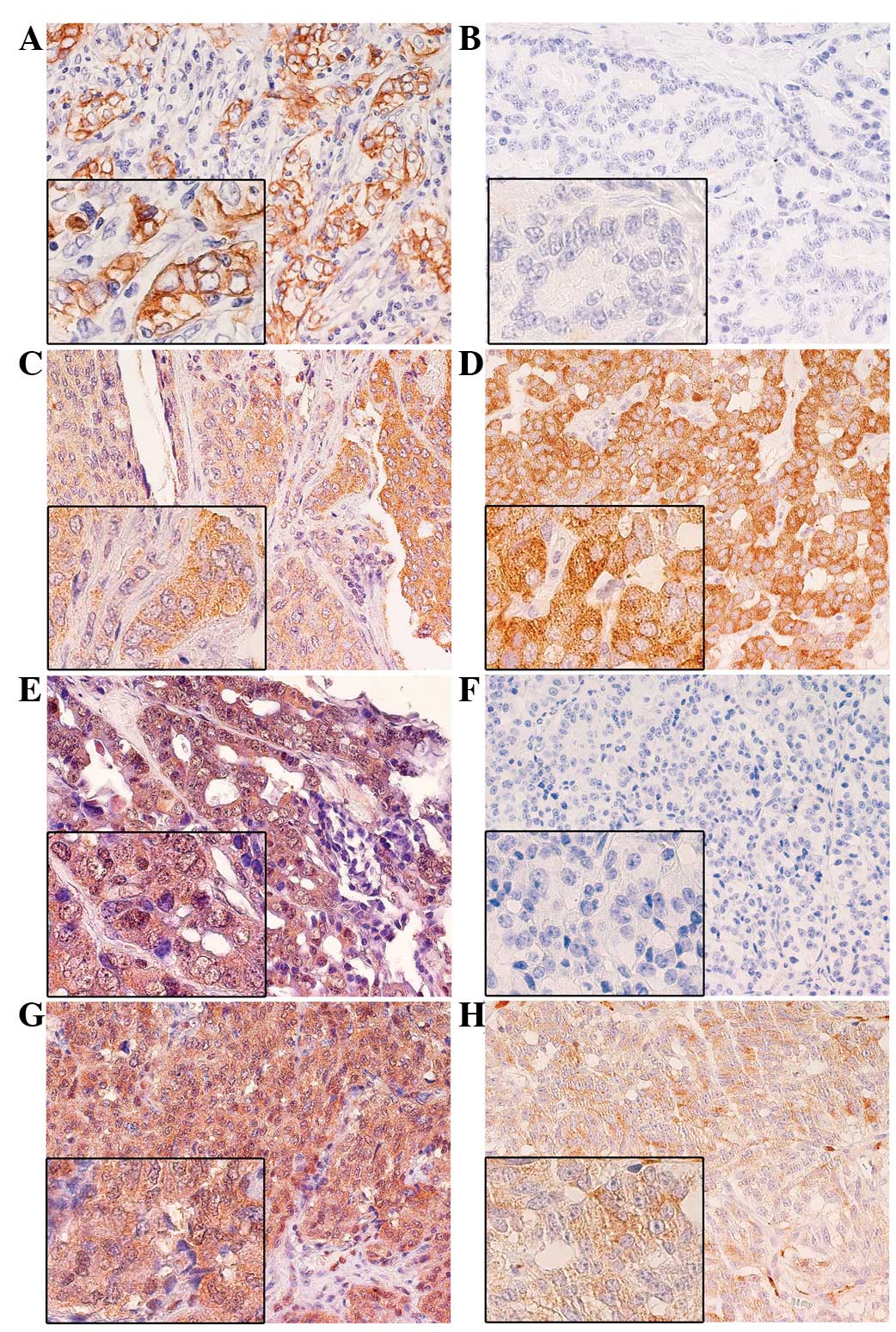
Immunoreactivity for HER1 was present mainly in the
tumor cell membranes. Reactivity for HER2 and p-HER2 receptors was
absent in all samples. Specific positive staining for HER3 and HER4
was exclusively found in the cytoplasm of the tumor cells (Fig. 1). p-HER1 and p-HER3 were mainly
detected in the cytoplasm. However, in association with cytoplasmic
expression, nuclear staining for p-HER1 and p-HER3 was also
observed in a small proportion of tumor cells [mean ± standard
error (SE), 6.3±4.1 and 2.3±1.0%, respectively]. For analytical
purposes, only cytoplasmic expression for p-HER1 and p-HER3 was
considered.
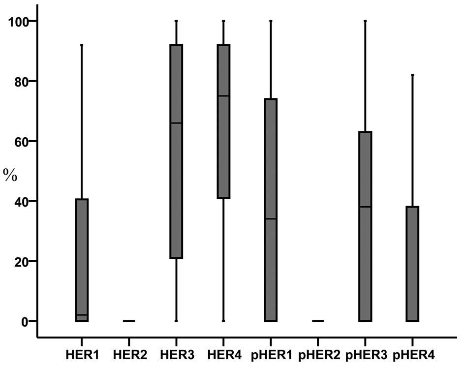
The analysis showed that 65.5% of TC and 50.0% of AC
tumors expressed HER1. HER3-positive tumors occurred in 79.3 and
100% of TC and AC cases, respectively, while HER4-positive tumors
occurred in 82.8 and 87.5%, respectively. In the cases of TC, the
following proportions of positive tumors were recorded; 51.7% for
p-HER1, 75.9% for p-HER3 and 48.3% for p-HER4. In the cases of AC,
the proportions of positive tumors were as follows: 62.5% for
p-HER1, 62.5% for p-HER3 and 37.5% for p-HER4. The distribution of
HER and p-HER expression is reported as a box-and-whisker plot in
Fig. 2.
The expression (mean % ± SE) of the HERs and their
activated forms, in all cases and in TC vs. AC tumor types, are
reported in Table I. As assessed by
independent-samples t-test, the TCs and ACs did not
significantly differ in terms of HER/p-HER expression.
 | Table I.Expression of HER markers and their
activated forms in pulmonary carcinoids. |
Table I.
Expression of HER markers and their
activated forms in pulmonary carcinoids.
| Markera | All cases (n=37) | Typical carcinoids
(n=29) | Atypical carcinoids
(n=8) | P-valueb |
|---|
| HER1 | 22.2±5.6 | 19.0±5.8 | 33.0±14.6 | 0.296 |
| HER2 |
0.0±0.0 |
0.0±0.0 | 0.0±0.0 |
| HER3 | 58.0±6.2 | 56.9±7.5 | 57.0±11.0 | 0.690 |
| HER4 | 62.5±5.9 | 62.6±7.0 | 62.0±11.1 | 0.966 |
| p-HER1 | 36.8±8.1 | 36.0±9.6 | 38.7±16.5 | 0.884 |
| p-HER2 |
0.0±0.0 |
0.0±0.0 | 0.0±0.0 |
| p-HER3 | 35.0±6.4 | 38.7±7.8 | 25.5±11.2 | 0.370 |
| p-HER4 | 19.4±5.4 | 20.1±6.2 | 17.7±11.5 | 0.849 |
By Spearman's correlation analysis of the expression
of HERs and p-HERs in PCs (Table
II), HER3 expression was found to be positively correlated with
that of HER4 (rho=0.493; P=0.002), p-HER3 (rho=0.687; P<0.001)
and p-HER4 (rho=0.495; P=0.014). The expression of HER4 was also
directly correlated with that of p-HER1 (rho=0.482; P=0.017),
p-HER3 (rho=0.508; P=0.005) and p-HER4 (rho=0.550; P=0.005). In
addition, significant direct correlations were found between p-HER3
and p-HER1 (rho=0.535; P=0.009), p-HER3 and p-HER4 (rho=0.728;
P<0.001), and p-HER4 and p-HER1 (rho=0.882; P<0.001)
(Table II).
| Table II.Correlationsa among HER and p-HER expression
(percentage values) in pulmonary carcinoids. |
Table II.
Correlationsa among HER and p-HER expression
(percentage values) in pulmonary carcinoids.
| Marker | HER1 | HER3 | HER4 | p-HER1 | p-HER3 | p-HER4 |
|---|
| HER1 |
|
|
|
|
|
|
|
rho | 1.000 | 0.003 | −0.088 | 0.367 | 0.067 | 0.292 |
|
P-value |
| 0.987 | 0.615 | 0.078 | 0.734 | 0.177 |
| HER3 |
|
rho | 0.003 | 1.000 | 0.493b | 0.392 | 0.687b | 0.495b |
|
P-value | 0.987 |
| 0.002b | 0.058 | 0.000b | 0.014b |
| HER4 |
|
rho | −0.088 | 0.493b | 1.000 | 0.482b | 0.508b | 0.550b |
|
P-value | 0.615 | 0.002b |
| 0.017b | 0.005b | 0.005b |
| p-HER1 |
|
rho | 0.367 | 0.392 | 0.482b | 1.000 | 0.535b | 0.882b |
|
P-value | 0.078 | 0.058 | 0.017b |
| 0.009b | 0.000b |
| p-HER3 |
|
rho | 0.067 | 0.687b | 0.508b | 0.535b | 1.000 | 0.728b |
|
P-value | 0.734 | 0.000b | 0.005b | 0.009b |
| 0.000b |
| p-HER4 |
|
rho | 0.292 | 0.495b | 0.550b | 0.882b | 0.728b | 1.000 |
|
P-value | 0.177 | 0.014b | 0.005b | 0.000b | 0.000b |
|
PC samples utilized in this study have previously
been characterized for the expression of menin and the mutational
status of the MEN1 gene (20).
Therefore, in the present study, possible correlations were
searched for among the expression results of the HERs/p-HERs and
that of menin, by Spearman's rho test. As reported in Table III, the cytoplasmic but not the
nuclear expression of menin was positively correlated with HER3
(rho=0.457; P=0.004), HER4 (rho=0.398; P=0.015), p-HER1 (rho=0.551;
P=0.005), p-HER3 (rho=0.641; P<0.001), and p-HER4 (rho=0.635;
P=0.001) expression.
| Table III.Correlationsa among menin and HER/p-HER
expression (percentage values) in pulmonary carcinoids. |
Table III.
Correlationsa among menin and HER/p-HER
expression (percentage values) in pulmonary carcinoids.
| Localization | n-menin | c-menin | HER1 | HER3 | HER4 | p-HER1 | p-HER3 | p-HER4 |
|---|
|
n-meninb |
|
|
|
|
|
|
|
|
|
rho | 1.000 | −0.059 | −0.046 | 0.073 | 0.167 | −0.066 | −0.233 | −0.073 |
|
P-value |
|
0.723 |
0.792 | 0.668 | 0.323 |
0.759 |
0.224 | 0.736 |
|
c-meninb |
|
|
|
|
|
|
|
|
|
rho | −0.059 | 1.000 | −0.094 | 0.457b | 0.398b |
0.551b |
0.641b | 0.635b |
|
P-value |
0.723 |
|
0.592 | 0.004b | 0.015b |
0.005b |
0.000b | 0.001b |
Table IV shows the
expression of HERs and their activated forms according to
MEN1 gene status. By independent-sample t-test, HER3 and
p-HER3 were found to be significantly more expressed in PCs with
MEN1 variants than in tumors with the MEN1 wild-type
(P=0.000 and P=0.025, respectively). This was true for the TCs and
the ACs. HER4, but not its activated form, was also found to be
more expressed in PC tumors (P=0.009) with MEN1 variants. No
statistical significances were found between MEN1 status and
the expression levels of HER1 and p-HER1 (Table IV).
| Table IV.Expression of HERs and their
activated forms in pulmonary carcinoids according to MEN1
gene status. |
Table IV.
Expression of HERs and their
activated forms in pulmonary carcinoids according to MEN1
gene status.
|
| All cases | Typical
carcinoids | Atypical
carcinoids |
|---|
|
|
|
|
|
|---|
| Markers | Mean ± SE | P-value | Mean ± SE | P-value | Mean ± SE | P-value |
|---|
| HER1 |
|
MEN1 variants | 18.6±9.1 | 0.648 | 18.4±10.9 | 0.949 | 19.0±19.0 | 0.379 |
|
MEN1 wild-type | 24.0±7.1 |
| 19.2±7.0 |
| 47.0±22.6 |
| HER3 |
|
MEN1 variants | 84.8±5.4 | 0.000a | 85.1±7.0 | 0.001a | 84.0±9.3 | 0.012a |
|
MEN1 wild-type | 43.5±7.7 |
| 44.2±9.1 |
| 40.0±8.1 |
|
| HER4 |
| MEN1
variants | 79.5±4.8 | 0.009a | 79.6±6.5 | 0.039a |
79.3±6.8 | 0.156 |
| MEN1
wild-type | 53.3±8.2 |
| 55.0±9.3 | | 44.8±18.3 |
|
| p-HER1 |
| MEN1
variants | 45.9±15.7 | 0.443 | 57.5±23.1 | 0.226 | 34.3±22.9 | 0.785 |
| MEN1
wild-type | 32.3±9.5 |
| 29.4±10.2 |
| 44.7±28.6 |
|
| p-HER3 |
| MEN1
variants | 54.5±8.5 | 0.025a | 59.7±9.5 | 0.045a | 46.8±16.7 | 0.046a |
| MEN1
wild-type | 24.8±7.9 |
| 30.3±9.5 |
|
4.3±2.5 |
|
| p-HER4 |
| MEN1
variants | 26.6±9.9 | 0.354 | 25.5±9.0 | 0.643 | 27.8±19.3 | 0.358 |
| MEN1
wild-type | 15.8±6.5 |
| 18.4±7.8 |
| 4.3±4.3 |
Discussion
HER1 is the first member of the HER family to be
identified in PCs, and the existence of an autocrine
growth-promoting circuit based on HER1 and transforming growth
factor-α has previously been documented (21,22). Lack
of HER2 expression in PCs was first observed by Wilkinson et
al (23). More recently, Rickman
et al (12) investigated a
series of PCs by IHC analysis performed in TMAs to detect the
expression of the HER family, and found that 46% of TCs and 28% of
ACs expressed HER1, while none expressed HER2 and 100% showed
intense to moderate staining for HER3 and HER4. The present study
is the first report including data on the expression of p-HER
forms. Notably PCs were found to express HER1, HER3 and HER4
receptors, as well as their phosphorylated forms.
Several malignancies, including lung, breast,
stomach, colorectal, head and neck, and pancreatic carcinomas, and
glioblastoma, melanoma and ovarian cancers, are associated with the
mutation or increased expression of members of the HER family. HER3
is the only member of this receptor tyrosine kinase family that
does not have intrinsic kinase activity, but is unexpectedly the
most robust signaling complex of the HER family (17). Different degrees of HER3 expression
have been observed in lung, breast, ovarian, prostate, gastric and
colorectal cancer, and high expression usually correlates with a
poor prognosis (24). HER2 is
unlikely to be the catalytic partner of HER3, as HER2 expression is
low or absent in the majority of melanomas. HER3 may function as an
allosteric activator of HER1 or HER4 in melanomas (25) and PCs, as suggested by the observation
that HER2 is absent in these tumors. In addition, the present study
found that the expression of activated HER3 was directly correlated
with that of activated HER1 and HER4. The role of HER4 in
tumorigenesis is complex and not completely understood. In a breast
cancer study, HER4 expression was found to be correlated with a
favorable prognosis (26). By
contrast, non-small cell lung cancer patients with tumors that
expressed HER4 experienced decreased survival times compared with
patients with tumors that did not express HER4, and a positive
correlation was also found with lymph node metastasis and HER4
expression (27).
The MEN1 gene is implicated in the
pathogenesis of hereditary and sporadic PCs, and MEN1 gene
mutations are associated with a poor prognosis in these tumors
(28). This association with poor
prognosis appears to be specific to PCs, as it is not observed in
pancreatic neuroendocrine tumors (29).
Menin, the protein encoded by the MEN1 gene,
is a component of histone methyltransferase complexes and is
ubiquitously expressed (18,30). We previously observed that PCs
displaying MEN1 nucleotide variants were characterized by
higher menin accumulation in the cytoplasm (disarrayed
distribution), but not in the nucleus, compared with those without
MEN1 variants (20). In the pancreas,
IHC staining for menin showed that normal islet cells exhibited
intense nuclear and extremely faint cytoplasmic staining, while 40%
of pancreatic endocrine tumors lacked nuclear immunostaining and
expressed high levels of cytoplasmic menin (31). The present study results showed that
the presence of the activated HER1, HER3 and HER4 forms was
directly correlated with the disarrayed cytoplasmic menin
expression, and more importantly, that higher proportions of
p-HER3-positive cells are present in PCs harboring MEN1
variants when compared with the wild-type counterpart. The
association between HERs and the aberrant cytoplasmic expression of
menin in tumors with MEN1 variants is noteworthy, and future
studies may address this observation. At present, several
HER3-targeting drugs are being tested in clinical trials (16)
Thus, the inhibition of HER signaling in these
tumors by monoclonal antibodies or small tyrosine kinase inhibitors
may represent a potential therapeutic strategy in the treatment of
patients with p-HER-positive tumors, particularly for those
patients whose cancer cannot be surgically resected, or in patients
with advanced disease. In particular, HER3 could constitute a
relevant therapeutic target in patients with MEN1 gene
variants. The present study findings may have clinical implications
for the treatment of patients with PCs.
Acknowledgements
This study was supported by the Italian Association
for Cancer Research, and by the Italian Ministry of Education,
University and Research.
References
|
1
|
Bini A, Brandolini J, Cassanelli N, Davoli
F, Dolci G, Sellitri F and Stella F: Typical and atypical pulmonary
carcinoids: Our institutional experience. Interact Cardiovasc
Thorac Surg. 7:415–418. 2008. View Article : Google Scholar : PubMed/NCBI
|
|
2
|
Grozinsky-Glasberg S and Pavel M:
Inhibition of mTOR in carcinoid tumors. Target Oncol. 7:189–195.
2012. View Article : Google Scholar : PubMed/NCBI
|
|
3
|
Travis WD, Rush W, Flieder DB, Falk R,
Fleming MV, Gal AA and Koss MN: Survival analysis of 200 pulmonary
neuroendocrine tumors with clarification of criteria for atypical
carcinoid and its separation from typical carcinoid. Am J Surg
Pathol. 22:934–944. 1998. View Article : Google Scholar : PubMed/NCBI
|
|
4
|
Cardillo G, Sera F, Di Martino M, Graziano
P, Giunti R, Carbone L, Facciolo F and Martelli M: Bronchial
carcinoid tumors: Nodal status and long-term survival after
resection. Ann Thorac Surg. 77:1781–1785. 2004. View Article : Google Scholar : PubMed/NCBI
|
|
5
|
Rugge M, Fassan M, Clemente R, Rizzardi G,
Giacomelli L, Pennelli G, Mescoli C, Segat D and Rea F:
Bronchopulmonary carcinoid: Phenotype and long-term outcome in a
single-institution series of italian patients. Clin Cancer Res.
14:149–154. 2008. View Article : Google Scholar : PubMed/NCBI
|
|
6
|
Thomas CF Jr, Tazelaar HD and Jett JR:
Typical and atypical pulmonary carcinoids: Outcome in patients
presenting with regional lymph node involvement. Chest.
119:1143–1150. 2001. View Article : Google Scholar : PubMed/NCBI
|
|
7
|
Chakravarthy A and Abrams RA: Radiation
therapy in the management of patients with malignant carcinoid
tumors. Cancer. 75:1386–1390. 1995. View Article : Google Scholar : PubMed/NCBI
|
|
8
|
Jodrell DI and Smith IE: Carboplatin in
the treatment of metastatic carcinoid tumours and paraganglioma: A
phase ii study. Cancer Chemother Pharmacol. 26:62–64. 1990.
View Article : Google Scholar : PubMed/NCBI
|
|
9
|
Saltz L, Lauwers G, Wiseberg J and Kelsen
D: A phase II trial of carboplatin in patients with advanced APUD
tumors. Cancer. 72:619–622. 1993. View Article : Google Scholar : PubMed/NCBI
|
|
10
|
Leotlela PD, Jauch A, Holgreve-Grez A and
Thakker RV: Genetics of neuroendocrine and carcinoid tumours.
Endocr Relat Cancer. 10:437–450. 2003. View Article : Google Scholar : PubMed/NCBI
|
|
11
|
Forde PM, Hooker CM, Boikos SA, Petrini I,
Giaccone G, Rudin CM, Yang SC, Illei PB, Hann CL, Ettinger DS, et
al: Systmic therapy, clinical outcomes, and overall survival in
locally advanced or metastatic pulmonary carcinoid: A brief report.
J Thorac Oncol. 9:414–418. 2014. View Article : Google Scholar : PubMed/NCBI
|
|
12
|
Rickman OB, Vohra PK, Sanyal B, Vrana JA,
Aubry MC, Wigle DA and Thomas CF Jr: Analysis of erbb receptors in
pulmonary carcinoid tumors. Clin Cancer Res. 15:3315–3324. 2009.
View Article : Google Scholar : PubMed/NCBI
|
|
13
|
Campbell MR, Amin D and Moasser MM: Her3
comes of age: New insights into its functions and role in
signaling, tumor biology, and cancer therapy. Clin Cancer Res.
16:1373–1383. 2010. View Article : Google Scholar : PubMed/NCBI
|
|
14
|
Lemmon MA and Schlessinger J: Cell
signaling by receptor tyrosine kinases. Cell. 141:1117–1134. 2010.
View Article : Google Scholar : PubMed/NCBI
|
|
15
|
Roskoski R Jr: ErbB/HER protein-tyrosine
kinases: Structures and small molecule inhibitors. Pharmacol Res.
87:42–59. 2014. View Article : Google Scholar : PubMed/NCBI
|
|
16
|
Kol A, van Terwisscha Scheltinga AG,
Timmer-Bosscha H, Lamberts LE, Bensch F, de Vries EG and Schröder
CP: HER3, serious partner in crime: Therapeutic approaches and
potential biomarkers for effect of HER3-targeting. Pharmacol Ther.
143:1–11. 2014. View Article : Google Scholar : PubMed/NCBI
|
|
17
|
Roskoski R Jr: The ErbB/her family of
protein-tyrosine kinases and cancer. Pharmacol Res. 79:34–74. 2014.
View Article : Google Scholar : PubMed/NCBI
|
|
18
|
Travis WD, Brambila E, Müller-Hermelink HK
and Harris CC: World Health Organization Classification of Tumours.
Pathology and Genetics. Tumours of the Lung, Pleura, Thymus and
Heart. IARC Press. (Lyon). 2004.
|
|
19
|
Lattanzio R, Marchisio M, La Sorda R,
Tinari N, Falasca M, Alberti S, Miscia S, Ercolani C, Di Benedetto
A, Perracchio L, et al: Overexpression of activated phospholipase
Cγ1 is a risk factor for distant metastases in T1-T2, N0 breast
cancer patients undergoing adjuvant chemotherapy. Int J Cancer.
132:1022–1031. 2013. View Article : Google Scholar : PubMed/NCBI
|
|
20
|
Veschi S, Lattanzio R, Aceto GM, Curia MC,
Magnasco S, Angelucci D, Cama A, Piantelli M and Battista P:
Alterations of MEN1 and E-cadherin/β-catenin complex in sporadic
pulmonary carcinoids. Int J Oncol. 41:1221–1228. 2012.PubMed/NCBI
|
|
21
|
Battista P, Pizzicannella G, Vitullo P,
Palmirotta R and Mariani-Costantini R: The epidermal growth factor
family in pulmonary carcinoids: Immunohistochemical evidence of
growth-promoting circuits. Mod Pathol. 6:162–166. 1993.PubMed/NCBI
|
|
22
|
Rusch VW, Klimstra DS and Venkatraman ES:
Molecular markers help characterize neuroendocrine lung tumors. Ann
Thorac Surg. 62:798–810. 1996. View Article : Google Scholar : PubMed/NCBI
|
|
23
|
Wilkinson N, Hasleton PS, Wilkes S and
Quigley A: Lack of C-erbB-2 protein expression in pulmonary
carcinoid tumours. J Clin Pathol. 44:3431991. View Article : Google Scholar : PubMed/NCBI
|
|
24
|
Amin DN, Campbell MR and Moasser MM: The
role of HER3, the unpretentious member of the HER family, in cancer
biology and cancer therapeutics. Semin Cell Dev Biol. 21:944–950.
2010. View Article : Google Scholar : PubMed/NCBI
|
|
25
|
Ueno Y, Sakurai H, Tsunoda S, Choo MK,
Matsuo M, Koizumi K and Saiki I: Heregulin-induced activation of
ErbB3 by EGFR tyrosine kinase activity promotes tumor growth and
metastasis in melanoma cells. Int J Cancer. 123:340–347. 2008.
View Article : Google Scholar : PubMed/NCBI
|
|
26
|
Sundvall M, Iljin K, Kilpinen S, Sara H,
Kallioniemi OP and Elenius K: Role of ErbB4 in breast cancer. J
Mammary Gland Biol Neoplasia. 13:259–268. 2008. View Article : Google Scholar : PubMed/NCBI
|
|
27
|
Starr A, Greif J, Vexler A,
Ashkenazy-Voghera M, Gladesh V, Rubin C, Kerber G, Marmor S,
Lev-Ari S, Inbar M, et al: ErbB4 increases the proliferation
potential of human lung cancer cells and its blockage can be used
as a target for anti-cancer therapy. Int J Cancer. 119:269–274.
2006. View Article : Google Scholar : PubMed/NCBI
|
|
28
|
Swarts DR, Scarpa A, Corbo V, Van
Criekinge W, van Engeland M, Gatti G, Henfling ME, Papotti M,
Perren A, Ramaekers FC, et al: Men1 gene mutation and reduced
expression are associated with poor prognosis in pulmonary
carcinoids. J Clin Endocrinol Metab. 99:E374–E378. 2014. View Article : Google Scholar : PubMed/NCBI
|
|
29
|
Swarts DR, Claessen SM, Jonkers YM, van
Suylen RJ, Dingemans AM, de Herder WW, de Krijger RR, Smit EF,
Thunnissen FB, Seldenrijk CA, et al: Deletions of 11q22.3-q25 are
associated with atypical lung carcinoids and poor clinical outcome.
Am J Pathol. 179:1129–1137. 2011. View Article : Google Scholar : PubMed/NCBI
|
|
30
|
Matkar S, Thiel A and Hua X: Menin: A
scaffold protein that controls gene expression and cell signaling.
Trends Biochem Sci. 38:394–402. 2013. View Article : Google Scholar : PubMed/NCBI
|
|
31
|
Corbo V, Dalai I, Scardoni M, van Suylen
RJ, Dingemans AM, de Herder WW, de Krijger RR, Smit EF, Thunnissen
FB, Seldenrijk CA, et al: Men1 in pancreatic endocrine tumors:
Analysis of gene and protein status in 169 sporadic neoplasms
reveals alterations in the vast majority of cases. Endocr Relat
Cancer. 17:771–783. 2010. View Article : Google Scholar : PubMed/NCBI
|