Introduction
Desmoid tumor, also known as aggressive
fibromatosis, is a rare, deep-seated, mesenchymal fibroblastic
tumor. This type of tumor does not metastasize, but is highly
locally invasive, and exhibits a propensity for recurrence, even
following aggressive surgery with free surgical margin (1). Surgery has been the mainstay of
treatment for desmoid tumors; however, there has been controversy
about the association between the microscopic margin status and
recurrence rates (2–9), and therefore, no definitive conclusion
has been reached regarding the significance of the histological
margin status.
A few studies have investigated the predictive value
of catenin β-1 (CTNNB1) mutation, which is harbored by the majority
of sporadic desmoid tumors, for the outcome of surgical treatment.
The results of these studies, however, have been controversial,
possibly due to the fact that they focused on retrospective cohorts
with inhomogeneous treatment modalities (i.e. with or without
radiotherapy) and different surgical margin statuses
[microscopically negative (R0) or positive (R1) margins] (10–12). In
previous studies by the present authors, patients with
extraperitoneal desmoid tumors were prospectively and consecutively
treated with meloxicam, a cyclooxygenase-2 (COX-2) inhibitor
(13,14), and a significant predictive value of
CTNNB1 for this conservative treatment was demonstrated (15).
The present authors hypothesized that simple
resections with positive surgical margins could be applicable to
patients with truncal desmoid tumors, and that the outcomes of
surgical treatment would be correlated with the mutational status
of CTNNB1. In the present study, the surgical outcome in patients
with extraperitoneal desmoid tumors mainly treated with meloxicam
followed by planned simple resection was analyzed, and the
mutational status of these tumors was evaluated.
Patients and methods
Treatment strategies for patients with
extraperitoneal desmoid tumors
Until 2003, patients with extraperitoneal desmoid
tumors were surgically treated with wide surgical margins; however,
an unsatisfactory recurrence rate, even in cases with
microscopically negative margins (9),
prompted a treatment change to meloxicam, a COX-2 inhibitor, from
2003 onwards, based on the results of a study using genetically
modified mice (16). Meloxicam
treatment for patients with extraperitoneal desmoid tumors was
approved by the Institutional Review Board of the Center for
Advanced Medicine and Clinical Research of Nagoya University
(Nagoya, Japan). Favorable short-term results were initially
reported (14,17); however, several patients subsequently
exhibited resistance to meloxicam treatment (18). Since 2009, the treatment modality was
formally organized with low-dose chemotherapy (methotrexate and
vinblastine) or planned simple surgery for patients with desmoid
tumors refractory to or stable to meloxicam treatment. In order to
select patients for planned simple resection, functional impairment
should not be anticipated. These selection criteria exhibited a
tendency to include truncal desmoid tumors, and exclude extremity
desmoid tumors. Since 2010, the mutational status has been
considered as one of the selection criteria based on the reports
describing poor surgical outcome of patients harboring a tumor with
S45F mutation (10–12). Informed consent for simple resection
and use of anonymized samples for research was obtained from all
patients in the present cohort. The study protocol, which included
the analysis of the mutational status of CTNNB1, was approved by
the Institutional Review Board of Nagoya University.
Mutation analysis for the CTNNB1
gene
In all patients enrolled in the present study since
August 2008, desmoid tumors were histologically diagnosed using
specimens obtained by incisional biopsy at the time of referral and
treated at Nagoya University Hospital. Part of the obtained
specimens were snap-frozen and stored at −80°C for mutation
analysis as previously described (15). Briefly, DNA was extracted from the
frozen tissue or 5-µm-thick formalin-fixed, paraffin-embedded
tissue using the High Pure PCR Template Preparation kit (Roche
Diagnostics GmbH, Mannheim, Germany), according to the
manufacturer's protocol. The extracted DNA was subjected to
polymerase chain reaction (PCR) analysis in order to determine the
existence of point mutations in codons 41 or 45 of CTNNB1 exon 3
using two pairs of primers spanning these mutation sites, as
previously described (15). Amplified
PCR products were subjected to direct sequencing. All sequencing
results were compared with β-catenin sequences in the National
Center for Biotechnology Information databases using The Basic
Local Alignment Search Tool to evaluate the mutational status of
the CTNNB1 gene.
Planned simple resection
Simple resection was planned to minimize the extent
of resection of surrounding normal tissue, including muscles,
fascia and other connective tissues, which reduced the rate of soft
tissue reconstruction. No major nerve injury occurred as a result
of simple resection in the present study. Desmoid tumors were
excised without the cuff of surrounding tissues. Muscles were
partially excised along with the tumors when detachment proved
challenging. Excised specimens were all subjected to pathological
evaluation. Two-directional surfaces through the midline of the
excised specimens were examined for microscopic margin status by
experienced pathologists. Patients with planned simple resection
were routinely evaluated for local recurrence with computed
tomography (CT) and/or magnetic resonance imaging (MRI) every 3
months until 1 year after surgery, and every 6 months
thereafter.
Statistical analysis
Data were analyzed using the Fisher's exact test for
dichotomous variables to determine correlations between the
mutational status of CTNNB1 and clinicopathological
characteristics. Continuous variables of age and tumor size were
compared between the two groups using the Student's t test.
All statistical analyses were performed using SPSS version 20 (IBM
SPSS, Armonk, NY, USA). P<0.05 was considered to indicate a
statistically significant difference.
Results
Patient demographics
Since 2003, there were 60 consecutive cases with
desmoid tumors of the abdominal or extra-abdominal wall
histologically diagnosed histologically diagnosed at Nagoya
University Hospital, Nagoya Memorial Hospital, and Konan Kosei
Hospital. Patients treated with meloxicam (10 mg/body daily)
received MRI and/or CT follow-ups at the outpatient unit of the
Department of Orthopedic Surgery of Nagoya University Graduate
School of Medicine (Nagoya, Japan) every 3–6 months. Since 2009, 9
patients with progressive disease (PD) who received meloxicam
treatment and 4 who refused to receive meloxicam treatment were
subjected to planned simple resection, according to the Response
Evaluation Criteria in Solid Tumors. In total. 2 out of the 9
patients with PD were initially treated with methotrexate and
vinblastine, followed by planned simple resection. Of the 13
patients, 11 were female and 2 were male, with a mean age of 39
years (age range, 19–70 years). A total of 6 tumors occurred in the
abdominal wall, 4 in the chest wall, 2 in the posterior and 1 in
the anterior neck region. The mean diameter of the tumors was 9.9
cm (range, 4.5–18.0 cm). The mean and median follow-up periods
after planned simple resection was 30 and 26 months, respectively
(range, 6–63 months; Table I).
 | Table I.Demographic data of 13 patients
prospectively treated with simple resection. |
Table I.
Demographic data of 13 patients
prospectively treated with simple resection.
| Age, years | Gender | Tumor site | Tumor size, cm | Follow-up duration,
months | Antecedent
treatment | Rec | Mutation |
|---|
| 30 | F | Abd | 18.0 | 63 | Meloxicam | − | WT |
| 19 | F | Abd | 13.0 | 54 | Meloxicam | + | S45F |
| 25 | F | Back | 5.0 | 48 | No | − | T41A |
| 45 | M | Back | 5.0 | 38 | No | − | T41A |
| 29 | F | Neck | 7.1 | 38 | Meloxicam | − | WT |
| 39 | F | Abd | 8.4 | 45 | Meloxicam | − | WT |
| 70 | F | Neck | 8.7 | 26 | Meloxicam, Chemo | − | T41A |
| 36 | F | Neck | 4.5 | 26 | No | − | WT |
| 39 | F | Back | 17.0 | 14 | Meloxicam, Chemo | − | T41A |
| 35 | F | Abd | 14.0 | 13 | Meloxicam | − | T41A |
| 40 | F | Abd | 12.0 | 10 | Meloxicam | − | WT |
| 62 | M | CW | 12.0 | 9 | Meloxicam | − | WT |
| 36 | F | Abd | 4.5 | 6 | No | − | T41A |
Mutational status of the CTNNB1
gene
Point mutations of CTNNB1 exon 3 were confirmed in
7/13 cases (54%), and 2 patterns of mutations were identified.
Replacement of threonine by alanine (T41A) in codon 41 was detected
in 6 cases, and replacement of serine by phenylalanine (S45F) in
codon 45 was detected in 1 case (Table
I). No mutation (wild-type) was detected in exon 3 in 3/6
desmoid tumors arising in the abdominal wall, while codon 41
mutation (T41A) was detected in 2 tumors and codon 45 mutation
(S45F) in 1 tumor. No significant differences were observed in the
tumor site (P=0.9100), size (P=0.7600), gender (P=1.000) or age
(P=0.9700) between the wild-type and mutation groups.
Outcome of surgical treatment and
mutational status
All 13 patients underwent simple desmoid tumor
excision. Soft tissue reconstruction following resection was
required in 2/13 cases. The desmoid tumor site in both cases was
the abdominal wall, and a small portion of an iliotibial band was
used to patch a rectus sheath defect. No patients had
surgery-related complications. Histological examination of the
excised specimens revealed the surgical margin to be
microscopically positive in all 13 cases. Only 1/13 cases (8%)
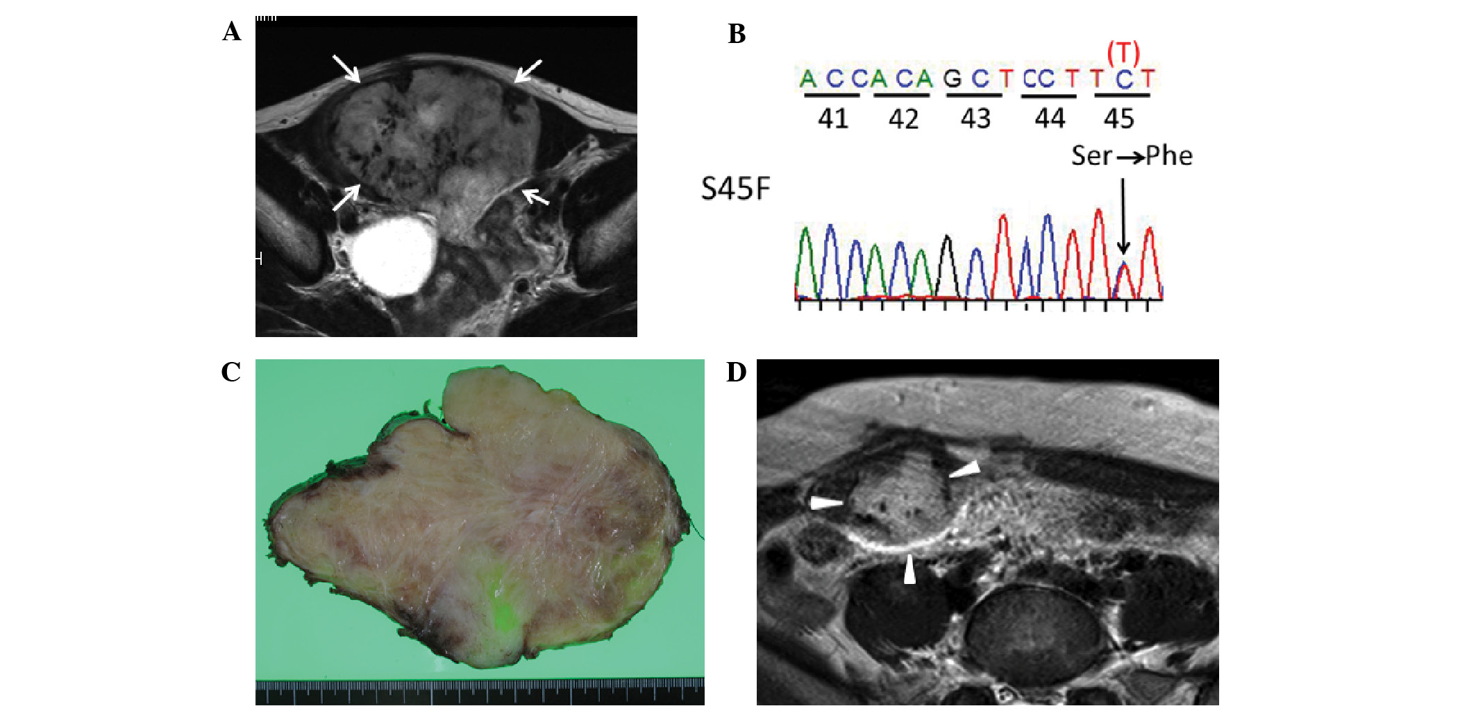
presented with recurrence 16 months after surgery, and this case
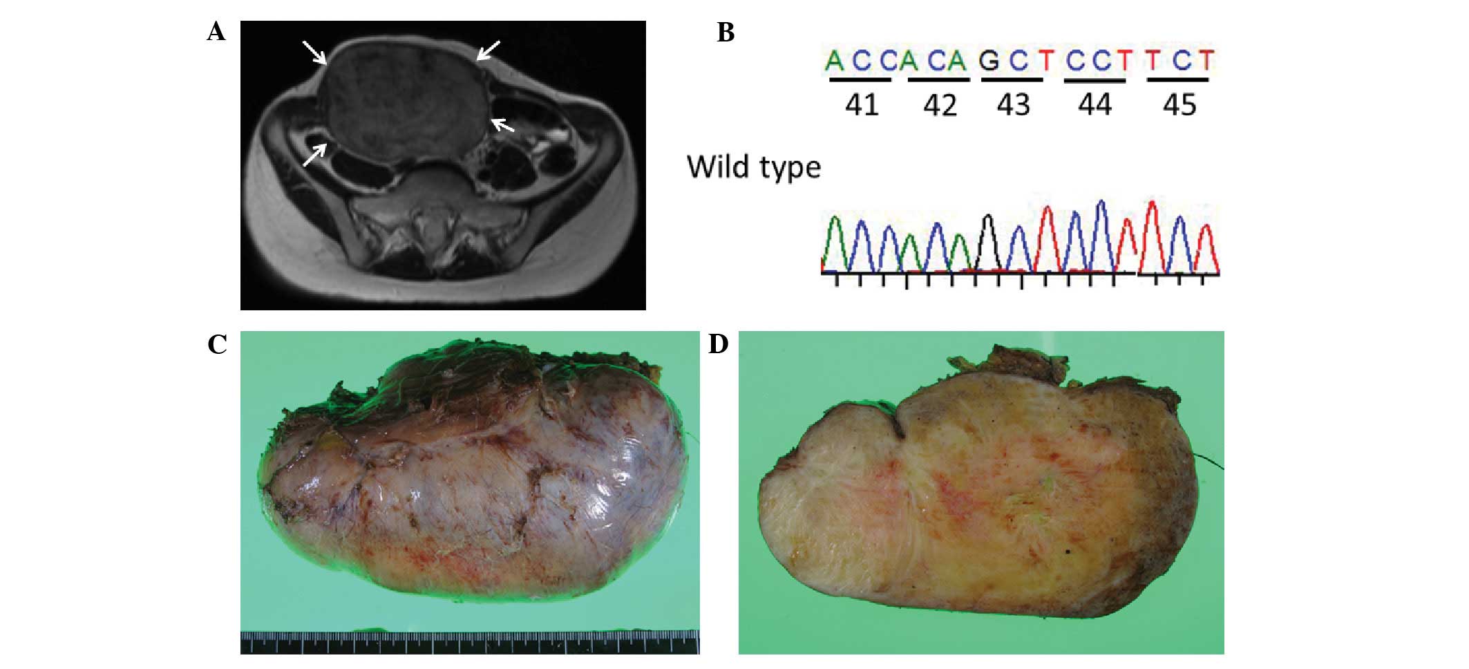
was characterized by an S45F mutation (Fig. 1). The other 12 cases, which did not
present with recurrence, had a T41A mutation or had no mutation
(wild-type) (Fig. 2). There was a
trend of recurrence in patients with S45F mutation (P=0.077).
Discussion
The present study demonstrated the feasibility of
simple resection in a cohort of truncal desmoid tumors
prospectively treated with meloxicam. Of note, although tumor sites
of the present cohort were limited in the trunk region (abdominal
wall, chest wall and neck), 12 tumors with either T41A or no
mutations (wild-type) did not recur with microscopic positive
margins. Several recent studies investigated the association
between CTNNB1 mutational status and clinical outcome of surgery
(10–12,19–21). Lazar
et al revealed with a single institution-based study that
desmoid tumors with an S45F mutation had a worse recurrence-free
survival following surgical treatment than patients with other
mutations (11). Subsequently,
Colombo et al reported their multicenter retrospective study
(10), including Lazar's study
cohort. The results of that study, which was based on 179 cases
that received surgical treatment, indicated that tumors with an
S45F mutation had a significantly higher recurrence rate compared
with those with other mutations or wild-type tumors; however, of
the 166 patients with a margin status that could be evaluated, 98
(59%) had microscopically negative (R0) resection and 68 (41%) had
microscopically positive (R1) resection, indicating that the margin
status was not identical in their cohort, making it difficult to
draw any definitive conclusions regarding the association between
recurrence and mutational status and/or margin status. Contrary to
the results of the above studies, Mullen et al reported a
slightly worse 5-year recurrence-free survival for patients with
CTNNB1 mutated tumors (58%) than for those with wild-type tumors in
115 cases treated with curative-intent surgical resection (12). In that study, radiation therapy was
delivered in an adjuvant manner at the discretion of the surgeon
and radiation oncologist when a high risk of recurrence was
predicted on clinical grounds, which may have masked the
correlation of the mutational status of CTNNB1 with local
recurrence. Dômont et al (20)
demonstrated a significant correlation (P=0.02) between higher risk
of recurrence and CTNNB1-mutated tumors; however, no significant
differences were observed among mutation types. Of note, after
analyzing patients with R0 resection, it was observed that the
recurrence rate was significantly higher in patients with mutated
tumors than in those with wild-type tumors (P=0.02) (20). Considering that R0 resection appears
to be more associated with functional impairment in patients with
desmoid tumors, the significance of a specific genotype, including
wild-type, in patients with R1 resection should be investigated.
The present study could suggest the possible favorable prognostic
value of wild-type and T41A mutation in patients with R1
resection.
The present study, however, had certain limitations.
Thus, although prospectively treated patients with identical cohort
(microscopic positive margins and no radiotherapy) were included in
the study, only a small number of cases could be enrolled. Desmoid
tumors arising in the extremities were not included in the present
study, since a previous study had indicated a significantly higher
incidence of the S45F mutation in desmoid tumors of the extremities
(P=0.005) than in tumors of other sites (22), and simple microscopic positive
resection could not be applied for the majority of patients with
desmoid tumors of the extremities.
In conclusion, the present study reported a case
series of successful planned simple resection even in cases with
microscopic positive margins, which aimed at reducing functional
impairment in patients with truncal desmoid tumors with wild-type
or T41A-mutated tumors. Accumulating larger numbers of patients
will help to clarify the significance of the results of the present
study more precisely with prospectively treated cohorts.
Acknowledgements
The authors would like to thank Ms. Eri Ishihara for
handling the study grant and purchasing experimental reagents, and
Dr Eisuke Arai for supporting the study by storing tumor specimens
in our department at Nagoya University Hospital. The present study
was partly funded by the Ministry of Education, Culture, Sports,
Science and Technology of Japan [Tokyo, Japan; grant-in-aid no.
262933341 for Scientific Research (B)] and Health Labour Sciences
Research Grant of Japan (grant no. H26-014).
References
|
1
|
Reitamo JJ, Scheinin TM and Häyry P: The
desmoid syndrome. New aspects in the cause, pathogenesis and
treatment of the desmoid tumor. Am J Surg. 151:230–237. 1986.
View Article : Google Scholar : PubMed/NCBI
|
|
2
|
Ballo MT, Zagars GK, Pollack A, Pisters PW
and Pollack RA: Desmoid tumor: Prognostic factors and outcome after
surgery, radiation therapy, or combined surgery and radiation
therapy. J Clin Oncol. 17:158–167. 1999.PubMed/NCBI
|
|
3
|
Gronchi A, Casali PG, Mariani L, Lo Vullo
S, Colecchia M, Lozza L, Bertulli R, Fiore M, Olmi P, Santinami M
and Rosai J: Quality of surgery and outcome in extra-abdominal
aggressive fibromatosis: A series of patients surgically treated at
a single institution. J Clin Oncol. 21:1390–1397. 2003. View Article : Google Scholar : PubMed/NCBI
|
|
4
|
Huang K, Fu H, Shi YQ, Zhou Y and Du CY:
Prognostic factors for extra-abdominal and abdominal wall desmoids:
A 20-year experience at a single institution. J Surg Oncol.
100:563–569. 2009. View Article : Google Scholar : PubMed/NCBI
|
|
5
|
Lev D, Kotilingam D, Wei C, Ballo MT,
Zagars GK, Pisters PW, Lazar AA, Patel SR, Benjamin RS and Pollock
RE: Optimizing treatment of desmoid tumors. J Clin Oncol.
25:1785–1791. 2007. View Article : Google Scholar : PubMed/NCBI
|
|
6
|
Merchant NB, Lewis JJ, Woodruff JM, Leung
DH and Brennan MF: Extremity and trunk desmoid tumors: A
multifactorial analysis of outcome. Cancer. 86:2045–2052. 1999.
View Article : Google Scholar : PubMed/NCBI
|
|
7
|
Nuyttens JJ, Rust PF, Thomas CR Jr and
Turrisi AT III: Surgery versus radiation therapy for patients with
aggressive fibromatosis or desmoid tumors: A comparative review of
22 articles. Cancer. 88:1517–1523. 2000. View Article : Google Scholar : PubMed/NCBI
|
|
8
|
Salas S, Dufresne A, Bui B, Blay JY,
Terrier P, Ranchere-Vince D, Bonvalot S, Stoeckle E, Guillou L, Le
Cesne A, et al: Prognostic factors influencing progression-free
survival determined from a series of sporadic desmoid tumors: A
wait-and-see policy according to tumor presentation. J Clin Oncol.
29:3553–3558. 2011. View Article : Google Scholar : PubMed/NCBI
|
|
9
|
Shido Y, Nishida Y, Nakashima H, Katagiri
H, Sugiura H, Yamada Y and Ishiguro N: Surgical treatment for local
control of extremity and trunk desmoid tumors. Arch Orthop Trauma
Surg. 129:929–933. 2009. View Article : Google Scholar : PubMed/NCBI
|
|
10
|
Colombo C, Miceli R, Lazar AJ, Perrone F,
Pollock RE, Le Cesne A, Hartgrink HH, Cleton-Jansen AM, Domont J,
Bovée JV, et al: CTNNB1 45F mutation is a molecular prognosticator
of increased postoperative primary desmoid tumor recurrence: An
independent, multicenter validation study. Cancer. 119:3696–3702.
2013. View Article : Google Scholar : PubMed/NCBI
|
|
11
|
Lazar AJ, Tuvin D, Hajibashi S, Habeeb S,
Bolshakov S, Mayordomo-Aranda E, Warneke CL, Lopez-Terrada D,
Pollock RE and Lev D: Specific mutations in the beta-catenin gene
(CTNNB1) correlate with local recurrence in sporadic desmoid
tumors. Am J Pathol. 173:1518–1527. 2008. View Article : Google Scholar : PubMed/NCBI
|
|
12
|
Mullen JT, DeLaney TF, Rosenberg AE, Le L,
Iafrate AJ, Kobayashi W, Szymonifka J, Yeap BY, Chen YL, Harmon DC,
et al: β-Catenin mutation status and outcomes in sporadic desmoid
tumors. Oncologist. 18:1043–1049. 2013. View Article : Google Scholar : PubMed/NCBI
|
|
13
|
Nishida Y, Tsukushi S, Shido Y, Urakawa H,
Arai E and Ishiguro N: Transition of treatment for patients with
extra-abdominal desmoid tumors: Nagoya university modality. Cancers
(Basel). 4:88–99. 2012. View Article : Google Scholar : PubMed/NCBI
|
|
14
|
Nishida Y, Tsukushi S, Shido Y, Wasa J,
Ishiguro N and Yamada Y: Successful treatment with meloxicam, a
cyclooxygenase-2 inhibitor, of patients with extra-abdominal
desmoid tumors: A pilot study. J Clin Oncol. 28:e107–e109. 2010.
View Article : Google Scholar : PubMed/NCBI
|
|
15
|
Hamada S, Futamura N, Ikuta K, Urakawa H,
Kozawa E, Ishiguro N and Nishida Y: CTNNB1 S45F mutation predicts
poor efficacy of meloxicam treatment for desmoid tumors: A pilot
study. PLoS One. 9:e963912014. View Article : Google Scholar : PubMed/NCBI
|
|
16
|
Poon R, Smits R, Li C, Jagmohan-Changur S,
Kong M, Cheon S, Yu C, Fodde R and Alman BA: Cyclooxygenase-two
(COX-2) modulates proliferation in aggressive fibromatosis (desmoid
tumor). Oncogene. 20:451–460. 2001. View Article : Google Scholar : PubMed/NCBI
|
|
17
|
Nishida Y, Tsukushi S, Urakawa H, Arai E
and Ishiguro N: Is it possible to identify clinically useful
prognostic groups for patients with desmoid tumors? J Clin Oncol.
30:1390author reply 1391. 2012. View Article : Google Scholar : PubMed/NCBI
|
|
18
|
Hamada S, Urakawa H, Kozawa E, Futamura N,
Ikuta K, Shimoyama Y, Nakamura S, Ishiguro N and Nishida Y: Nuclear
expression of β-catenin predicts the efficacy of meloxicam
treatment for patients with sporadic desmoid tumors. Tumour Biol.
35:4561–4566. 2014. View Article : Google Scholar : PubMed/NCBI
|
|
19
|
Bo N, Wang D, Wu B, Chen L and Ruixue Ma:
Analysis of β-catenin expression and exon 3 mutations in pediatric
sporadic aggressive fibromatosis. Pediatr Dev Pathol. 15:173–178.
2012. View Article : Google Scholar : PubMed/NCBI
|
|
20
|
Dômont J, Salas S, Lacroix L, Brouste V,
Saulnier P, Terrier P, Ranchère D, Neuville A, Leroux A, Guillou L,
et al: High frequency of beta-catenin heterozygous mutations in
extra-abdominal fibromatosis: A potential molecular tool for
disease management. Br J Cancer. 102:1032–1036. 2010. View Article : Google Scholar : PubMed/NCBI
|
|
21
|
Huss S, Nehles J, Binot E, Wardelmann E,
Mittler J, Kleine MA, Künstlinger H, Hartmann W, Hohenberger P,
Merkelbach-Bruse S, et al: β-catenin (CTNNB1) mutations and
clinicopathological features of mesenteric desmoid-type
fibromatosis. Histopathology. 62:294–304. 2013. View Article : Google Scholar : PubMed/NCBI
|
|
22
|
Le Guellec S, Soubeyran I, Rochaix P,
Filleron T, Neuville A, Hostein I and Coindre JM: CTNNB1 mutation
analysis is a useful tool for the diagnosis of desmoid tumors: A
study of 260 desmoid tumors and 191 potential morphologic mimics.
Mod Pathol. 25:1551–1558. 2012. View Article : Google Scholar : PubMed/NCBI
|