Introduction
Colorectal cancer (CRC) is the third most common
type of cancer worldwide, accounting for the diagnosis of ~1.4
million novel cases in 2012 (1).
Early diagnosis and prompt treatment substantially improve outcome
in CRC patients (2). At present,
studies are being conducted in order to identify biological
substances that may be involved in cancer development (3). The identification of specific cancer
biomarkers is expected to improve early cancer diagnosis in the
asymptomatic stage of the disease (4). In humans, galanin (GAL) is a 30-amino
acid neuroendocrine peptide, originally isolated from the
intestine, which is widely distributed in the central nervous
system, endocrine system and autonomic nervous system, including
the enteric nervous system (ENS) (5).
GAL exhibits various functions, including regulation of feeding and
learning, and is involved in responses to nerve injury and pain
(5). In the gastrointestinal tract,
GAL is involved with intestinal contraction (6). It regulates gastric acid secretion
(7) and inhibits the release of
pancreatic peptides (8). Notably,
increased levels of GAL have been identified within
neoplastically-altered tissues of neuroectodermic origin, such as
pheochromocytoma (9), pituitary
adenoma (10), neuroblastoma
(11) and squamous cell carcinoma
(12), as well as in malignant
melanoma (13). In a previous study,
DNA microarray analysis of gene expression in CRC tumors of 51
Korean patients revealed GAL mRNA levels ten times higher than that
in normal colon tissues. In addition, GAL expression was observed
in colon adenocarcinoma cell lines (LOVO, HCT116, SW480, SW260),
whereas GAL mRNA was not detected in the cell lines of lung cancer,
ovarian carcinoma or testicular carcinoma (14). Kim et al (14) also reported that the CRC patient group
exhibited an increased GAL serum concentration when compared with
healthy controls, however, the source of the increased GAL serum
levels was not determined, and the peptide was not identified in
CRC tumors (14).
Due to the fact that colorectal cancer incidence
rates for Asian individuals are lower that the rates for Caucasian
and African-descended individuals (15), GAL serum concentrations were measured
in a cohort of European CRC patients and healthy subjects in the
present study. Since our previous study revealed that the
expression of neuropeptides (16),
including GAL (17), in the ENS
ganglia was altered when in close proximity to CRC invasion, the
present study additionally aimed to determine the GAL levels in the
homogenates of CRC tumors and two dissected sections of the colon
wall (mucosa with submucosa and the muscularis externa) both
proximal to the tumor and distant from the tumor, which served as
the control. Since a reduction in myenteric plexus size and
decomposition has been observed in the colon wall of CRC patients
(18), the GAL-immunoreactive
(GAL-Ir) myenteric plexuses in the vicinity of and distant from CRC
invasion in the colon wall were quantitatively assessed.
Materials and methods
Patients
A total of 68 CRC patients (38 men and 30 women)
with a mean age [±standard deviation (SD)] of 68.91±10.95 years
(range 34–87 years) that underwent surgery at the Warmia and Mazury
Oncological Center (Olsztyn, Poland) between October 2012 and
November 2013 were included in the present study. Full blood
samples were collected preoperatively from all CRC patients and
postoperative colorectal samples from resected tissues for the
dissection of particular sections of the colon wall were obtained
from 22 patients (15 men and 7 women) with a mean age (±SD) of
68.79±10.15 years (range, 34–87 years). None of the CRC patients
suffered from inflammatory bowel disease (IBD) or other
gastrointestinal diseases and no patients reported a family history
of malignancy. Patients that had undergone neoadjuvant radiotherapy
or chemotherapy were excluded from the study.
Blood samples were also obtained from a control
group of 39 healthy volunteers (9 men and 30 women) with a mean age
(±SD) of 55.76±5.47 years (range, 43–69 years). The control
subjects exhibited no acute or chronic diseases at the time of the
study and reported no family history of cancer or IBD. None of the
control patients were on medication at the time of study.
The study was approved by the Bioethical Commission
of the University of Warmia and Mazury (Olsztyn, Poland) (approval
no. 43/2011), and written consent was obtained from all
participants.
Collection of serum, tumor samples and
colon wall samples
Blood samples were obtained from all patients and
healthy volunteers by venipuncture (~7 ml) and centrifuged at 2000
× g for 10 min at room temperature. The collected serum (3–4 ml)
was then aliquoted and stored at −20°C until further use.
Tissue samples were obtained from 22 CRC patients.
Immediately after resection of the cancerous section of the large
intestine, two small (~1–2 cm in diameter), full-thickness (~3–4
mm) intestinal wall samples were collected: one sample was obtained
directly from the section of the colon wall adjacent to the tumor
and the second sample (control sample) was obtained from the
proximal end of the intestine, ≥5 cm from the tumor margin.
The sample of colon wall obtained adjacent from the
tumor was carefully dissected into two sections: AM and AMM. The
‘AM’ section consisted of tumor-adjacent mucosa and submucosa
(weight, 0.1–0.2 g) containing submucosal plexuses, whereas the
‘AMM’ section consisted of tumor-adjacent muscularis externa
(weight, 0.1–0.3 g) containing circular and longitudinal muscle
layers and the myenteric plexuses localized between them. In
addition, a 2×2×2 mm sample from the periphery of the tumor
(weight, 0.1–0.3 g) was also resected. Using the same method,
individual sections of the colon were obtained from the
non-cancerous section of the colon wall and referred to as the
control mucosa and submucosa (DM; distant mucosa and submucosa) and
the control muscularis externa (DMM, distant muscularis). The
tissue samples intended for the analysis of GAL content in the
tissue homogenates were immediately frozen in liquid nitrogen and
stored at −80°C. Immediately after dissection, the colon wall
sections (~3–4 mm thick) obtained adjacent to or distant from the
CRC tumor were fixed in 4% formaldehyde for 24 h, dehydrated in an
ethanol series, embedded in paraffin and cut into 5-µm
sections.
Measurement of GAL concentration in
blood serum, CRC tumor and dissected sections of the colon
wall
The concentration of GAL in the sera of 68 patients
and 39 healthy volunteers and the tissue homogenates of 22 CRC
patients was measured using an ELISA kit for human GAL (#CEB084Hu;
USCN Life Science Inc., Wuhan, China). Cryopreserved tissues were
thawed and rinsed in ice-cold phosphate-buffered saline (PBS; pH
7.4) and weighed. Following separation by the use of surgical
scalpel and tweezers, 100 mg tissue was homogenized (Ultra-Turrax
T10 homogenizer; IKA -Werke GmbH & Co. KG, Staufen, Germany) in
5 ml PBS on ice three times for 20 sec. The homogenates were then
centrifuged at 5,000 × g for 5 min at 4°C and the supernatants were
stored at −80°C for further processing. All analysis was performed
in duplicate according to the manufacturer's instructions. The
optical density was measured using a microplate reader (Infinite
M200 Pro; Tecan Group Ltd., Männedorf, Switzerland). Standard
curves were generated for GAL concentrations of 1000.00, 333.33,
111.11, 37.04 and 12.35 pg/ml. The intra- and inter-assay
coefficients of variation for the GAL assays were <12%. The data
were linearized by plotting the log of GAL concentration vs. the
log of the optical density, and the results were expressed in terms
of pg of GAL per ml of serum and as pg of GAL per g of tissue.
Immunohistochemistry (IHC)
IHC, defined as positive immunoreactivity for GAL,
was performed as described previously by Godlewski et al
(19) with certain modifications. The
sections were subjected to antigen retrieval by microwaving for 20
min in retrieval solution buffer (pH 6.0; Leica, Wetzlar, Germany)
and incubated with 3% H2O2 in methanol for 10
min followed by 2.5% normal horse serum (Vector Laboratories, Inc.,
Burlingame, CA, USA) in PBS for 30 min. The sections were incubated
overnight at 4°C with rabbit polyclonal anti-human GAL primary
antibody (1:400; #HPA 049864; Sigma-Aldrich; Merck Millipore,
Darmstadt, Germany). Sections were then rinsed three times with PBS
and incubated with ImmPRESS™ universal reagent anti-mouse/rabbit
immunoglobulin G (commercially diluted; #MP-7500; Vector
Laboratories, Inc.) for 30 min at room temperature. To visualize
the immunoreaction, the sections were immersed in DAB (Dako,
Glostrup, Denmark) and counterstained in Mayer's hematoxylin. The
slides were dehydrated in ethanol, cleared in xylene and mounted.
The specificity of the IHC staining was determined by omitting the
primary antibody and replacing it with the same dilution of horse
serum. Images of the labeled tissues were captured using a XC-50
camera (Olympus Corporation, Tokyo, Japan) mounted on a light
microscope (BX-41; Olympus Corporation). Hematoxylin and eosin
staining was performed on separate sections to assess tissue
morphology.
Morphometric analysis of the area
occupied by myenteric plexuses expressing GAL immunoreactivity
Since the submucosal plexuses occupy a markedly
smaller area in the human colon than the myenteric plexuses
(20), the latter were selected to
assess the area occupied by GAL-Ir structures in the muscularis
layer of the colon wall proximal to or distant from the CRC tumor
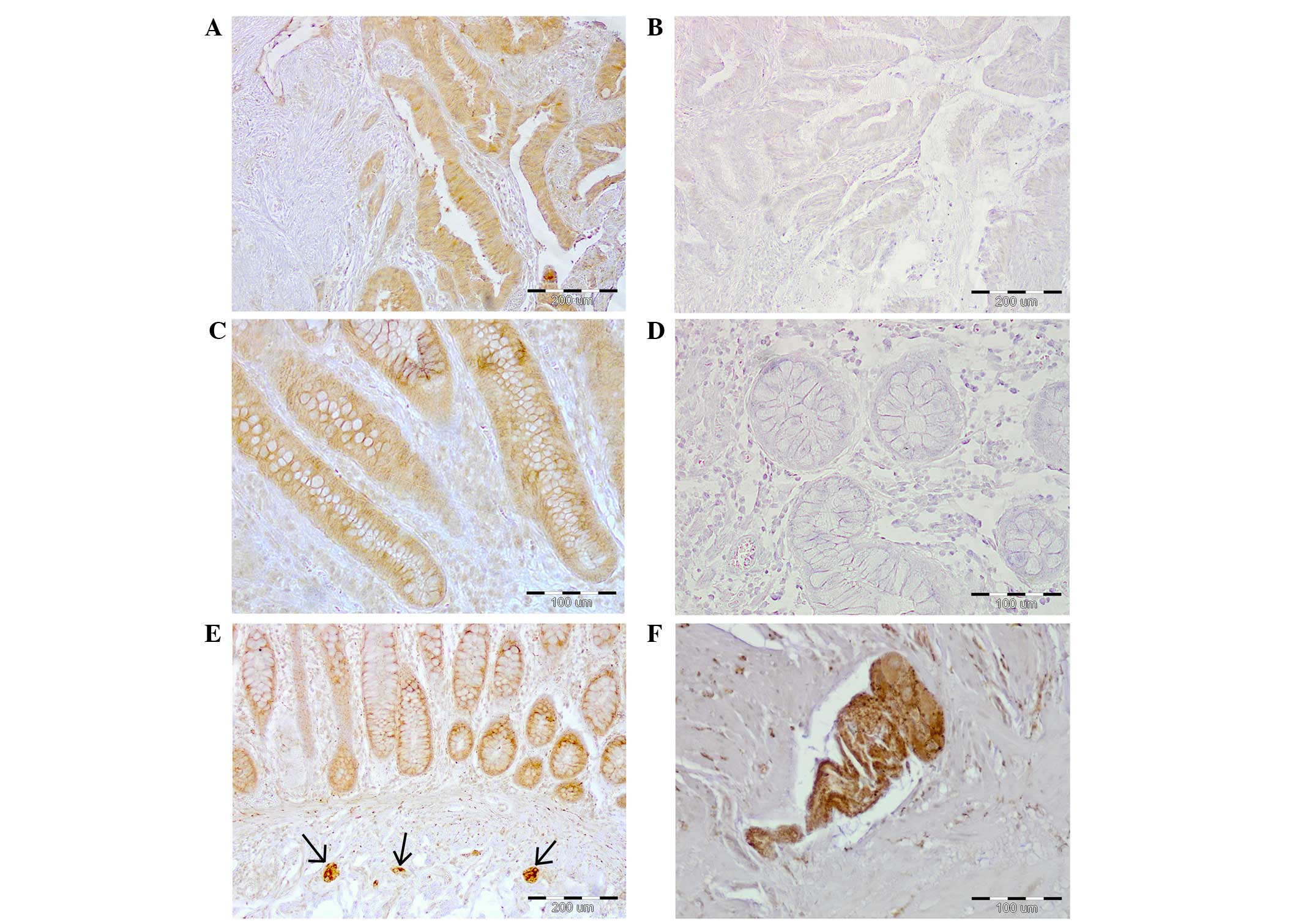
(Fig. 1). In each microscopic field
of view (magnification, ×4), a large rectangular area with visible
GAL-Ir structures and the area of the GAL-Ir myenteric plexuses was
determined using Cell^B Imaging Software for Life Science
Microscopy version 3.2 (Olympus Corporation). In each patient, the
total area occupied by the GAL-Ir neurons in the myenteric plexuses
located in the vicinity of and distantly from the tumor was
measured in 5–10 fields of view, depending on the size of the
section, to encompass the whole length of the border between the
circular and longitudinal layers of the muscularis externa. Thus,
22 sections of the large intestinal wall located within close
proximity to the tumor and 22 matched sections located distant from
the tumor were analyzed to yield the total area of GAL-Ir plexuses,
and the ratio of this to the total measured area in a selected
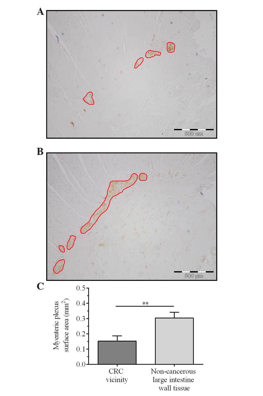
histological section of the colon wall was determined (Fig. 2).
Statistical analysis
The concentrations of GAL in the serum of CRC
patients and healthy controls and in the tissue homogenates of the
patients were analyzed using the Mann-Whitney U-test. The results
of morphometric measurement of the area occupied by GAL-Ir
myenteric plexuses were analyzed using the Wilcoxon signed-rank
test. All statistical analysis was performed using Statistica
software version 12.5 (StatSoft, Inc., Tulsa, OK, USA). The results
were expressed as the mean ± standard error of the mean (SEM).
P<0.05 was considered to indicate a statistically significant
difference.
Results
Patient clinicopathological
characteristics
Patient clinicopathological data, including gender,
age, tumor location, depth of invasion, lymph node metastasis and
TNM stage are presented in Table
I.
 | Table I.Clinicopathological characteristics of
68 colorectal cancer patients according to the TNM staging
system. |
Table I.
Clinicopathological characteristics of
68 colorectal cancer patients according to the TNM staging
system.
| Parameter | n (%) |
|---|
| Gender |
|
| Male | 38 (55.9) |
|
Female | 30 (44.1) |
| Age, years |
|
| ≤67 | 33 (48.5) |
|
>67 | 35 (51.5) |
| Tumor
localization |
|
| Cecum,
ascending and transverse colon | 18 (26.5) |
|
Descending, sigmoid colon,
rectum | 50 (73.5) |
| Depth of invasion (pT
status) |
|
| T1 | 0
(0) |
| T2 | 11 (16.2) |
| T3 | 44 (64.7) |
| T4 | 13 (19.1) |
| Lymph node metastases
(pN status) |
|
| N0 | 33 (48.5) |
| N1 | 18 (26.5) |
| N2 | 17 (25.0) |
| Metastasis (pM
status) |
|
| M0 | 61 (89.7) |
| M1 | 7
(10.3) |
| TNM stage |
|
| I | 9
(13.2) |
| II | 23 (33.8) |
|
III | 29 (42.7) |
| IV | 7
(10.3) |
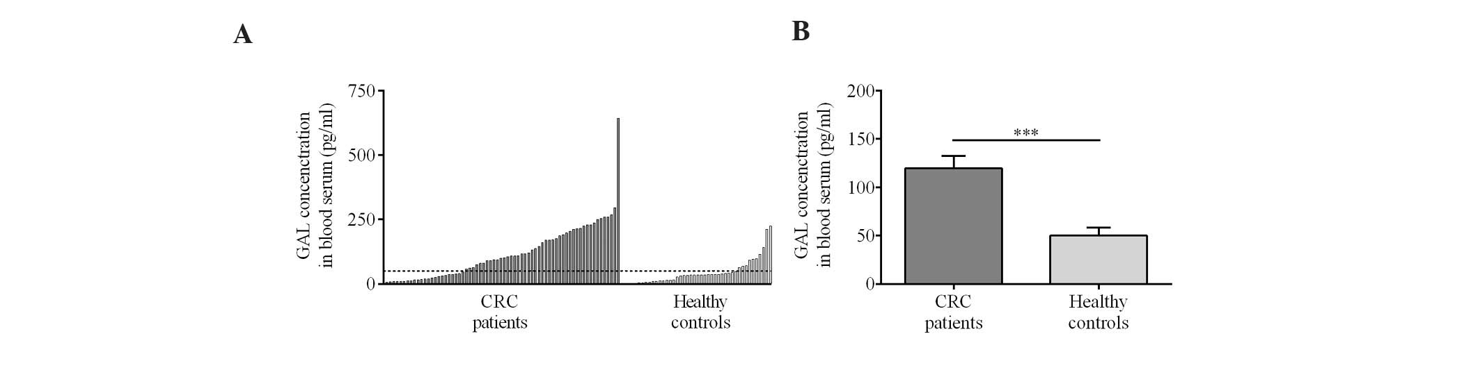
CRC patients exhibit higher GAL serum
concentrations than healthy controls
Analysis of the sera of 68 CRC patients revealed
significantly higher mean GAL concentrations than in the 39 healthy
volunteers (119.47±12.87 vs. 50.14±8.13 pg/ml, respectively)
(Fig. 3A and B).
Statistical analysis revealed no associations
between GAL concentration in the sera of the CRC patients and
clinicopathological features, including gender, age, tumor
location, depth of invasion, lymph node metastasis and TNM stage
(data not shown).
GAL content of CRC tumor tissue
homogenates is higher than that of the muscularis externa
The highest levels of GAL were identified in the
tumor and mucosa obtained from the non-cancerous section of the
intestinal wall (Fig. 4). The mucosal
tissues adjacent to the CRC tumor expressed significantly lower
levels of GAL (306.63±27.60 pg/g) than the tumor tissues
(408.71±30.28 pg/g; P=0.019) and mucosa from the section of the
colon wall distant from the tumor (462.73±28.54 pg/g). The GAL
concentration of the colon wall muscularis externa obtained
adjacent to the tumor (286.66±13.43 pg/g) was significantly higher
than that of the muscularis distant from the tumor (175.23±15.33
pg/g; P<0.001). The GAL concentration in the CRC tumor was
significantly higher than that in either muscularis externa
location (Fig. 4; P=0.012 in CRC vs.
AMM; P<0.001 in CRC vs. DMM).
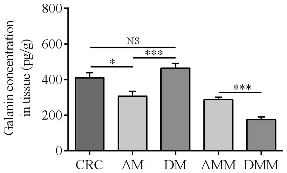 | Figure 4.Mean GAL content in the homogenates of
tissues obtained from CRC patients. CRC, colorectal cancer; AM,
mucosa in the vicinity of the CRC tumor; DM, mucosa located
distantly from tumor; AMM, muscularis externa in the vicinity of
the CRC tumor; DMM, muscularis externa located distantly from
tumor; NS, not significant. *P=0.019, CRC vs. AM; P=0.187, CRC vs.
DM (NS); ***P<0.001, AM vs. DM, and AMM vs. DMM. |
CRC tumor and colon wall tissues
exhibit strong GAL immunoreactivity in CRC patients
IHC revealed strong GAL immunoreactivity within the
cells of the intestinal epithelium, cancer cells and the myenteric
plexuses (Fig. 1). The
immunoreactivity of GAL in cancer cells, enterocytes and goblet
(mucous) cells was predominantly identified in the cytoplasm.
GAL-Ir cells were also observed in the components of
the submucosal plexuses and were demonstrated to be smaller in size
than the myenteric plexuses from the samples obtained from the
colon wall located in the vicinity of and distantly from the tumor
(Fig. 1E).
GAL-Ir myenteric plexuses in close
proximity to CRC tumors are smaller than those distant from CRC
tumors
The examination of the GAL-Ir structures in the
myenteric plexuses revealed that the plexuses located in close
proximity to the CRC tumor were smaller in size than those located
distantly from the cancer (Fig. 2A and
B). To assess atrophy, morphometry was used to measure the area
occupied by the GAL-Ir myenteric plexuses in the sections of the
colon wall of 22 CRC patients.
The mean area of GAL-Ir myenteric plexuses in the
intestinal wall proximal to the CRC tumor was ~50% smaller than
that of the distant section of the colon wall (0.15±0.03
mm2 vs. 0.30±0.03 mm2, respectively)
(Fig. 2).
This finding was confirmed by comparing the relative
areas occupied by the GAL-Ir myenteric plexuses in the sections of
colon wall close to and distant from the CRC, which were 1.23±0.85
and 1.85±0.56%, respectively. No significant associations were
identified between the reduced size of the myenteric plexuses in
the vicinity of the tumor and clinicopathological parameters of CRC
patients (data not shown).
Discussion
To the best of our knowledge, this is the first
study to demonstrate strong immunoreactivity of GAL in CRC tumors.
These morphological findings were validated by the analysis of GAL
concentration in neoplastic CRC tissue and two dissected sections
of the colon wall: the mucosa and submucosa and the circular and
longitudinal layers of muscularis externa with the myenteric
plexuses present between them. Additionally, using morphometry, the
atrophy of myenteric plexuses was identified in the vicinity of CRC
tumors. Thus, we hypothesize that the elevated concentration of GAL
in the serum of CRC patients may be caused by the secretion of this
neuropeptide by cancer cells, however, the possibility that other
cell types or tissues may be involved cannot be excluded.
The present study is the first to report that CRC
patients exhibit a statistically significant higher serum
concentration of GAL than healthy patients, as the marked increase
in serum GAL levels reported previously in 66 CRC Korean patients
was not statistically significant (14). Kim et al (14) postulated that the CRC tumor was the
source of the increased serum GAL concentration, as the levels of
GAL mRNA were markedly increased in the CRC tumor; however, the
concentration of the neuropeptide, GAL, was not measured (14). Similarly, Nagayoshi et al
(21) reported that GAL mRNA levels
in 112 tumor samples obtained from CRC patients were significantly
higher than that in 27 normal colon mucosa samples; however, the
concentration of GAL was not determined.
In the present study, GAL concentration was measured
in both the homogenates of the CRC tumor and two major sections of
the colon wall by ELISA assay, which provided notable results. High
GAL levels were observed not only in the CRC tumor, but also in the
homogenates of mucosa and submucosa from the non-cancerous section
of the colon wall. The GAL concentration of the tumor was
significantly higher than that in the homogenates of the muscularis
layer of the colon wall, which are known to contain GAL in the
myenteric plexuses (6,17), and intense GAL immunoreactivity was
observed in the cytoplasm of CRC tumor cells, which indicates that
the CRC tumor is an important source of GAL in the sera of CRC
patients. GAL immunoreactivity has also been observed in the cells
of neuroendocrine tumors, including neuroblastomas (11), pituitary adenomas (10) and paragangliomas (22), as well as in a variety of
non-neuroendocrine tumors, such as melanomas (13), glioblastomas (23) and embryonic carcinomas (24).
Furthermore, in the present study IHC demonstrated,
for the first time, the presence of GAL-like immunoreactivity in
the epithelium of human colon mucosa, which had only been
demonstrated in human submucosal (17) and myenteric nervous plexuses to date
(6,17). The presence of cytoplasmic GAL
immunoreactivity in the neoplastic cells of CRC tumors and in the
epithelial cells of the mucosa of the large intestine may be
associated with the function of GAL as a regulator of epithelial
secretion and proliferation (25,26). This
hypothesis is supported by the fact that GAL has been identified in
the human epidermis and the ductal cells of sweat glands (27), which are epithelial tissues of
different embryonic origin than the intestine.
The presence of cytoplasmic GAL immunoreactivity in
neoplastic CRC cells may be associated with the function of GAL as
a regulator of cellular proliferation under pathological
conditions. In the model HCT116 CRC cell line, silencing of the GAL
receptor 1 (GaLR1) or silencing of GAL, induces caspase 8-dependent
apoptosis, which suggests that GaLR1/GAL may present a potential
drug target for CRC chemotherapy (28). Additionally, in pancreatic cancer cell
lines (29), small-cell lung cancer
(30) and rat pituitary tumors
(31), GAL interacts with GaLR1 to
activate the mitogen-activated protein kinase signaling pathway and
induces mitogenic activity of these cancer cells. However, it has
been also suggested that GAL may exhibit a protective function in
head and neck squamous cell cancer (HNSCC), since GaLR1 and 2
inhibit the proliferation of HNSCC cells via extracellular
signal-regulated kinase 1/2-mediated effects on cell-cycle control
proteins (32).
The morphometric analysis in the present study
provided a quantitative aspect to the observations of Godlewski
(18), who reported alterations in
the structure of the ENS components in the vicinity of the CRC
tumor. Notably, in the present study, the muscularis externa in the
vicinity of the CRC invasion contained higher GAL levels than that
in the distant section, which indicates the increased synthesis of
GAL or the release of this neuropeptide from the atrophic myenteric
plexuses into the surrounding tissue. Thus, this indicates that the
myenteric plexuses located close to the CRC tumor undergo
morphological and thus, functional alterations that may also affect
the function of the colon wall.
In conclusion, the present study comprehensively
investigated the presence of GAL in the colon wall of CRC patients.
To the best of our knowledge, this study is the first to
demonstrate GAL immunoreactivity in the epithelium of the human
colon and CRC tumor cells, as well as high GAL levels in CRC
tumors. In addition, increased GAL levels were identified in the
muscularis externa, despite the atrophy of the myenteric plexuses
in the vicinity of the CRC tumor, when compared with the muscularis
located distantly from the tumor. The results indicate that serum
and tissue GAL levels may present useful potential biomarkers in
CRC patients. However, the broad range of GAL serum levels
identified in both CRC patients and controls and reported by Kim
et al (14) indicate that
further studies using larger patient cohorts are required to
establish the clinical significance of these findings.
Acknowledgments
This study was supported by The University of Warmia
and Mazury (Olsztyn, Poland; grant no. 1507-0881). The authors
would like to thank Dr. Ewa Wędrowska for performing the
statistical analysis for the study.
References
|
1
|
Ferlay J, Soerjomataram I, Ervik M,
Dikshit R, Eser S, Mathers C, Rebelo M, Parkin DM, Forman D and
Bray F: GLOBOCAN 2012 v1.0, Cancer Incidence and Mortality
Worldwide: IARC CancerBase No. 11 [Internet]. Lyon, France:
International Agency for Research on Cancer; 2013 http://globocan.iarc.frJanuary 16–2015
|
|
2
|
Keane MG and Johnson GJ: Early diagnosis
improves survival in colorectal cancer. Practitioner. 256:15–18.
2012.PubMed/NCBI
|
|
3
|
Chen H, Zucknick M, Werner S, Knebel P and
Brenner H: Head-to-head comparison and evaluation of 92 plasma
protein biomarkers for early detection of colorectal cancer in a
true screening setting. Clin Cancer Res. 21:3318–3326. 2015.
View Article : Google Scholar : PubMed/NCBI
|
|
4
|
Gonzalez-Pons M and Cruz-Correa M:
Colorectal cancer biomarkers: Where are we now? Biomed Res Int.
2015:1490142015. View Article : Google Scholar : PubMed/NCBI
|
|
5
|
Mitsukawa K, Lu X and Bartfai T: Galanin,
galanin receptorss, and drug targetsGalanin. Hökfelt T: 102.
Springer; Basel: pp. 7–9. 2010, View Article : Google Scholar
|
|
6
|
Burleigh DE and Furness JB: Distribution
and actions of galanin and vasoactive intestinal peptide in the
human colon. Neuropeptides. 16:77–82. 1990. View Article : Google Scholar : PubMed/NCBI
|
|
7
|
von Rosenvinge EC and Raufman JP:
Gastrointestinal peptides and regulation of gastric acid secretion.
Curr Opin Endocrinol Diabetes Obes. 17:40–43. 2010. View Article : Google Scholar : PubMed/NCBI
|
|
8
|
Barreto SG, Carati CJ, Toouli J and
Saccone GT: The islet-acinar axis of the pancreas: More than just
insulin. Am J Physiol Gastrointest Liver Physiol. 299:G10–G22.
2010. View Article : Google Scholar : PubMed/NCBI
|
|
9
|
Bauer FE, Hacker GW, Terenghi G, Adrian
TE, Polak JM and Bloom SR: Localization and molecular forms of
galanin in human adrenals: Elevated levels in pheochromocytomas. J
Clin Endocrinol Metab. 63:1372–1378. 1986. View Article : Google Scholar : PubMed/NCBI
|
|
10
|
Grenbäck E, Bjellerup P, Wallerman E,
Lundblad L, Anggård A, Ericson K, Aman K, Landry M, Schmidt WE,
Hökfelt T and Hulting AL: Galanin in pituitary adenomas. Regul
Pept. 117:127–139. 2004. View Article : Google Scholar : PubMed/NCBI
|
|
11
|
Tuechler C, Hametner R, Jones N, Jones R,
Iismaa TP, Sperl W and Kofler B: Galanin and galanin receptor
expression in neuroblastoma. Ann NY Acad Sci. 863:438–441. 1998.
View Article : Google Scholar : PubMed/NCBI
|
|
12
|
Sugimoto T, Seki N, Shimizu S, Kikkawa N,
Tsukada J, Shimada H, Sasaki K, Hanazawa T, Okamoto Y and Hata A:
The galanin signaling cascade is a candidate pathway regulating
oncogenesis in human squamous cell carcinoma. Genes Chromosomes
Cancer. 48:132–142. 2009. View Article : Google Scholar : PubMed/NCBI
|
|
13
|
Gilaberte Y, Vera J, Coscojuela C, Roca
MJ, Parrado C and González S: Expression of galanin in melanocytic
tumors. Actas Dermosifiliogr. 98:24–34. 2007.(In Spanish).
View Article : Google Scholar : PubMed/NCBI
|
|
14
|
Kim KY, Kee MK, Chong SA and Nam MJ:
Galanin is up-regulated in colon adenocarcinoma. Cancer Epidemiol
Biomarkers Prev. 16:2373–2378. 2007. View Article : Google Scholar : PubMed/NCBI
|
|
15
|
Ries LA, Wingo PA, Miller DS, Howe HL,
Weir HK, Rosenberg HM, Vernon SW, Cronin K and Edwards BK: The
annual report to the nation on the status of cancer, 1973–1997,
with a special section on colorectal cancer. Cancer. 88:2398–2424.
2000. View Article : Google Scholar : PubMed/NCBI
|
|
16
|
Godlewski J and Łakomy IM: Changes in
vasoactive intestinal peptide, pituitary adenylate cyclase-
activating polypeptide and neuropeptide Y-ergic structures of the
enteric nervous system in the carcinoma of the human large
intestine. Folia Histochem Cytobiol. 48:208–216. 2010. View Article : Google Scholar : PubMed/NCBI
|
|
17
|
Godlewski J and Pidsudko Z: Characteristic
of galaninergic components of the enteric nervous system in the
cancer invasion of human large intestine. Ann Anat. 194:368–372.
2012. View Article : Google Scholar : PubMed/NCBI
|
|
18
|
Godlewski J: Morphological changes in the
enteric nervous system caused by carcinoma of the human large
intestine. Folia Histochem Cytobiol. 48:157–162. 2010. View Article : Google Scholar : PubMed/NCBI
|
|
19
|
Godlewski J, Krazinski BE, Kiezun J,
Kwiatkowski P, Sulik M, Tenderenda M, Biernat W and Kmiec Z: PLAGL1
protein is differentially expressed in the nephron segments and
collecting ducts in human kidney. Folia Histochem Cytobiol.
53:96–104. 2015. View Article : Google Scholar : PubMed/NCBI
|
|
20
|
Furness JB: Structure of the enteric
nervous systemThe Enteric Nervous System. Wiley-Blackwell; Hoboken,
NJ: pp. 62006
|
|
21
|
Nagayoshi K, Ueki T, Tashiro K, Mizuuchi
Y, Manabe T, Araki H, Oda Y, Kuhara S and Tanaka M: Galanin plays
an important role in cancer invasiveness and is associated with
poor prognosis in stage II colorectal cancer. Oncol Rep.
33:539–546. 2015.PubMed/NCBI
|
|
22
|
Tadros TS, Strauss RM, Cohen C and Gal AA:
Galanin immunoreactivity in paragangliomas but not in carcinoid
tumors. Appl Immunohistochem Mol Morphol. 11:250–252. 2003.
View Article : Google Scholar : PubMed/NCBI
|
|
23
|
Berger A, Santic R, Almer D,
Hauser-Kronberger C, Huemer M, Humpel C, Stockhammer G, Sperl W and
Kofler B: Galanin and galanin receptors in human gliomas. Acta
Neuropathol. 105:555–560. 2003.PubMed/NCBI
|
|
24
|
Skotheim RI, Lind GE, Monni O, Nesland JM,
Abeler VM, Fosså SD, Duale N, Brunborg G, Kallioniemi O, Andrews PW
and Lothe RA: Differentiation of human embryonal carcinomas in
vitro and in vivo reveals expression profiles relevant to normal
development. Cancer Res. 65:5588–5598. 2005. View Article : Google Scholar : PubMed/NCBI
|
|
25
|
Benya RV, Marrero JA, Ostrovskiy DA,
Koutsouris A and Hecht G: Human colonic epithelial cells express
galanin-1 receptors, which when activated cause Cl- secretion. Am J
Physiol. 276:G64–G72. 1999.PubMed/NCBI
|
|
26
|
Lang R, Gundlach AL, Holmes FE, Hobson SA,
Wynick D, Hökfelt T and Kofler B: Physiology, signaling and
pharmacology of galanin peptides and receptors: Three decades of
emerging diversity. Pharmacol Rev. 67:118–175. 2015. View Article : Google Scholar : PubMed/NCBI
|
|
27
|
Kofler B, Berger A, Santic R, Moritz K,
Almer D, Tuechler C, Lang R, Emberger M, Klausegger A, Sperl W and
Bauer JW: Expression of neuropeptide galanin and galanin receptors
in human skin. J Invest Dermatol. 122:1050–1053. 2004. View Article : Google Scholar : PubMed/NCBI
|
|
28
|
Stevenson L, Allen WL, Turkington R,
Jithesh PV, Proutski I, Stewart G, Lenz HJ, Van Schaeybroeck S,
Longley DB and Johnston PG: Identification of galanin and its
receptor GalR1 as novel determinants of resistance to chemotherapy
and potential biomarkers in colorectal cancer. Clin Cancer Res.
18:5412–5426. 2012. View Article : Google Scholar : PubMed/NCBI
|
|
29
|
Tjomsland V and El-Salhy M: Effects of
single, double or triple combinations of octreotide, galanin and
serotonin on a human pancreatic cancer cell line. Histol
Histopathol. 20:537–541. 2005.PubMed/NCBI
|
|
30
|
Seufferlein T and Rozengurt E: Galanin,
neurotensin, and phorbol esters rapidly stimulate activation of
mitogen-activated protein kinase in small cell lung cancer cells.
Cancer Res. 56:5758–5764. 1996.PubMed/NCBI
|
|
31
|
Hammond PJ, Smith DM, Akinsanya KO, Mufti
WA, Wynick D and Bloom SR: Signaling pathways mediating secretory
and mitogenic responses to galanin and pituitary adenylate
cyclase-activating polypeptide in the 235-1 clonal rat lactotroph
cell line. J Neuroendocrinol. 8:457–464. 1996. View Article : Google Scholar : PubMed/NCBI
|
|
32
|
Kanazawa T, Misawa K, Misawa Y, Uehara T,
Fukushima H, Kusaka G, Maruta M and Carey TE: G-protein-coupled
receptors: Next generation therapeutic targets in head and neck
cancer? Toxins (Basel). 7:2959–2984. 2015. View Article : Google Scholar : PubMed/NCBI
|