Introduction
Colorectal cancer (CRC) is the third most common
type of cancer and the fourth most common cause of cancer-related
mortality worldwide (1). Therefore,
scientists have been searching for specific biomarkers that could
be applied in the diagnosis and treatment monitoring of CRC
patients. However, no such indices have yet been found. Neoplastic
biomarkers that may have clinical implications in CRC patients have
to go through a number of qualifying stages. At the initial stage,
the neoplastic biomarker is usually detected and analyzed
scientifically. In order to be clinically valuable, the biomarker
has to be assessed in a large retrospective group and then
confirmed through prospective randomized research (2). Only when all the aforementioned criteria
have been fulfilled can the biomarker be used in clinical practice.
The difficulty with obtaining tumor-specific markers is also
associated with neoplasia complexity involving proliferative
signaling, evading growth suppressors, resisting cell death,
enabling replicative immortality, angiogenesis, activating invasion
and metastases (3). One of the major
processes determining cell homeostasis and the development of the
immune system is apoptosis, i.e., programmed cell death. In
neoplastic diseases, the two signaling pathways are disturbed and
the expression of the proteins involved is impaired. Apoptosis is
induced via two main routes: The extrinsic or death receptor
pathway and the intrinsic B-cell lymphoma 2-dependent pathway
(4,5).
Thus far, it has been confirmed that cancer cells of the colon show
overexpression of FasR and FasL (6,7).
Upregulation of FasR expression on cancer cells promotes compatible
ligand binding on the surface of T lymphocytes, i.e., cells engaged
in the immune response (8). Death
domain receptors make the first link of the extrinsic apoptosis
signal, activating caspase family proteins and inducing apoptosis.
The process of programmed cell death is regulated at several stages
by a number of inhibitor of apoptosis proteins (IAPs), of which the
best known are livin, X-linked inhibitor of apoptosis (XIAP) and
survivin (9).
Survivin is a 16.5-kDa protein consisting of an
N-terminal Zn2+-binding baculovirus inhibitor of
apoptosis protein repeat domain connected with a 65 Å amphipathic
C-terminal α-helix (10). It was
initially believed that survivin, together with XIAP, regulated the
caspase-dependent apoptotic pathway, blocked the activity of
caspases-3, −7 and −9, and caused their degradation. However,
further research has confirmed that survivin does not have a
specific motif to bind caspases, and only with the involvement of
XIAP may it inhibit caspase-9 (11).
Moreover, the protein takes part in the process of mitosis, forming
a chromosomal passenger complex with aurora-B kinase, inner
centromere protein and Borealin. In contrast to the other IAPs,
survivin does not occur in normal differentiated tissue; its
presence has been observed in transformed cell types and various
types of cancer (12). The function
of survivin may vary depending on its location in the cell. The
expression of survivin in the nucleus may be responsible for cell
proliferation control, while its presence in the cytoplasm
regulates cell survival (13).
Therefore, the study objective was the immunohistochemical
assessment of survivin expression and its serum level in CRC
patients.
Materials and methods
Patients
The study group consisted of 55 patients (20 women
and 35 men) treated surgically in the Second Department of General
and Gastroenterological Surgery in the Medical University of
Białystok (Białystok, Poland). The patients ranged in age from
34–86 years (mean, 67.11±1.89 years). The pathological diagnosis
confirmed CRC and its stage (tumor-node-metastasis) according to
World Health Organization classification (14). Adenocarcinoma was diagnosed in 48
individuals, whereas adenocarcinoma with a mucous component was
identified in 7 individuals. The investigated tumors were
classified as moderately-differentiated (G2) in 48 patients and
poorly-differentiated (G3) in 7 patients. A pT1 tumor was observed
in 1 case, a pT2 tumor in 3 patients, a pT3 tumor in 49 patients
and a pT4 tumor in 2 patients. At the time of the diagnosis,
metastases to the local lymph nodes were observed in 29 out of 55
cases, whereas the presence of metastases to distant organs was
noted in 25 out of the 55 studied cases. The control group
consisted of 22 healthy volunteers (12 males and 10 females, aged
45–75 years).
Study material consisted of venous blood samples (6
ml) obtained from healthy controls and from the colorectal
carcinoma patients prior to the surgery. Blood serum was stored at
−80°C immediately after centrifugation (704 × g) until assays were
performed.
The study received the approval of the local
Bioethics Committee (Nr. R-I-002-/40/2009). All the participants
(study group and controls) signed informed consent forms prior to
the examination.
Immunohistochemical assay
The immunohistochemistry (IHC) method was performed
in 38 out of the 55 patients with CRC. Formalin-fixed and
paraffin-embedded tissue specimens were cut on a microtome into
4-µm sections. The sections were deparaffinized in xylene and
hydrated in alcohol (Chempur, Piekary Śląskie, Poland). To
visualize the antigens of survivin, the sections were heated in a
microwave oven (Laznie WLS084; Adverti, Lodz, Poland) for 20 min in
a citrate buffer (pH, 6.0) (Sigma-Aldrich, St. Louis, MO, USA).
Next, they were incubated with 3% hydrogen peroxide solution for 5
min in order to block endogenous peroxidase (Chempur). Incubation
was then performed with polyclonal rabbit anti-human survivin
antibody (cat. no. SAB4501459; dilution 1:200; Sigma-Aldrich) and
incubated at room temperature for 60 min. The reaction was
performed using the Novocastra Novolink Polymer Detection system
(NCL; Novocastra; Leica Biosystems, Milton Keynes, UK). A color
reaction for peroxidase was developed with chromogene
diaminobenzidine. Positive and negative controls were performed
according to the manufacturer's protocols (Novocastra; Leica
Biosystems). Counterstaining was performed with hematoxylin.
Immunohistochemical staining was evaluated by two
independent pathologists who were blinded to the clinical
information. Protein expression was found in the nuclei and the
cytoplasm of the tumor cells, and graded separately in an identical
manner. Expression was determined using the semiquantitative method
and defined with regard to the intensity of staining (0, absent; 1,
weak; 2, moderate; and 3, strong) and the percentage of positive
tumor cells. The H-score was derived by summing up the percentages
of cell staining at each intensity and then multiplied by the
weighted intensity of staining. Score values ranged from 0–300. The
study group was divided into negative cases (H-score <150) and
positive cases (H-score >150).
Enzyme-linked immunosorbent assay
(ELISA)
Survivin level was analyzed in 55 patients using the
Human Survivin Quantikine ELISA kit (cat no. DSV00; R&D Systems
Inc., Minneapolis, MN, USA). Serum samples were prepared according
to the manufacturer's protocols. Prior to the assay, the samples
were 100-fold diluted with Calibrator Diluents. A monoclonal
antibody specific for survivin had been pre-coated onto a
microplate and incubated with the serum samples. After the first
washing, an enzyme-linked polyclonal antibody specific for survivin
was added to the wells. Following the second wash, a substrate
solution was added. Next, the color development was stopped. All
products were enclosed within ELISA kit. The reaction measurement
was based on the intensity of the sample color and automatically
measured using an Epoch Microplate Spectrophotometer (Biotek
Instruments, Inc., Winooski, VT, USA) at an absorbance of 450 nm.
The measurement of optical density was converted to survivin
concentration (pg/ml) based on a standard curve, which was
established using Gen5 Data Secure (BioTek Instruments, Inc.). All
the specimens were assayed twice. No statistically significant
differences were found between the measurements. The minimum
detectable dose (MDD) of survivin ranged from 1.58–9.96 pg/ml. The
mean MDD was 4.44 pg/ml.
Statistical analysis
Statistical analysis was conducted based on the
STATISTICA 10.0 program (Dell Statistica, Tulsa, OK, USA). In order
to compare the two groups, the Mann-Whitney U test was applied.
Correlations between the parameters were calculated by the
Pearson's correlation coefficient tests. P<0.05 was considered
to indicate a statistically significant difference. The missing
data was removed in pairs. The analysis of the receiver operating
characteristic (ROC) curve was performed using MedCalc statistical
software (MedCalc Software, Ostend, Belgium).
Results
Immunohistochemical localization of
survivin in CRC tissues and its correlation with
clinicopathological features
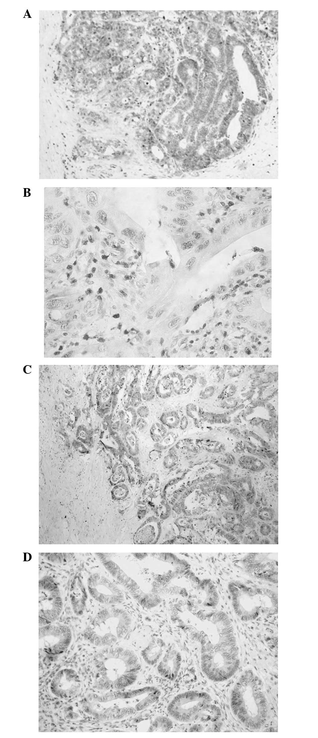
The expression of survivin was observed in the
nuclei and cytoplasm of the tumor cells. A positive immunoreaction
in the tumor tissue was observed in 84.2% (32/38) of patients with
CRC, including nuclear expression in 63.2% (24/38) and cytoplasmic
expression in 81.6% (31/38). Moreover, the detailed
immunohistochemical analysis confirmed 23 subjects as
Nuc+/Cyt+, 8 subjects as
Nuc−/Cyt+, 6 subjects as
Nuc−/Cyt− and 1 subject as
Nuc+/Cyt− (Fig.
1). There was a positive correlation between the nuclear and
cytoplasmic expression of survivin in the CRC tissues (P=0.020,
R=0.374). The statistical analysis of survivin expression showed
that a positive protein reaction in the tumor nuclei was associated
with tumor mass localization and the presence of distant metastasis
(P=0.048, R=0.323 and P=0.026, R=0.360, respectively) (Table I). The nuclear immunoreactivity of
survivin was frequent in tumors located in the rectum and in cases
with distant metastasis.
 | Table I.Correlations between survivin
expression in the tumor tissue and the clinicopathological
parameters in patients with colorectal cancer. |
Table I.
Correlations between survivin
expression in the tumor tissue and the clinicopathological
parameters in patients with colorectal cancer.
|
| Survivin
expression |
|---|
|
|
|
|---|
|
| Nuclear | Cytoplasmic |
|---|
|
|
|
|
|---|
| Parameter | Negative, n (%) | Positive, n (%) | P-value | Negative, n (%) | Positive, n (%) | P-value |
|---|
| Age, years |
|
| 0.464 |
|
| 0.657 |
| ≤60 | 6 (15.8) | 6 (15.8) |
| 3 (7.9) | 9 (23.7) |
|
|
>60 | 18 (47.4) | 8 (21.1) |
| 9 (23.7) | 17 (44.7) |
|
| Gender |
|
| 0.587 |
|
| 0.337 |
| Male | 10 (26.3) | 5 (13.2) |
| 6 (15.8) | 9 (23.7) |
|
|
Female | 14 (36.8) | 9 (23.7) |
| 6 (15.8) | 17 (44.7) |
|
| Localization |
|
| 0.048 |
|
| 0.838 |
|
Colon | 18 (47.4) | 6 (15.8) |
| 8 (21.1) | 16 (42.1) |
|
|
Rectum | 6 (15.8) | 8 (21.1) |
| 4 (10.5) | 10 (26.3) |
|
| Adenocarcinoma
type |
|
| 0.156 |
|
| 0.293 |
|
Non-mucinous | 10 (26.3) | 5 (13.2) |
| 6 (15.8) | 9 (23.7) |
|
|
Mucinous | 14 (36.8) | 9 (23.7) |
| 6 (15.8) | 17 (44.7) |
|
| Grade of
malignancies |
|
| 0.866 |
|
| 0.959 |
| 2 | 22 (57.9) | 11 (28.9) |
| 11 (28.9) | 22 (57.9) |
|
| 3 | 3 (7.9) | 2 (5.3) |
| 2 (5.3) | 3 (7.9) |
|
| Tumor size, cm |
|
| 0.446 |
|
| 0.958 |
|
<5 | 14 (36.8) | 8 (21.0) |
| 6 (15.8) | 14 (36.8) |
|
|
>5 | 9 (23.7) | 7 (18.4) |
| 5 (13.2) | 13 (34.2) |
|
| pT stage |
|
| 0.289 |
|
| 0.888 |
| 1 | 1 (2.6) | 0 (0.0) |
| 0 (0.0) | 1 (2.6) |
|
| 2 | 0 (0.0) | 4 (10.5) |
| 1 (2.6) | 3 (7.9) |
|
| 3 | 22 (57.9) | 10 (26.3) |
| 11 (28.9) | 21 (55.3) |
|
| 4 | 1 (2.6) | 0 (0.0) |
| 0 (0.0) | 1 (2.6) |
|
| Lymph node
metastasis |
|
| 0.552 |
|
| 0.168 |
|
Absent | 8 (21.0) | 9 (23.7) |
| 6 (15.8) | 10 (26.3) |
|
|
Present | 16 (42.1) | 5 (13.2) |
| 6 (15.8) | 16 (42.1) |
|
| Distant
metastasis |
|
| 0.026 |
|
| 0.546 |
|
Absent | 17 (44.7) | 5 (13.2) |
| 9 (23.7) | 13 (34.2) |
|
|
Present | 7 (18.4) | 9 (23.7) |
| 3 (7.9) | 13 (34.2) |
|
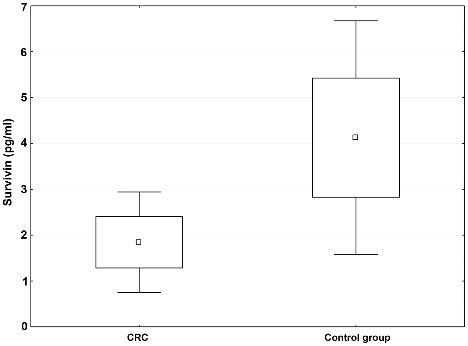
Serum survivin level in CRC and its
correlation with clinicopathological and morphological blood
parameters
Survivin antibodies were detected in the sera of
38.2% (21/55) of CRC patients and in 81.8% (18/22) of healthy
individuals. The positive cases were defined as having a protein
level >0 pg/ml. The mean value of survivin levels in the sera of
the patients with CRC was 1.844 ng/ml (range, 0–11.746 ng/ml),
which was significantly lower than that in the healthy control
group (mean, 4.954; range, 0–23.415 ng/ml) (P=0.016) (Fig. 2). Moreover, positive survivin
concentration was observed in the sera of T3 and T4 CRC patients.
The results are shown in Table II.
The sensitivity and specificity of the serum survivin level were 57
and 82.6%, at a cutoff value of 13.042 pg/ml. The area under the
ROC curve (AUC) for survivin showed that the protein exhibited
moderate diagnostic power (AUC, 0.729; P=0.0002).
| Table II.Correlations between the survivin
levels in serum and the clinicopathological parameters in patients
with colorectal cancer. |
Table II.
Correlations between the survivin
levels in serum and the clinicopathological parameters in patients
with colorectal cancer.
|
| Survivin
(pg/ml) |
|---|
|
|
|
|---|
| Parameter | n | Mean | Range | Coefficient | P-value |
|---|
| Age, years |
|
|
≤60 | 19 | 1.402 | 0.000–7.856 | 0.081 | 0.412 |
|
>60 | 36 | 2.116 | 0.000–23.415 |
|
|
| Gender |
|
|
Male | 35 | 1.969 | 0.000–23.415 | 0.028 | 0.304 |
|
Female | 20 | 1.723 | 0.000–10.449 |
|
|
| Localization |
|
|
Colon | 33 | 1.616 | 0.000–11.746 | 0.040 | 0.132 |
|
Rectum | 22 | 1.946 | 0.000–23.415 |
|
|
| Adenocarcinoma
type |
|
|
Non-mucinous | 48 | 1.727 | 0.000–23.415 | 0.014 | 0.981 |
|
Mucinous | 7 | 1.896 | 0.000–7.856 |
|
|
| Grade of
malignancies |
|
| 2 | 48 | 1.759 | 0.000–23.415 | −0.072 | 0.984 |
| 3 | 7 | 0.793 | 0.000–3.967 |
|
|
| Tumor size, cm |
|
|
<5 | 20 | 2.102 | 0.000–23.415 | −0.134 | 0.682 |
|
>5 | 18 | 1.003 | 0.000–7.856 |
|
|
| pT stage |
|
| 1 | 1 | 0.000 | 0.000 | 0.058 | 0.661 |
| 2 | 3 | 0.000 | 0.000 |
|
|
| 3 | 49 | 1.817 | 0.000–23.415 |
|
|
| 4 | 2 | 0.686 | 0.000–0.970 |
|
|
| Lymph node
metastasis |
|
|
Absent | 17 | 1.271 | 0.000–11.746 | 0.101 | 0.897 |
|
Present | 21 | 2.085 | 0.000–23.415 |
|
|
| Distant
metastasis |
|
|
Absent | 30 | 1.060 | 0.000–7.856 | 0.221 | 0.892 |
|
Present | 25 | 2.901 | 0.000–23.415 |
|
|
| IHC expression of
survivin in cancer cells |
|
|
Nuc+/Cyt+ | 23 | 2.010 | 0.000–0.856 | −0.310 | 0.440 |
|
Nuc+/Cyt− | 1 | 0.000 | 0.000 |
|
|
|
Nuc−/Cyt− | 6 | 0.025 | 0.000–0.077 |
|
|
|
Nuc−/Cyt+ | 8 | 0.228 | 0.000–1.373 |
|
|
Survivin level in the sera of the CRC patients was
not found to correlate with any clinicopathological parameters
(Table II). However, the serum
protein level was associated with the morphological blood
parameters as follows: Hematocrit (P=0.035, R=0.404), hemoglobin
(P=0.008, R=0.487) and albumin (P=0.045, R=−0.477) (Table III).
| Table III.Correlations between the level of
survivin protein in the serum and the morphological blood
parameters. |
Table III.
Correlations between the level of
survivin protein in the serum and the morphological blood
parameters.
|
| Survivin |
|---|
|
|
|
|---|
| Parameter | n | Coefficient | P-value |
|---|
| Red blood cell
count | 29 |
0.262 |
0.177 |
| White blood cell
count | 29 | −0.199 |
0.309 |
| Platelets | 29 | −0.190 |
0.333 |
| Hematocrit | 29 |
0.404 |
0.035 |
| Hemoglobin | 29 |
0.487 |
0.008 |
| Sodium | 29 | −0.038 | <0.01 |
| Potassium | 29 |
0.063 | <0.01 |
| Prothrombin
time | 29 | −0.083 |
0.678 |
| INR | 28 |
0.022 |
0.916 |
| Total proteins | 20 | −0.290 |
0.228 |
| Albumin | 18 | −0.477 |
0.045 |
| Aspartate
transaminase | 22 |
0.065 |
0.780 |
| Alanine
transaminase | 22 |
0.166 |
0.471 |
| Glucose | 13 | −0.316 |
0.292 |
| Urea | 26 | −0.159 |
0.447 |
| Creatinine | 23 | −0.144 |
0.523 |
Discussion
Survivin is overexpressed in malignancies located in
organs such as the lungs, liver, ovaries, stomach, breasts and
prostate (12). The present study
observed the positive expression of survivin in 84.2% of CRC
patients. These findings are consistent with the observations
reported in the studies by Kalliakmanis et al (15), and Choi and Chang (16), which noted the positive expression of
survivin in 88.3 and 83.3% of CRC cases, respectively. Hernandez
et al (17) confirmed the
presence of this protein in 93% of patients with colon cancer,
while Xi et al (18) observed
its expression in 60.7% of CRC patients. Survivin has different
functions depending on its location. In the nucleus, the protein
regulates cell growth, whereas its distribution to the cytoplasm
determines the viability of cancer cells (13). In the present study, the positive
expression of survivin was more common in the cell cytoplasm (81.5%
of cases) compared with the cell nucleus (63.1% of cases) in the
CRC patients. Ponnelle et al (19) also used IHC and confirmed a higher
incidence of positive survivin expression in the cytoplasm (41% of
cases) compared with its localization in the nucleus (39%) of colon
adenocarcinoma cells. The data are, however, inconsistent with the
results published in studies by Shintani et al (20) and Qi et al (21), which found that positive survivin
expression was more frequent in the nucleus than in the cytoplasm
of cancer cells in CRC patients.
Survivin as an apoptotic protein may condition tumor
aggressiveness and tumor cell invasiveness, including lymph node
involvement and distant metastases (22). It has been shown that survivin may
determine, via different signaling pathways, the capacity of
prostate and breast cancer cells to metastasize (22,23).
Single studies have confirmed this potential of survivin in CRC
patients. Chu et al (24) and
Xiaoyuan et al (25) observed
a positive correlation between survivin overexpression and lymph
node metastasis in CRC patients. Furthermore, Shen et al
(26) found that recombinant
adenovirus reduced survivin expression and when linked with
fluorouracil, blocked the cancer cell metastasis of CRC. Lymph node
involvement is associated with a poor prognosis in CRC patients,
since the risk of invasion into distant organs is increased. Li
et al (27) noted that the
immunohistochemical reaction of survivin was associated with the
presence of metastases and disease relapse in CRC patients.
Moreover, Lee et al (28)
showed that the positive expression of survivin was closely
associated with primary tumor and distant metastasis categories,
and with tumor stage. The present detailed analysis revealed a
positive correlation between survivin located in the nucleus of
cancer cells in CRC patients and distant metastases and tumor
location. The findings contradict those reported in the study by Qi
et al (21), which noted that
nuclear survivin expression was associated with a lower incidence
of distant metastasis. Nuclear survivin location is responsible for
cell cycle progression. It has been proven that the nuclear
overexpression of survivin increases the activity of cell growth
and the passage of the cells to the S phase of the cell cycle, and
results in a decline in the percentage of the G0/G1 phase cells
(29). The positive nuclear survivin
expression in CRC cells appears to indicate the growth of tumor
mitotic activity and increases the risk of developing metastatic
foci, including those to distant organs.
The serum level of survivin was measured in CRC
patients using ELISA in the present study. The protein was found
only in ~38% of CRC cases and in 81% of healthy subjects. Moreover,
the protein level was statistically significantly lower in the CRC
patients than in the healthy controls. Yagihashi et al
(30) assessed serum survivin in
various cancers of the digestive system. Survivin antibodies were
noted in 39.7% of all cancers studied, including 33% cases of CRC,
hepatoma and esophageal cancer, 40% of gastric cancer cases, 42% of
pancreatic ductal adenocarcinoma cases and 43% of biliary-tract
cancer cases. Similar results were reported in the study by Wang
et al (31), which confirmed
the presence of survivin in the sera of 31.5% of CRC patients. In
turn, Rohayen et al (32)
observed purified recombinant survivin reactions only in 8.2% of
CRC patients. The present study failed to confirm any correlations
between serum survivin level and the chosen clinicopathological
parameters in the CRC patients. However, the serum level of
survivin in the CRC patients was found to positively correlate with
hematocrit and hemoglobin in the peripheral blood, and negatively
correlate with albumin level. The aforementioned observations,
together with the present findings, suggest that the serum level of
survivin in various digestive system malignancies, particularly
CRC, exhibits little diagnostic value and will not have any
clinical implication in the future.
In conclusion, the immunohistochemical assessment of
survivin in the tissues of CRC patients revealed a major role of
protein localization in cancer cells. A positive reaction for
survivin in cancer cell nuclei may condition their proliferative
potential, which is associated with a higher risk of developing
metastatic foci. The assessment of survivin expression requires
further detailed studies as it may aid in the diagnosis of CRC in
the future. However, in our opinion, the serum level of survivin in
CRC patients appears to be diagnostically ineffectual for clinical
use.
References
|
1
|
Ferlay J, Shin HR, Bray F, Forman D,
Mathers C and Parkin DM: Estimates of worldwide burden of cancer in
2008: GLOBOCAN 2008. Int J Cancer. 127:2893–2917. 2010. View Article : Google Scholar : PubMed/NCBI
|
|
2
|
Pepe MS, Etzioni R, Feng Z, Potter JD,
Thompson ML, Thornquist M, Winget M and Yasui Y: Phases of
biomarker development for early detection of cancer. J Natl Cancer
Inst. 93:1054–1061. 2001. View Article : Google Scholar : PubMed/NCBI
|
|
3
|
Hanahan D and Weinberg RA: Hallmarks of
cancer: The next generation. Cell. 144:646–674. 2011. View Article : Google Scholar : PubMed/NCBI
|
|
4
|
Walczak H and Krammer PH: The CD95
(APO-1/Fas) and the TRAIL (APO-2L) apoptosis systems. Exp Cell Res.
256:58–66. 2000. View Article : Google Scholar : PubMed/NCBI
|
|
5
|
Thomadaki H and Scorilas A: BCL2 family of
apoptosis-related genes: Functions and clinical implications in
cancer. Crit Rev Clin Lab Sci. 43:1–67. 2006. View Article : Google Scholar : PubMed/NCBI
|
|
6
|
Sträter J, Hinz U, Hasel C, Bhanot U,
Mechtersheimer G, Lehnert T and Möller P: Impaired CD95 expression
predisposes for recurrence in curatively resected colon carcinoma:
Clinical evidence for immunoselection and CD95L mediated control of
minimal residual disease. Gut. 54:661–665. 2005. View Article : Google Scholar : PubMed/NCBI
|
|
7
|
Korkolopoulou P, Saetta AA, Levidou G,
Gigelou F, Lazaris A, Thymara I, Scliri M, Bousboukea K,
Michalopoulos NV, Apostolikas N, et al: c-FLIP expression in
colorectal carcinomas: Association with Fas/FasL expression and
prognostic implications. Histopathology. 51:150–156. 2007.
View Article : Google Scholar : PubMed/NCBI
|
|
8
|
Strand S, Hofmann WJ, Hug H, Müller M,
Otto G, Strand D, Mariani SM, Stremmel W, Krammer PH and Galle PR:
Lymphocyte apoptosis induced by CD95 (APO-1/Fas) ligand-expressing
tumor cells-a mechanism of immune evasion? Nat Med. 2:1361–1366.
1996. View Article : Google Scholar : PubMed/NCBI
|
|
9
|
Deveraux QL, Stennicke HR, Salvesen GS and
Reed JC: Endogenous inhibitors of caspases. J Clin Immunol.
19:388–398. 1999. View Article : Google Scholar : PubMed/NCBI
|
|
10
|
Chantalat L, Skoufias DA, Kleman JP, Jung
B, Dideberg O and Margolis RL: Crystal structure of human survivin
reveals a bow tie-shaped dimer with two unusual alpha-helical
extensions. Mol Cell. 6:183–189. 2000. View Article : Google Scholar : PubMed/NCBI
|
|
11
|
Dohi T, Okada K, Xia F, Wilford CE, Samuel
T, Welsh K, Marusawa H, Zou H, Armstrong R, Matsuzawa S, et al: An
IAP-IAP complex inhibits apoptosis. J Biol Chem. 279:34087–34090.
2004. View Article : Google Scholar : PubMed/NCBI
|
|
12
|
Ambrosini G, Adida C and Altieri DC: A
novel anti-apoptosis gene, survivin, expressed in cancer and
lymphoma. Nat Med. 3:917–921. 1997. View Article : Google Scholar : PubMed/NCBI
|
|
13
|
Mahotka C, Wenzel M, Springer E, Gabbert
HE and Gerharz CD: Survivin-deltaEx3 and survivin-2B: Two novel
splice variants of the apoptosis inhibitor survivin with different
antiapoptotic properties. Cancer Res. 59:6097–6102. 1999.PubMed/NCBI
|
|
14
|
Hamilton SR and Aaltonen LA: World Health
Organization Classification of TumoursPathology and Genetics of
Tumours of the Digestive System. IARC Press; Lyon: pp. 1042000
|
|
15
|
Kalliakmanis JG, Kouvidou Ch, Latoufis C,
Kouvatseas G, Anagnostakis D, Papatheodoridis G, Koskinas J and
Archimandritis A: Survivin expression in colorectal carcinomas:
Correlations with clinicopathological parameters and survival. Dig
Dis Sci. 55:2958–2964. 2010. View Article : Google Scholar : PubMed/NCBI
|
|
16
|
Choi J and Chang H: The expression of MAGE
and SSX, and correlation of COX2, VEGF, and survivin in colorectal
cancer. Anticancer Res. 32:559–564. 2012.PubMed/NCBI
|
|
17
|
Hernandez JM, Farma JM, Coppola D, Hakam
A, Fulp WJ, Chen DT, Siegel EM, Yeatman TJ and Shibata D:
Expression of the antiapoptotic protein survivin in colon cancer.
Clin Colorectal Cancer. 10:188–193. 2011. View Article : Google Scholar : PubMed/NCBI
|
|
18
|
Xi RC, Biao WS and Gang ZZ: Significant
elevation of survivin and livin expression in human colorectal
cancer: Inverse correlation between expression and overall
survival. Onkologie. 34:428–432. 2011. View Article : Google Scholar : PubMed/NCBI
|
|
19
|
Ponnelle T, Chapusot C, Martin L, Bouvier
AM, Plenchette S, Faivre J, Solary E and Piard F: Cellular
localisation of survivin: Impact on the prognosis in colorectal
cancer. J Cancer Res Clin Oncol. 131:504–510. 2005. View Article : Google Scholar : PubMed/NCBI
|
|
20
|
Shintani M, Sangawa A, Yamao N and
Kamoshida S: Immunohistochemical expression of nuclear and
cytoplasmic survivin in gastrointestinal carcinoma. Int J Clin Exp
Pathol. 6:2919–2927. 2013.PubMed/NCBI
|
|
21
|
Qi G, Tuncel H, Aoki E, Tanaka S, Oka S,
Kaneko I, Okamoto M, Tatsuka M, Nakai S and Shimamoto F:
Intracellular localization of survivin determines biological
behavior in colorectal cancer. Oncol Rep. 22:557–562.
2009.PubMed/NCBI
|
|
22
|
McKenzie JA, Liu T, Goodson AG and
Grossman D: Survivin enhances motility of melanoma cells by
supporting Akt activation and {alpha}5 integrin upregulation.
Cancer Res. 70:7927–7937. 2010. View Article : Google Scholar : PubMed/NCBI
|
|
23
|
Zhang M, Coen JJ, Suzuki Y, Siedow MR,
Niemierko A, Khor LY, Pollack A, Zhang Y, Zietman AL, Shipley WU
and Chakravarti A: Survivin is a potential mediator of prostate
cancer metastasis. Int J Radiat Oncol Biol Phys. 78:1095–1103.
2010. View Article : Google Scholar : PubMed/NCBI
|
|
24
|
Chu XY, Chen LB, Wang JH, Su QS, Yang JR,
Lin Y, Xue LJ, Liu XB and Mo XB: Overexpression of survivin is
correlated with increased invasion and metastasis of colorectal
cancer. J Surg Oncol. 105:520–528. 2012. View Article : Google Scholar : PubMed/NCBI
|
|
25
|
Xiaoyuan C, Longbang C, Jinghua W,
Xiaoxiang G, Huaicheng G, Qun Z and Haizhu S: Survivin: A potential
prognostic marker and chemoradiotherapeutic target for colorectal
cancer. Ir J Med Sci. 179:327–335. 2010. View Article : Google Scholar : PubMed/NCBI
|
|
26
|
Shen W, Tu JK, Wang XH and Fu ZX:
Oncolytic adenovirus mediated Survivin RNA interference and
5-fluorouracil synergistically suppress the lymphatic metastasis of
colorectal cancer. Oncol Rep. 24:1285–1290. 2010.PubMed/NCBI
|
|
27
|
Li JH, He WJ and He YJ: Expression and
clinical significance of Survivin and Livin in DukesoB colorectal
cancer. Ai Zheng. 26:547–551. 2007.(In Chinese). PubMed/NCBI
|
|
28
|
Lee YY, Yu CP, Lin CK, Nieh S, Hsu KF,
Chiang H and Jin JS: Expression of survivin and cortactin in
colorectal adenocarcinoma: Association with clinicopathological
parameters. Dis Markers. 26:9–18. 2009. View Article : Google Scholar : PubMed/NCBI
|
|
29
|
Suzuki A, Hayashida M, Ito T, Kawano H,
Nakano T, Miura M, Akahane K and Shiraki K: Survivin initiates cell
cycle entry by the competitive interaction with Cdk4/p16(INK4a) and
Cdk2/cyclin E complex activation. Oncogene. 19:3225–3234. 2000.
View Article : Google Scholar : PubMed/NCBI
|
|
30
|
Yagihashi A, Asanuma K, Nakamura M, Araya
J, Mano Y, Torigoe T, Kobayashi D and Watanabe N: Detection of
anti-survivin antibody in gastrointestinal cancer patients. Clin
Chem. 47:1729–1731. 2001.PubMed/NCBI
|
|
31
|
Wang YQ, Zhang HH, Liu CL, Xia Q, Wu H, Yu
XH and Kong W: Correlation between auto-antibodies to survivin and
MUC1 variable number tandem repeats in colorectal cancer. Asian Pac
J Cancer Prev. 13:5557–5562. 2012. View Article : Google Scholar : PubMed/NCBI
|
|
32
|
Rohayem J, Diestelkoetter P, Weigle B,
Oehmichen A, Schmitz M, Mehlhorn J, Conrad K and Rieber EP:
Antibody response to the tumor-associated inhibitor of apoptosis
protein survivin in cancer patients. Cancer Res. 60:1815–1817.
2000.PubMed/NCBI
|