Introduction
Although the early detection, prevention and
treatment of cancer has significantly improved in recent decades,
it remains the most prevalent and lethal disease, worldwide
(1). Based on GLOBOCAN estimates,
~14.1 million new cancer cases and 8.2 million cancer-asssociated
mortalities occurred in 2012 worldwide (2). Surgery remains the most effective method
of curing or controlling the majority of cancers, however, advances
in chemotherapy, radiotherapy and specific cancer targeting
therapies have significantly improved patient survival, quality of
life and prognosis (3). Thus, the
development of more potent agents with fewer side effects is key to
the effective control of cancer. At present, ongoing studies aim to
identify novel and effective methods and novel therapeutics that
may be used to combat cancer (1).
Recently, studies investigating medicinal fungi have
become an important potential source of novel anticancer drugs
(4,5).
Lentinula edodes (Shiitake) is a type of edible fungus that
has been considered medicinal for hundreds of years (6). Since 1991, increasing evidence has
indicated that Lentinula edodes possesses a variety of
antitumor, antivirus and immunomodulatory properties (7–9). In
vivo and in vitro studies have confirmed that compounds
produced by Lentinula edodes induce tumor cell apoptosis.
Among these compounds, lentinan is considered to exhibit the most
effective suppression of tumor cell proliferation (10–12).
Following decades of study, a specialized strain of Lentinula
edodes (Lentinula edodes C91-3) was generated
and fermented protein derived from this strain has been
demonstrated to induce apoptosis in numerous tumor cell types, such
as A549, S180 and H22 cells (13–15).
In our previous study (16), the functional genomics of Lentinula
edodes were investigated by obtaining high-quality DNA
sequences (>4 four billion bases) using Illumina/Solexa
technology. The data demonstrated the suitability of short-read DNA
sequences for de novo assembly and annotation of genes
expressed in eukaryotes without prior existing genome information.
Our previous study also established a conventional naming system
for Lentinula edodes transcriptomic proteins as Lentinula
edodes transcriptome protein-(Number) (Latcripin-(Number),
LP-(Number) and this nomenclature has been submitted and received
by National Center for Biotechnology Information (NCBI) GenBank. In
our previous study, the Latcripin-1 gene was cloned and
expressed in Pichia pastoris and its antitumor properties
were analyzed (13). In the present
study, Latcripin-15 (LP-15) regulator of chromosome condensation 1
(RCC1) domain protein was identified, cloned, expressed, purified
and re-folded from Lentinula edodes C91-3 and the
potential of this protein as a novel anti-tumor agent was
investigated in vitro.
Materials and methods
The shiitake strain, Lentinula edodes
C91-3, was obtained from The Chinese General
Microbiological Culture Collection Center (Beijing, China).
Escherichia coli (E. coli) JM109, which was maintained in
our laboratory, was used to amplify plasmids for cloning the LP-15
gene. E. coli Rosetta-gami (DE3) cells, used to facilitate
expression of LP-15-RCC1 protein, were obtained from Novagen (Merck
Millipore, Darmstadt, Germany). The pMD20-T cloning vector was
purchased from Takara (Shiga, Japan), while the expression vector
pET-32a(+) was purchased from Novagen (Merck Millipore).
The 3′-full rapid amplification of cDNA ends (RACE)
core set (Version 2.0), 5′-full RACE kit, miniBEST plasmid
purification kit (Version 3.0), miniBEST agarose gel DNA extraction
kit (Version 3.0), EcoRI, XhoI, primers, competent
cell preparation kit and in-fusion™ advantage polymerase chain
reaction (PCR) cloning kit were also obtained from Takara. TRIzol
reagent, mouse anti-His tag monoclonal antibody (catalog no.
66005-1), peroxidase-conjugated affinipure goat anti-mouse
immunoglobulin (Ig)G (catalog no. SA00001-1) and nitrocellulose
filter membranes were purchased from Invitrogen Life Technologies
(Thermo Fisher Scientific, Inc., Waltham, MA, USA). Carbenicillin,
chloramphenicol, kanamycin, tetracycline and isopropyl
β-D-thiogalactopyranoside (IPTG) were purchased from Tiangen
Biotech Co., Ltd., (Beijing, China). The 6xHis-tagged protein
purification kit was purchased from CWBIO (Beijing, China). The
bicinchoninic acid (BCA) Protein Assay Kit, Annexin V-fluorescein
isothiocyanate (FITC)/propidium iodide (PI) kit and apoptotic cell
Hoechst 33258 detection kit were obtained from Keygen Biotech Co.,
Ltd., (Nanjing, China). The integrated potato culture medium and
other common laboratory reagents were prepared in our
laboratory.
Cloning of LP-15 RCC1 functional
domain cDNA and vector construction
Lentinula edodes C91-3 mycelium
was cultured in integrated potato culture medium for 18 days at
room temperature. Total RNA was isolated from mycelium culture
using TRIzol reagent according to the manufacturer's instructions.
Isolated RNA samples were then used as the RACE template. Briefly,
primers for 3′-RACE and 5′-RACE experiments were designed using
Oligo 6.0 software (Molecular Biology Insights, Inc., Colorado
Springs, CO, USA) and Lentinula edodes C91-3
transcriptome sequences, as previously described (16). Experiments were performed using the
abovementioned kits, according to the manufacturer's instructions,
as previoulsy described (16). The
resulting products were DNA sequenced. Full-length cDNA was
amplified using PCR and the DNA was sequenced and compared using
the NCBI database (http://blast.ncbi.nlm.nih.gov/Blast.cgi). The
functional domain sequence of LP-15 was selected and predicted by
online tools from Sanger Pfam (http://pfam.xfam.org/). A pair of primers was designed
to amplify the functional domain of LP-15 by PCR: Forward,
5′-ATCGGATCCGAATTCCATGGCAATGTCTACACCTG-3′ and reverse,
5′-GTGGTGGTGCTCGAGCAATCCTACGACATGCTGAC-3′. The PCR conditions were
as follows: 30 cycles of denaturing at 98°C for 10 sec, primer
annealing at 55°C for 10 sec, extension at 72°C for 1 min and a
final extension at 72°C for 5 min. After PCR amplification, a
6xHis-tag was added to the 3′end of the PCR product for
characterization and identification (17,18).
EcoRI and XhoI restriction sites were added to the N-
and C-terminal, respectively, to enable in-fusion ligation
(19).
The DNA-sequence-confirmed PCR products were
purified using the miniBEST agarose gel DNA extraction kit (Version
3.0) and inserted into pET-32a(+) EcoRI and XhoI sites using the
in-fusion™ advantage PCR cloning kit and E. coli JM109
competent cells. The complete construct was amplified in E.
coli DE3 cells followed by DNA sequencing. The amino acid
composition of the LP-15-RCC1 functional domain was analyzed using
the ExPASy database (http://www.expasy.org/). The secondary structure and
physicochemical properties of the LP-15-RCC1 functional domain were
analyzed using DNAStar software (Version 5.07; DNASTAR Inc.,
Madison, WI, USA). The tertiary structure of the LP-15-RCC1
functional domain was predicted using the Swiss-Model database
(https://swissmodel.expasy.org/interactive).
Expression and purification of the
LP-15 RCC1 functional domain protein
pET-32a(+)-LP-15-RCC1 plasmid was transferred into
E. coli DE3 cells for protein expression using an in
vitro protein expression and purification kit. Briefly, a
single bacterial colony was selected and cultured in 10 ml lysogeny
broth (LB) medium containing 50 µg/ml carbenicillin, 34 µg/ml
chloramphenicol, 15 µg/ml kanamycin and 12.5 µg/ml tetracycline at
37°C in a shaking incubator at 190 rpm overnight. The next day, the
culture was transferred to 1 l LB medium with the same composition
and grown at 37°C with shaking at 190 rpm until the optical density
(OD), measured at a wavelength of 600 nm, was between 0.5 and 0.6.
Next, 1.0 mM IPTG was added to the culture and incubated for an
additional 6 h at 37°C. pET-32a(+) only was used as a negative
control. Finally, the bacterial culture was collected and separated
by centrifugation at 6,000 × g for 20 min at 4°C.
For protein purification, a portion of the bacterial
pellet was subjected to western blot analysis with 12% sodium
dodecyl sulfonate polyacrylate gel electrophoresis (SDS-PAGE) and a
primary mouse monoclonal anti-His-antibody at a dilution of 1:250,
followed by incubation with secondary peroxidase-conjugated goat
anti-mouse IgG (H+L) antibody at a dilution of 1:5,000. Western
blot analysis confirmed the presence of LP-15-RCC1 protein in the
bacteria culture. Next, 100 mg bacteria pellets were lysed using 3
ml bacteria extraction reagent solution [1 ml bacteria extraction
reagent, 1 µl DNase I (1,000 U/ml), 2 µl Lysozyme (50 mg/ml) and 10
µl phenylmethylsulfonyl fluoride] and incubated at room temperature
for 30 min. After incubation, cell lysate was centrifuged at 10,000
x g for 20 min at 4°C to remove the supernatant. Next, the
pellet was resuspended and dissolved in binding buffer (pH 7.9; 8 M
urea, 5 mM imidazole, 0.5 M NaCl and 20 mM Tris-HCl; Keygen Biotech
Co., Ltd.) at 4°C for 1 h. Solubilized protein was then separated
from the insoluble cellular debris by centrifugation at 10,000 ×
g for 20 min at 4°C. The solubilized protein was purified
using a nickel-chelated column and washed 15 times with binding
buffer prior to elution of the bound protein with elution buffer
(pH 7.9; 8 M urea, 500 mM imidazole, 0.5 M NaCl and 20 mM
Tris-HCl). Each eluted fraction was analyzed by 12% SDS-PAGE to
measure the purity of the protein.
Refolding the LP-15 RCC1 functional
domain protein
The purified LP-15 RCC1 functional domain protein
was refolded by gradient dialysis using four types of the refolding
buffer at 4°C. The four dialysis buffers were: Dialysate 1 [4 M
urea, 2 mM reduced glutathione (GSH) and 0.2 mM oxidized
glutathione (GSSG) in phosphate-buffered saline (PBS)]; Dialysate 2
(2 M urea, 2 mM GSH and 0.2 mM GSSG in PBS); Dialysate 3 (2 mM GSH
and 0.2 mM GSSG in PBS); and Dialysate 4 (PBS). Dialysate was
replaced every 8 h and each concentration of dialysate was used
three times. Finally, the refolded protein was concentrated using
PEG 12000 (Sigma-Aldrich, Merck Millipore, Darmstadt, Germany) and
quantified using the BCA protein assay kit.
Cell culture
The human lung cancer A549 cell line was obtained
from the Shanghai Cell Bank, Chinese Academy of Sciences (Shanghai,
China) and cultured in RPMI 1640 medium (HyClone Laboratories; GE
Healthcare, Logan, UT, USA) supplemented with 10% fetal bovine
serum (FBS; TransGen Biotech, Inc., Beijing, China) in a humidified
atmosphere of 5% CO2 at 37°C (to 80% confluence). To
assess the effects of the LP-15 RCC1 functional domain protein on
the regulation of cellular behavior and gene expression, A549 cells
were seeded in culture plates or flasks at a density of
1×10⁵ cells/ml and cultured for 24 h. The growth medium
was replaced with fresh RPMI 1640 medium with 10% FBS containing
various concentrations (12.5, 25, 50, 100 and 200 µg) of the LP-15
RCC1 functional domain protein for the indicated periods of time
(24 and 48 h) and cellular behavior was subsequently assessed.
3-(4,5-dimethylthiazol-2-yl)-2,5-diphenyltetrazolium bromide (MTT)
cell viability assay
The effect of the LP-15 RCC1 functional domain
protein on the regulation of cell growth was determined using cell
viability MTT assays. Briefly, LP-15 RCC1 functional domain protein
with different concentrations (12.5, 25, 50, 100 and 200 µg) was
added to cell culture for 24 or 48 h, then 10 µl of MTT solution
was added to the cell culture medium (Sigma-Aldrich; Merck
Millipore) and the cells cultured at 37°C in a 5% CO2
incubator for an additional 4 h. Next, the growth medium was
replaced with 150 µl dimethyl sulfoxide (Sigma-Aldrich; Merck
Millipore). Finally, the absorbance of the cell culture plates was
measured at a wavelength of 589 nm using an ELISA microplate reader
(Thermo Fisher Scientific Inc.). Cell viability was determined by
the following equation: Cell viability (%) = [(control group OD -
experimental group OD)/control group OD] × 100.
Transmission electron microscopy
(TEM)
Changes in cellular ultrastructure were assessed
following treatment with LP-15 RCC1 functional domain protein using
TEM. Following treatment with 200 µg LP-15 RCC1 functional domain
protein for 48 h, A549 cells were harvested and fixed in 2.5%
glutaraldehyde solution (Sigma-Aldrich; Merck Millipore) at 4°C
overnight. The next day, the cells were washed with 0.1 M PBS and
postfixed with 1% (w/v) osmium tetroxide (Sigma-Aldrich; Merck
Millipore) for 2 h at room temperature. After dehydration using a
graded ethanol series (70–100%), cells were embedded in propylene
oxide and epon (Sigma-Aldrich; Merck Millipore) and solidified at
60°C for 48 h in 100% epoxy resin (Sigma-Aldrich, Merck Millipore).
Ultrathin sections (70 nm) were prepared, stained with uranyl
acetate and lead citrate (Sigma-Aldrich) Merck Millipore and viewed
under a TEM (JEOL Ltd., Tokyo, Japan).
Flow cytometry apoptosis assay
Flow cytometry was performed using FITC-labeled
annexin V and PI to detect phosphatidylserine expression as an
endpoint indicator of early apoptosis (20). Following 24 or 48 h treatment with 200
µg LP-15 RCC1 functional domain protein, A549 cells were collected
and washed in cooled PBS and centrifuged at 111 × g for 5
min at room temperature. The pellets were then resuspended in 400
µl binding buffer and 5 µl FITC-labeled annexin V and 5 µl PI were
added to the suspension. The cells were gently mixed, then
incubated at the room temperature in the dark for 15 min.
Fluorescence intensity was detected using a flow cytometer and
analyzed using Cell Quest software (Version 3.3; Cell Quest, Inc.,
Tokyo, Japan).
Hoechst 33258 staining
Cells were cultured on coverslips and treated with
50 µg or 100 µg LP-15 RCC1 functional domain protein for 48 h.
Cells were then stained with 5 µg Hoechst 33258 (Keygen Biotech
Co., Ltd.) in PBS at room temperature in the dark for 10 min. After
staining, the cells were washed twice with PBS and images were
captured using a fluorescence microscope (Nikon Corporation, Tokyo,
Japan) with an excitation wavelength of 330–380 nm.
Statistical analysis
All experiments were performed in duplicate and
repeated at least three times. Data are expressed as the mean ±
standard deviation. Treatment groups were compared using one-way
analysis of variance with SPSS 13.0 (SPSS, Inc., Chicago, IL, USA).
P<0.05 was considered to indicate a statistically significant
difference.
Results
Cloning, expression and purification
of LP-15 RCC1 functional domain protein
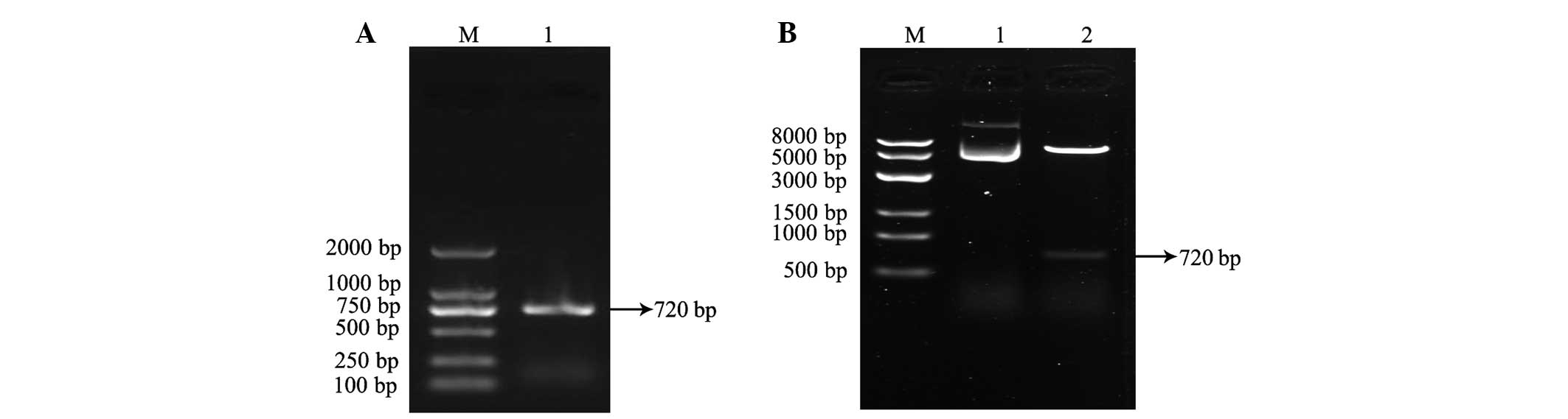
LP-15 RCC1 functional domain cDNA was cloned from
Lentinula edodes C91-3 using 3′-RACE and 5′-RACE
(Fig. 1A). DNA sequencing confirmed
that the correct 720 base pair (bp) fragment had been cloned
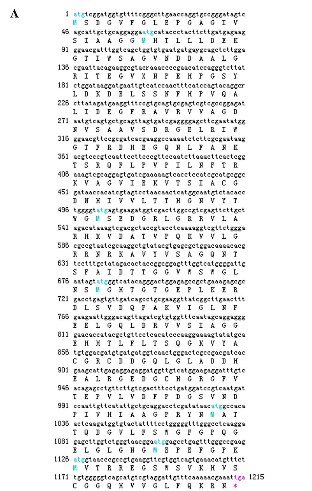
(Fig. 1B). The amino acid sequence of
the LP-15 full-length cDNA coding sequence region has been
submitted to GenBank (Accession Number, KF709447; Fig. 2A). The amino acid sequence of the
LP-15 functional domain was analyzed using the Pfam database
(http://pfam.xfam.org/). The result shown in the
Pfam-A database matched the RCC1 domains: A 720 bp-long open
reading frame encoding 240 amino acids (Fig. 2B). The amino acid composition of the
LP-15 functional domain was analyzed using the ExPASy database
(http://www.expasy.org/) (Fig. 2C). The secondary structure of the
LP-15 functional domain indicated that it contained ordinary α, β,
turn and coil regions (Fig. 2D). The
tertiary structure of the LP-15-RCC1 functional domain is shown in
Fig. 2E with a predicted molecular
weight (MW) of 25.80 kDa and isoelectric point (pI) of 6.33. The
predicted MW and pI of the pET-32a(+)-LP-15-RCC1 recombinant
protein were 44.85 kDa and 6.09, respectively. After completing
this analysis, the LP-15 RCC1 functional domain protein, which
appeared at approximately 45 kDa on a 12% SDS-PAGE gel, was
expressed, purified and refolded in vitro (Fig. 3).
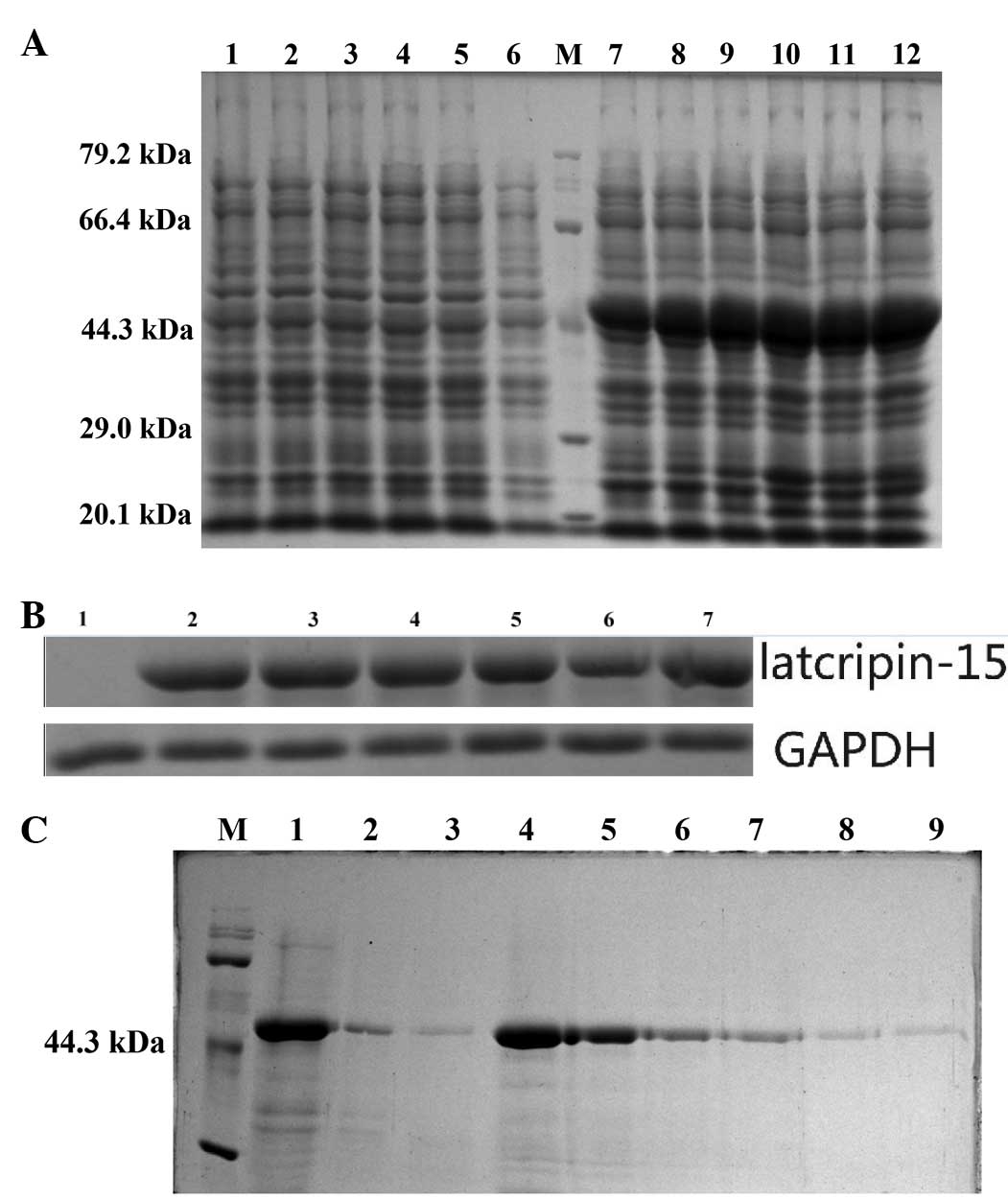 | Figure 3.Expression, purification and
identification of the LP-15 RCC1 functional domain protein. (A) 12%
SDS-PAGE of the cell lysate. Lanes 1–6, total cell lysate from DE3
induced using 1 mM IPTG for 6, 5, 4, 3, 2 and 1 h, respectively.
pET-32a(+) served as a negative control. M, molecular weight
marker. Lanes 7–12, total cell lysate from DE3 with
pET-32a(+)-LP-15-RCC1 induced using 1 mM IPTG for 1, 2, 3, 4, 5 and
6 h, respectively. (B) Western blot of pET-32a(+)-LP-15-RCC1. Lane
1, total cell lysate from DE3 induced with 1 mM IPTG for 6 h.
pET-32a(+) served as a negative control. Lanes 2–7, total cell
lysate from DE3 cells with pET-32a(+)-LP-15-RCC1 induced using 1 mM
IPTG for 1, 2, 3, 4, 5 and 6 h, respectively. (C) 12% SDS-PAGE of
purified protein. M, protein molecular weight marker. Lane 1, total
cell lysate after solubilization with buffer containing 8 M urea.
Lane 2, liquid flow through from the Ni2+ column
following addition of pET-32a(+)-LP-15-RCC1 sample. Lane 3, first
phase eluent. Lane 4, second phase eluent. Lane 5, third phase
eluent. Lane 6, fourth phase eluent. Lane 7, fifth phase eluent.
Lane 8, sixth phase eluent. Lane 9, seventh phase eluent. RCC1,
regulator of chromosome condensation 1; LP-15, Latcripin-15; IPTG,
isopropyl β-D-thiogalactopyranoside; SDS-PAGE, sodium dodecyl
sulfonate polyacrylate gel electrophoresis; GAPDH, glyceraldehyde
3-phosphate dehydrogenase; DE3, Rosetta-gami. |
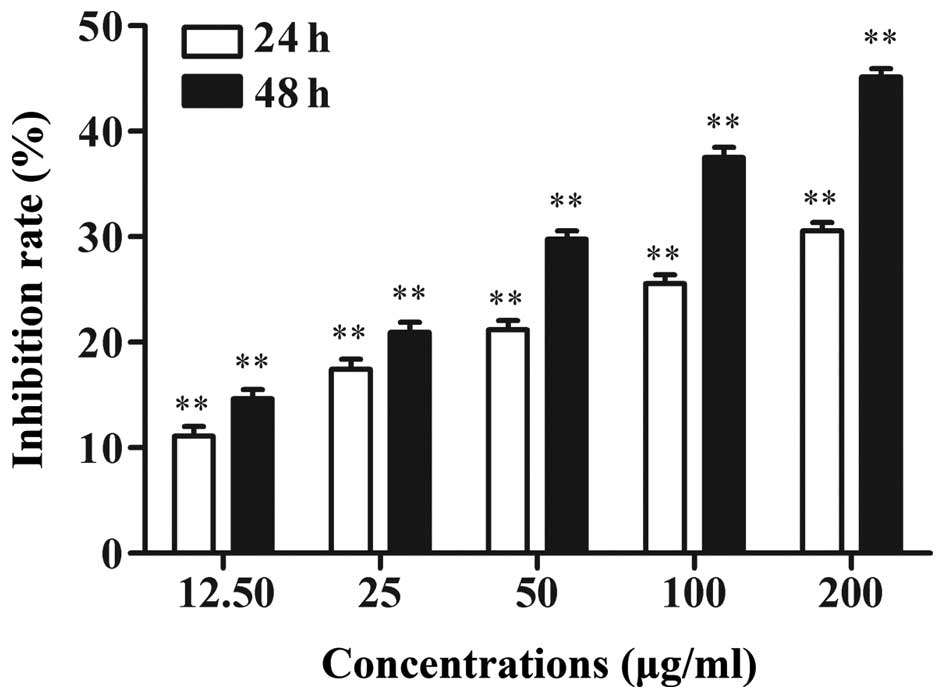
LP-15 RCC1 functional domain protein
reduces A549 cell viability
The effect of the LP-15 RCC1 functional domain
protein on the regulation of A549 cell viability was assessed.
Following tumor cell incubation with various concentrations of
LP-15 RCC1 functional domain protein for 24 h or 48 h, tumor cell
viability was significantly reduced in a time- and dose-dependent
manner (P<0.001; Fig. 4). In
comparison to the controls, cell viability inhibition rates were
29.75±5.8, 37.50±4.2 and 45.16±1.85 after 48 h treatment with 50,
100 and 200 µg RCC1 functional domain protein, respectively.
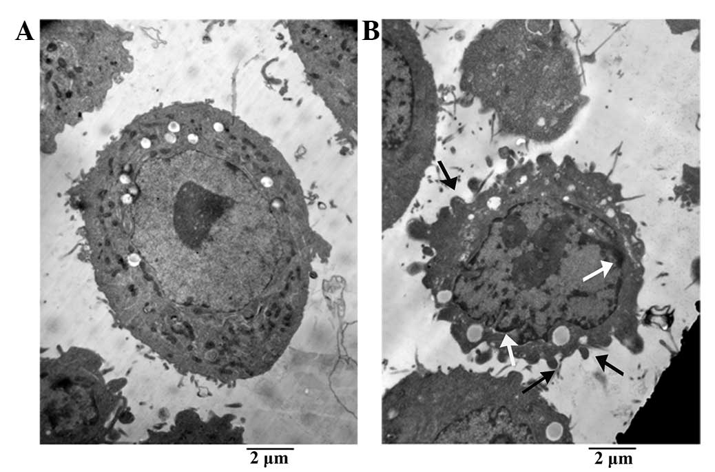
Treatment with LP-15 RCC1 functional
domain protein induces morphological changes in A549 cells
The morphological changes in A549 cells following
treatment with 200 µg LP-15 RCC1 functional domain protein for 48 h
was assessed using TEM (Fig. 5). A549
cells in the control group maintained regular morphology, with a
large nucleus, numerous mitochondria and membrane phase structural
integrity (Fig. 5A). By contrast,
after LP-15 RCC1 functional domain treatment, tumor cells exhibited
apoptotic bodies, chromatin condensation, nuclear fragmentation and
decreased numbers of mitochondria (Fig.
5B).
Treatment with LP-15 RCC1 functional
domain protein induces apoptotic changes in A549 cells
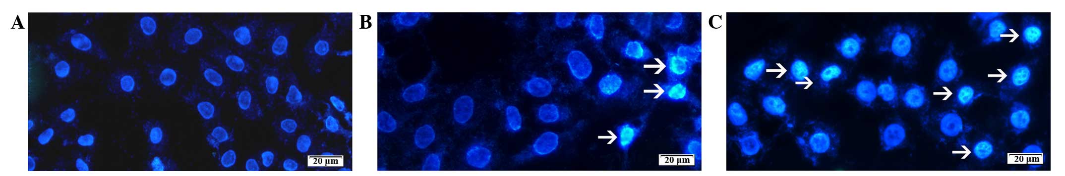
Apoptotic A549 cells were identified using Hoechst
33258 staining following treatment with 50 and 100 µg LP-15 RCC1
functional domain protein for 48 h. Analysis of nuclear morphology
identified cellular changes associated with apoptosis, such as
chromatin condensation and fragmentation. Following 48 h treatment
with 50 and 100 µg LP-15 RCC1 functional domain protein,
fluorescence microscopy revealed apoptotic tumor cells exhibiting
chromatin condensation (Fig. 6).
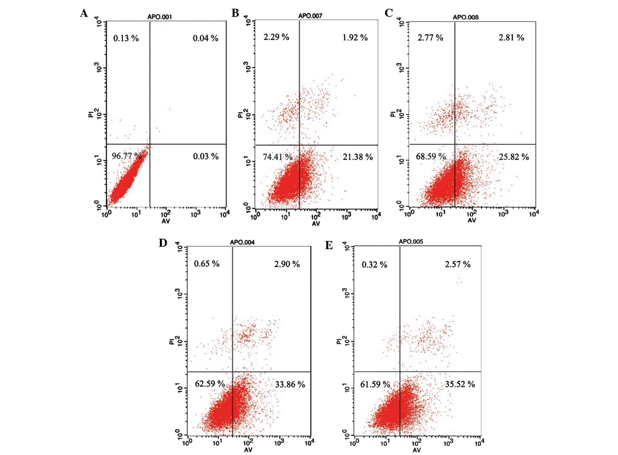
A459 cells were also analyzed using Annexin
V/PI-flow cytometry following treatment with 100 and 200 µg LP-15
RCC1 functional domain protein for 24 and 48 h. After 24 h
treatment with 100 and 200 µg LP-15 RCC1 functional domain protein,
the early and advanced apoptosis rates of A549 cells were 1.92 and
21.38% and 2.81 and 25.82%, respectively. After 48 h treatment with
100 and 200 µg LP-15 RCC1 functional domain protein, the early and
advanced apoptosis rates were 2.90 and 33.86% and 2.57 and 35.52%,
respectively. These results indicate that treatment with LP-15 RCC1
functional domain protein increases the apoptosis rate of A549
cells in a dose-dependent manner (Fig.
7).
Discussion
The identification of novel anticancer drugs has
progressed from a strategy that primarily focused on agents that
kill tumor cells to a more mechanistic strategy focused on
understanding the molecular targets involved in tumor signaling and
tumor cell transformation (21). In
the present study, a novel LP-15 RCC1 functional domain protein,
obtained from Lentinula edodes C91-3, was
identified, cloned, expressed, purified and refolded to assess the
antitumor activity of the protein in vitro. The treatment of
A549 cells with the LP-15 RCC1 functional domain protein
significantly reduced tumor cell viability and induced tumor cells
to undergo apoptosis. Thus, this protein presents a potential novel
anticancer therapeutic agent, however, further in vivo
studies are required.
The RCC1 domain of the LP-15 protein may be critical
to its antitumor properties. RCC1 is a RAs-related nuclear protein
(Ran) guanine nucleotide exchange factor for Ran guanosine
triphosphate (GTP)ase, which exhibits a critical function in
mitosis, nucleocytoplasmic transport and nuclear envelope assembly
(22). RCC1 catalyzes the exchange of
RanGDP to RanGTP, which is essential for nucleocytoplasmic
transport (23) and spindle and
nuclear envelope assembly during mitosis (24,25).
Cáceres-Gorriti et al (26)
demonstrated that the RAN network, including RAN's
nucleocytoplasmic transport and mitotic functions, is dysregulated
in serous ovarian carcinomas and this dysregulation affects tumor
progression and patient survival. Wong et al (27) reported that nuclear RanGTP levels
exhibit a crucial function in apoptosis initiation and RCC1 reads
the histone code generated by caspase-activated Mst1 to initiate
apoptosis by reducing RanGTP levels in the nucleus. Additionally,
Zhou and Münger (28) reported that
the chronic lymphocytic leukemia deletion gene 7 (Clld7)
gene, which encodes an evolutionarily conserved protein carrying an
RCC1 domain in addition to broad complex, tramtrack, bric-a-brac
and POxvirus and Zinc finger domains, reduces cell viability.
Clld7 depletion in normal human epithelial cells confers
resistance to DNA damage-induced apoptosis (28). The present current study provides
further evidence to support the hypothesis that the RCC1 domain
protein possesses pro-apoptotic or programmed cell death
activity.
Latcripin-1, a similar apoptosis-related protein
isolated from the Lentinula edodes C91-3
transcriptome, has been expressed using a eukaryotic expression
system (13). In our previous study,
the Latcripin-13 RCC1 and plant homeodomain proteins were
successfully expressed in a prokaryotic expression system (E.
coli DE3) (14). As a result of
our previous study, and due to the extensive amount of time and
effort required to generate sufficient protein concentrations using
the eukaryotic expression system, a prokaryotic expression system
was selected for the present study. The advantages of the
prokaryotic expression system include speed, high capacity, ease of
operation and low cost (29–31). To date, few studies investigating the
antitumor activity of RCC1 domain proteins have been reported in
the literature. Thus, although the antitumor mechanism of RCC1
remains to be determined, the results of the present study
represent a novel and significant step towards understanding the
antitumor activity of this protein domain. However, the current
study has a number of limitations, which must be considered. While
the present study successfully demonstrated the antitumor activity
of the LP-15 RCC1 domain protein in vitro, future studies
are required to focus on understanding its molecular antitumor
mechanisms. Additionally, in the future we aim to develop an in
vivo expression system to express this protein directly in
tumor cells and assess its antitumor activity and underlying
mechanisms in vivo.
Acknowledgements
This study was supported in part by The National
Natural Science Foundation of China (grant no. 81301995).
Glossary
Abbreviations
Abbreviations:
|
DE3
|
Rosetta-gami
|
|
RACE
|
rapid amplification of cDNA ends
|
|
Clld7
|
chronic lymphocytic leukemia deletion
gene 7
|
|
MTT
|
3-(4,5-dimethylthiazol-2-yl)-2,
5-diphenyltetrazolium bromide
|
|
TEM
|
transmission electron microscopy
|
|
RCC1
|
regulator of chromosome condensation
1
|
References
|
1
|
Wang J, Zhao B, Zhang W, Wu X, Wang R,
Huang Y, Chen D, Park K, Weimer BC and Shen Y: Mycoepoxydiene, a
fungal polyketide, induces cell cycle arrest at the G2/M phase and
apoptosis in HeLa cells. Bioorg Med Chem Lett. 20:7054–7058. 2010.
View Article : Google Scholar : PubMed/NCBI
|
|
2
|
Torre LA, Bray F, Siegel RL, Ferlay J,
Lortet-Tieulent J and Jemal A: Global cancer statistics, 2012. CA
Cancer J Clin. 65:87–108. 2015. View Article : Google Scholar : PubMed/NCBI
|
|
3
|
Bernier J and Cooper JS: Chemoradiation
after surgery for high-risk head and neck cancer patients: How
strong is the evidence? Oncologist. 10:215–224. 2005. View Article : Google Scholar : PubMed/NCBI
|
|
4
|
Sharvit LE, Wasser SP and Fares F: The
effect of culture liquid ethyl acetate mycelium extracts of
medicinal mushrooms on the viability of human pancreatic cancer
cells. Int J Med Mushrooms. 14:169–179. 2012. View Article : Google Scholar : PubMed/NCBI
|
|
5
|
Petrova RD: New scientific approaches to
cancer treatment: Can medicinal mushrooms defeat the curse of the
century? Int J Med Mushrooms. 14:1–20. 2012. View Article : Google Scholar : PubMed/NCBI
|
|
6
|
Jeff IB, Li S, Peng X, Kassim RM, Liu B
and Zhou Y: Purification, structural elucidation and antitumor
activity of a novel mannogalactoglucan from the fruiting bodies of
Lentinus edodes. Fitoterapia. 84:338–346. 2013. View Article : Google Scholar : PubMed/NCBI
|
|
7
|
Finimundy TC, Gambato G, Fontana R,
Camassola M, Salvador M, Moura S, Hess J, Henriques JA, Dillon AJ
and Roesch-Ely M: Aqueous extracts of Lentinula edodes and
Pleurotus sajor-caju exhibit high antioxidant capability and
promising in vitro antitumor activity. Nutr Res. 33:76–84. 2013.
View Article : Google Scholar : PubMed/NCBI
|
|
8
|
Hearst R, Nelson D, McCollum G, Millar BC,
Maeda Y, Goldsmith CE, Rooney PJ, Loughrey A, Rao JR and Moore JE:
An examination of antibacterial and antifungal properties of
constituents of Shiitake (Lentinula edodes) and Oyster (Pleurotus
ostreatus) mushrooms. Complement Ther Clin Pract. 15:5–7. 2009.
View Article : Google Scholar : PubMed/NCBI
|
|
9
|
Liu Y, Yuan Y, Lei XY, Yang H, Ibrahim SA
and Huang W: Purification and characterisation of two enzymes
related to endogenous formaldehyde in Lentinula edodes. Food Chem.
138:2174–2179. 2013. View Article : Google Scholar : PubMed/NCBI
|
|
10
|
Jeff IB, Yuan X, Sun L, Kassim RM, Foday
AD and Zhou Y: Purification and in vitro anti-proliferative effect
of novel neutral polysaccharides from Lentinus edodes. Int J Biol
Macromol. 52:99–106. 2013. View Article : Google Scholar : PubMed/NCBI
|
|
11
|
Suzuki N, Takimoto Y, Suzuki R, Arai T,
Uebaba K, Nakai M, Strong JM and Tokuda H: Efficacy of oral
administration of Lentinula eododes mycelia extract for breast
cancer patients undergoing postoperative hormone therapy. Asian Pac
J Cancer Prev. 14:3469–3472. 2013. View Article : Google Scholar : PubMed/NCBI
|
|
12
|
Tanaka K, Matsui Y, Ishikawa S, Kawanishi
T and Harada M: Oral ingestion of Lentinula edodes mycelia extract
can restore the antitumor T cell response of mice inoculated with
colon-26 cells into the subserosal space of the cecum. Oncol Rep.
27:325–332. 2012.PubMed/NCBI
|
|
13
|
Liu B, Zhong M, Lun Y, Wang X, Sun W, Li
X, Ning A, Cao J, Zhang W, Liu L and Huang M: A novel apoptosis
correlated molecule: Expression and characterization of protein
Latcripin-1 from Lentinula edodes C(91-3). Int J Mol Sci.
13:6246–6265. 2012. View Article : Google Scholar : PubMed/NCBI
|
|
14
|
Wang J, Zhong M, Liu B, Sha L, Lun Y,
Zhang W, Li X, Wang X, Cao J, Ning A and Huang M: Expression and
functional analysis of novel molecule-Latcripin-13 domain from
Lentinula edodes C91-3 produced in prokaryotic expression system.
Gene. 555:469–475. 2015. View Article : Google Scholar : PubMed/NCBI
|
|
15
|
Li X, Zhong M, Liu B, Wang X, Liu L, Zhang
W and Huang M: Antiproliferative protein from the culture
supernatant of Lentinula edodes C91-3 mycelia. J Agric Food Chem.
62:5316–5320. 2014. View Article : Google Scholar : PubMed/NCBI
|
|
16
|
Zhong M, Liu B, Wang X, Liu L, Lun Y, Li
X, Ning A, Cao J and Huang M: De novo characterization of Lentinula
edodes C91-3 transcriptome by deep Solexa sequencing. Biochem
Biophys Res Commun. 431:111–115. 2013. View Article : Google Scholar : PubMed/NCBI
|
|
17
|
Bu D, Zhou Y, Tang J, Jing F and Zhang W:
Expression and purification of a novel therapeutic single-chain
variable fragment antibody against BNP from inclusion bodies of
Escherichia coli. Protein Expr Purif. 92:203–207. 2013. View Article : Google Scholar : PubMed/NCBI
|
|
18
|
Guo L, Liu K, Zhao W, Li X, Li T, Tang F,
Zhang R, Wu W and Xi T: Immunological features and efficacy of the
reconstructed epitope vaccine CtUBE against Helicobacter
pyloriinfection in BALB/c mice model. Appl Microbiol Biotechnol.
97:2367–2378. 2013. View Article : Google Scholar : PubMed/NCBI
|
|
19
|
Ganguly A, Malabadi RB, Das D, Suresh MR
and Sunwoo HH: Enhanced prokaryotic expression of dengue virus
envelope protein. J Pharm Pharm Sci. 16:609–621. 2013. View Article : Google Scholar : PubMed/NCBI
|
|
20
|
Vermes I, Haanen C, Steffens-Nakken H and
Reutelingsperger C: A novel assay for apoptosis. Flow cytometric
detection of phosphatidylserine expression on early apoptotic cells
using fluorescein labelled Annexine V. J Immunol Methods.
184:39–51. 1995. View Article : Google Scholar : PubMed/NCBI
|
|
21
|
Dai QS, Liu W, Wang XB, Lu N, Gong DD,
Kong LY and Guo QL: NCPMF-60 induces G2/M cell cycle arrest and
apoptosis in human hepatocellular carcinoma HepG2 cells. Anticancer
Drugs. 22:46–57. 2011. View Article : Google Scholar : PubMed/NCBI
|
|
22
|
Chen T, Muratore TL, Schaner-Tooley CE,
Shabanowitz J, Hunt DF and Macara IG: N-terminal alpha-methylation
of RCC1 is necessary for stable chromatin association and normal
mitosis. Nat Cell Biol. 9:596–603. 2007. View Article : Google Scholar : PubMed/NCBI
|
|
23
|
Izaurralde E, Kutay U, von Kobbe C, Mattaj
IW and Görlich D: The asymmetric distribution of the constituents
of the Ran system is essential for transport into and out of the
nucleus. EMBO J. 16:6535–6547. 1997. View Article : Google Scholar : PubMed/NCBI
|
|
24
|
Hetzer M, Gruss OJ and Mattaj IW: The Ran
GTPase as a marker of chromosome position in spindle formation and
nuclear envelope assembly. Nat Cell Biol. 4:E177–E184. 2002.
View Article : Google Scholar : PubMed/NCBI
|
|
25
|
Quimby BB and Dasso M: The small GTPase
Ran: Interpreting the signs. Curr Opin Cell Biol. 15:338–344. 2003.
View Article : Google Scholar : PubMed/NCBI
|
|
26
|
Cáceres-Gorrit KY, Carmona E, Barrès V,
Rahimi K, Létourneau IJ, Tonin PN, Provencher D and Mes-Masson AM:
Ran nucleo-cytoplasmic transport and mitotic spindle assembly
partners XPO7 and TPX2 are new prognostic biomarkers in serous
epithelial ovarian cancer. PLoS One. 9:e910002014. View Article : Google Scholar : PubMed/NCBI
|
|
27
|
Wong CH, Chan H, Ho CY, Lai SK, Chan KS,
Koh CG and Li HY: Apoptotic histone modification inhibits nuclear
transport by regulating RCC1. Nat Cell Biol. 11:36–45. 2009.
View Article : Google Scholar : PubMed/NCBI
|
|
28
|
Zhou X and Münger K: Clld7, a candidate
tumor suppressor on chromosome 13q14, regulates pathways of DNA
damage/repair and apoptosis. Cancer Res. 70:9434–9443. 2010.
View Article : Google Scholar : PubMed/NCBI
|
|
29
|
Bachran C, Abdelazim S, Fattah RJ, Liu S
and Leppla SH: Recombinant expression and purification of a
tumor-targeted toxin in Bacillus anthracis. Biochem Biophys Res
Commun. 430:150–155. 2013. View Article : Google Scholar : PubMed/NCBI
|
|
30
|
Jeong TH, Son YJ, Ryu HB, Koo BK, Jeong
SM, Hoang P, Do BH, Song JA, Chong SH, Robinson RC and Choe H:
Soluble expression and partial purification of recombinant human
erythropoietin from E. coli. Protein Expr Purif. 95:211–218. 2014.
View Article : Google Scholar : PubMed/NCBI
|
|
31
|
Li R, Dan X and Li A: Siganus oramin
recombinant L-amino acid oxidase is lethal to Cryptocaryon
irritans. Fish Shellfish Immunol. 35:1867–1873. 2013. View Article : Google Scholar : PubMed/NCBI
|