Introduction
Trousseau's syndrome is a well-known
malignancy-associated hypercoagulative state, which leads to venous
or arterial thrombosis (1). Since its
original description in 1865 (1),
numerous studies have confirmed the common association between
cancer and thromboembolism. Approximately 15% of cancer patients
suffer a thromboembolic event during their clinical course, and as
many as 50% exhibit evidence of venous thromboembolism on
postmortem examinations (2).
Trousseau's syndrome occasionally occurs in patients with
carcinomas of the pancreas, ovaries or lungs (3). Trousseau syndrome is usually diagnosed
clinically as no specific biochemical markers exist for this
syndrome. With regard to the treatment of Trousseau syndrome, it is
important to control the causative tumor. Adequate anti-coagulant
therapies are also required for treatment (4). At present, Trousseau syndrome has a poor
prognosis as controlling thrombosis remains difficult (5). To the best of our knowledge, the present
study is the first reported case of Trousseau's syndrome occurring
in a patient with bladder cancer. The patient also presented with
other paraneoplastic disorders, including leukocytosis caused by
granulocyte colony-stimulating factor (G-CSF) production, and
hypercalcemia caused by parathyroid hormone-related protein (PTHrP)
production. Leukocytosis and hypercalcemia are paraneoplastic
syndromes associated with high-grade aggressive tumors (6,7). In
addition, the present report discusses paraneoplastic syndrome,
including leukocytosis and hypercalcemia. This study was approved
by the ethics committee of Toyama University Hospital (Toyama,
Japan).
Case report
A 38-year-old Japanese female patient was admitted
to Toyama University Hospital in March 2014, due to a sudden onset
of visual field defects and lower limb weakness from an unknown
cause. She had a history of surgical procedures for spina bifida
and hydrocephalus following birth; this had led to neurogenic
bladder in the patient, requiring intermittent catheterization for
30 years. Cerebral magnetic resonance imaging revealed
multi-regional hypointense areas compatible with acute cerebral
infarction, in addition to the known hydrocephalus. At the time of
admission, laboratory examination results revealed a high platelet
count (41.6×10⁴/µl; normal range,
16.3–34×104/µl) and elevated fibrinogen level (611
mg/dl; normal range, 200–400 mg/dl). Other coagulation study
results were within the normal ranges: D-dimer, 1.0 µg/ml (normal
range, 0–1.0 µg/ml); prothrombin time, 12.3 sec (normal range,
11.8–14.2 sec); activated partial thromboplastin time, 35.0 sec
(normal range, 26.5–41.8 sec); antithrombin III activity, 104%
(normal range, 80–120%). Anticoagulation therapy with low molecular
weight heparin was initiated immediately; however, despite
continuous antithrombotic therapy, repeated episodes of cerebral
infarction occurred. Venous Doppler ultrasonography revealed no
thrombi formation in the bilateral lower limbs, and Holter
electrocardiogram showed no signs of atrial fibrillation. Moyamoya
disease, collagenosis, sarcoidosis, vasculitis and coagulation in
response to infection were suspected as potential underlying
diseases, but no etiology could be determined. Trousseau's syndrome
was also considered in the differential diagnosis, and several
tumor markers were measured, several of which were found to be
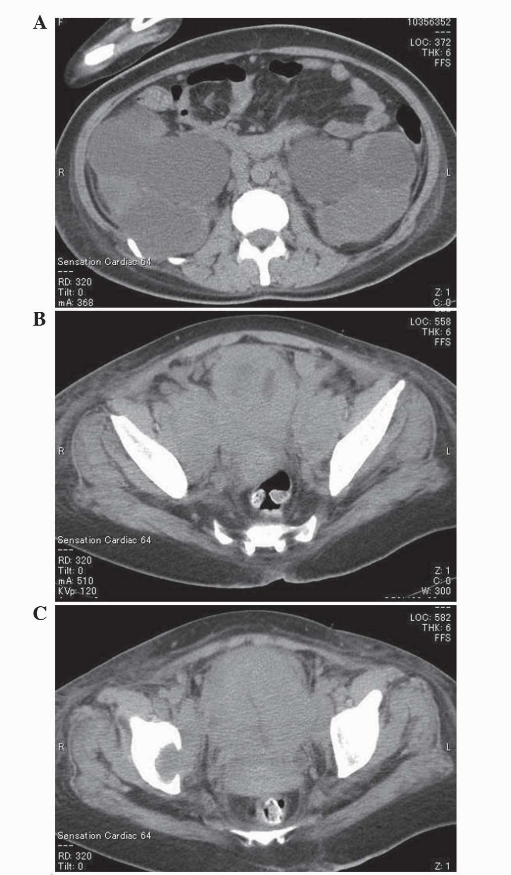
elevated, as described below. Consequently, computed tomography
scans were performed, revealing a massive bladder tumor, multiple
bone metastases (Th 9 spine, right coxal bone and left proximal
femur), multiple enlarged lymph nodes in the retroperitoneum and
pelvis, and bilateral hydronephrosis (Fig. 1).
The levels of serum albumin had decreased to 2.0
g/dl (normal range, 3.8–5.0 g/dl), while those of alkaline
phosphatase had increased to 581 IU/l (normal range, 100–325 IU/l).
C-reactive protein was 15.2 mg/dl (normal value, <0.3 mg/dl).
Laboratory data also indicated marked leukocytosis and
hypercalcemia [white blood cell count, 107,000/mm3
(neutrophils, 91.5%; lymphocytes, 2.0%) (normal range,
3,300–9,000/mm3); serum calcium, 14.4 mg/dl (normal
range, 8.5–10.5 mg/dl)]. The serum levels of G-CSF and PTHrP were
found to be elevated [G-CSF, 77.1 pg/ml (normal range, 0–39.0
pg/ml); PTHrP, 11.1 pmol/l (normal value, <1.1 pmol/l)].
Controlling the calcium level was challenging, despite the
administration of bisphosphonates and calcitonin.
The following tumor markers were found to be
elevated: Serum carbohydrate antigen (CA)19–9, 63 U/ml (normal
value, <37 U/ml); CA125, 376 U/ml (normal value, <35 U/ml);
carcinoembryonic antigen, 6.3 ng/ml (normal range, 0–3.4 ng/ml);
cytokeratin 19 fragment, 12.0 ng/ml (normal range, 0–2.8 ng/ml);
squamous cell carcinoma (SCC) antigen, 95 ng/ml (normal range,
0–1.5 ng/ml). The levels of α-fetoprotein, neuron-specific enolase
and pro-gastrin-releasing peptide levels were within the normal
limits.
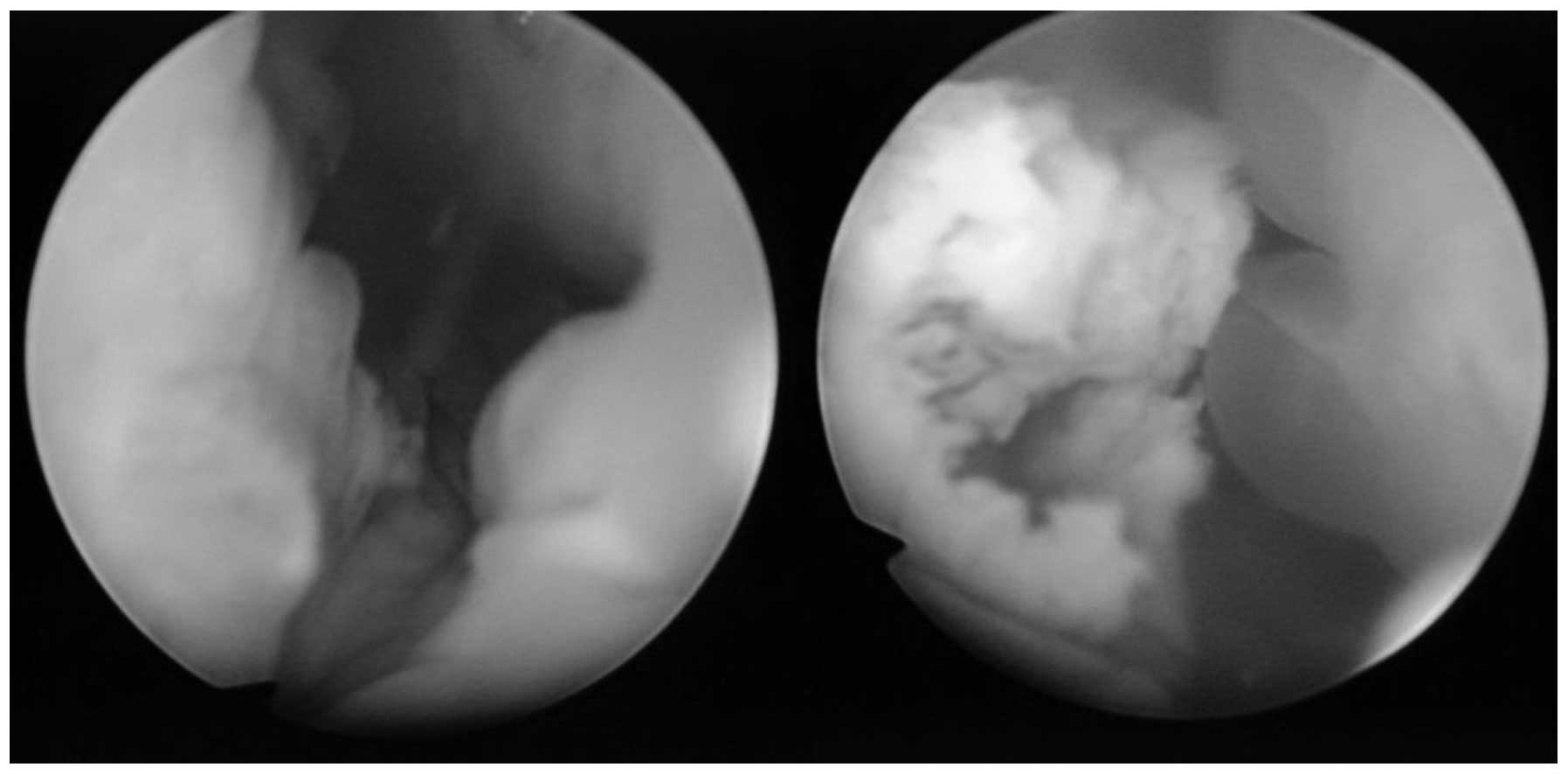
Subsequently, transurethral resection of the bladder
tumor was performed. According to cystoscopic findings, the tumor
appeared to be a predominantly non-papillary submucosal tumor with
a partially papillary appearance. The lumen area had become
constricted by the submucosal tumor, which occupied most of the
inner surface of the bladder (Fig.
2).
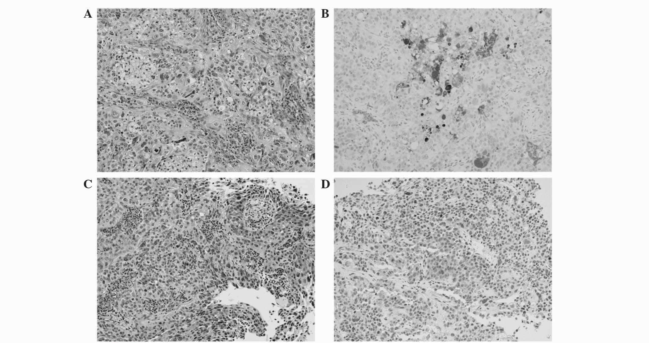
Serial sections of 4 µm were cut from
formalin-fixed, paraffin-embedded blocks of resected tissue.
Pathological examination findings from the resected specimens
revealed necrotic, invasive, poorly-differentiated urothelial
carcinoma (Fig. 3A and C). For
immunohistochemical analysis, sections were deparaffinized in
xylene, rehydrated through decreasing grades of alcohol to water,
and subjected to heat in target retrieval buffered solution (Dako
Japan Co., Ltd., Tokyo, Japan) in a microwave oven for 10 min.
Endogenous peroxidase activity was quenched in 3% hydrogen peroxide
solution in distilled water and sections were incubated with the
following primary antibodies in a wet chamber and irradiated
intermittently for 15 min: Mouse monoclonal anti-granulocyte-colony
stimulating factor (1:100; Immuno-Biological Laboratories Co.,
Ltd., Gunma, Japan) and rabbit polyclonal PTHrP (1:50; Atlas
Antibodies, Stockholm, Sweden). After rinsing in Tris-buffered
saline (TBS) containing 0.1% Tween 20, the sections were incubated
with Envision+/horseradish peroxidase (Dako Japan Co., Ltd.), and
irradiated intermittently for 15 min. After rinsing in TBS,
3,3′-diaminobenzidine was applied as a substrate for the secondary
antibody. Finally, sections were counterstained with hematoxylin
for 1 min, dehydrated through graded alcohols, and cleared in
xylene. Omission of the primary antibody was used as a negative
control. Immunohistochemical testing revealed positive staining for
G-CSF and PTHrP. Fig. 3B shows
immunohistochemical staining for G-CSF, which was focally or
diffusely positive. Scattered strong immunoreactivity was observed
with PTHrP (Fig. 3D).
The disease developed rapidly and the patient's
consciousness deteriorated, presumably due to severe hypercalcemia.
Subsequently, gemcitabine/cisplatin-based systemic chemotherapy was
administered. Although temporary, the patient's serum calcium level
decreased to 8.0 mg/dl and the white blood cell count to
1,160/mm3, and she seemed to regain her consciousness
following chemotherapy; however, 9 days from the initiation of
chemotherapy, the patient developed sudden oculogyric crisis due to
hemorrhagic cerebral infarction. Heparin therapy was inevitably
terminated to control cerebral hemorrhage and, thereafter, her
neurological disturbance advanced rapidly. The patient succumbed to
respiratory failure due to brainstem infarction 15 days from the
initiation of chemotherapy.
Discussion
Trousseau's syndrome is a well-known paraneoplastic
syndrome in which thromboembolism is caused by a malignancy; it was
first reported by Armand Trousseau in 1865 (1). Although no standard definition of the
syndrome has been established, certain scientists have defined it
as ‘hypercoagulability syndrome associated with cancer’ or
‘malignancy-related thromboembolism’ (1,2).
Trousseau's syndrome occasionally occurs in patients with
carcinomas of the pancreas, ovaries or lungs (3); however, to the best of our knowledge,
this is the first reported case of Trousseau's syndrome occurring
in a patient with bladder cancer. It may have been overlooked
partly due to the lack of biochemical markers for this syndrome and
partly due to the fact that it has not yet been conclusively
defined. The development of a novel marker to diagnose Trousseau's
syndrome is required.
Several mechanisms have been proposed to explain the
occurrence of hypercoagulation in patients with malignancy,
including thromboplastin-like substances, fibrin deposition, direct
factor X activation by tumor proteases, tissue factor, cysteine
protease, tumor hypoxia, tumor-induced inflammatory cytokines and
carcinoma-associated mucins (1,8–10). Among them, mucins are thought to
trigger this syndrome (9,10). The serum tumor markers CA19-9 (also
known as MUC-1/Y), CA125 (also known as MUC-16) and CA15-3 (also
known as MUC-1) are well known tumor-associated mucins; these are
large, heavily glycosylated molecules that are resistant to
denaturation, boiling and proteases (11). Carcinoma-associated mucins are thought
to induce platelet-rich microthrombi with increasing tissue factor
and selectins, resulting in intravascular hypercoagulation
(1,12). Indeed, Trousseau's syndrome is
occasionally associated with mucin-producing adenocarcinoma
(1,9,13).
Although the present case lacks the typical findings of
mucin-producing carcinoma, such as intracytoplasmic mucin or
extracellular mucin pools, the serum tumor markers CA19-9 and CA125
were found to be markedly elevated.
The expression CA125 and CA15-3 is believed to be
correlated with the incidence of thromboembolism in cancer patients
(4,14). In the present case, in addition to the
elevated CA125 levels, the elevated CA19-9 levels may also have
been involved in the formation of thromboembolism. In undiagnosed
patients with occult cancer who develop thromboembolism, mucin
tumor markers may indicate the origin of mucin-producing
carcinoma.
Controlling the causative tumor and providing
immediate systemic anticoagulation is crucial for the treatment of
Trousseau's syndrome (15). Although
there is no established evidence regarding the effects of
anticoagulant treatment on Trousseau's syndrome, heparin appears to
be more effective than vitamin K antagonists (1,3,16). In addition to the activation of
antithrombin, heparin cofactor II and protein C inhibitor, heparin
interrupts the binding of selectins, which mediates tumor-mucin
interactions (1). In the present
case, even though anticoagulant therapies with heparin were
performed, the thromboembolic events could not be adequately
controlled and mortality could not be avoided. In addition to the
use of anticoagulation, it is also very important to treat the
underlying malignancy.
Leukocytosis and hypercalcemia are paraneoplastic
syndromes associated with high-grade aggressive tumors.
Hypercalcemia associated with bladder cancer is not uncommon.
Humoral hypercalcemia of malignancy, first described by Albright in
1941 (6), is the most common
paraneoplastic syndrome and is caused by excessive production of
PTHrP, which is released from the tumor cells. PTHrP, whose
amino-terminal is identical to that of parathyroid hormone, binds
to the parathyroid hormone receptor and may stimulate osteoclastic
bone resorption and produce hypercalcemia (7).
Certain aggressive tumors have been reported to
produce G-CSF, resulting in marked leukocytosis without any
infection (17,18). G-CSF is a hematopoietic growth factor
that stimulates granulocyte proliferation and differentiation
(17). Leukocytosis in association
with bladder cancer is relatively rare, with ~35 cases of
leukocytosis in bladder cancer due to G-CSF production reported in
the literature. G-CSF-producing bladder cancer has a poor prognosis
(17).
A few bladder tumors simultaneously producing G-CSF
and PTHrP have been reported (18).
The majority of tumors producing G-CSF and PTHrP are SCCs, possibly
due to the fact that they are both produced by keratinocytes.
Although the present case lacked the typical findings of SCC in the
resected specimen, the patient's serum SCC antigen was markedly
elevated.
In summary, the present study reported the rare case
of a patient presenting with various paraneoplastic syndromes,
including Trousseau's syndrome, which were associated with a
bladder cancer. Occult visceral malignancy should be considered in
patients with unexplained thromboembolism.
Glossary
Abbreviations
Abbreviations:
|
G-CSF
|
granulocyte colony-stimulating
factor
|
|
PTHrP
|
parathyroid hormone-related
protein
|
References
|
1
|
Varki A: Trousseau's syndrome: Multiple
definitions and multiple mechanisms. Blood. 110:1723–1729. 2007.
View Article : Google Scholar : PubMed/NCBI
|
|
2
|
Bick RL: Cancer-associated thrombosis. N
Engl J Med. 349:109–111. 2003. View Article : Google Scholar : PubMed/NCBI
|
|
3
|
el-Shami K, Griffiths E and Streiff M:
Nonbacterial thrombotic endocarditis in cancer patients:
Pathogenesis, diagnosis and treatment. Oncologist. 12:518–523.
2007. View Article : Google Scholar : PubMed/NCBI
|
|
4
|
Tesselaar ME, Romijn FP, Van Der Linden
IK, Prins FA, Bertina RM and Osanto S: Microparticle-associated
tissue factor activity: A link between cancer and thrombosis? J
Thromb Haemost. 5:520–527. 2007. View Article : Google Scholar : PubMed/NCBI
|
|
5
|
Masubuchi H, Maeno T, Uchida M, Kono S,
Suzuki M, Takemura M, Yamaguchi A, Yamaguchi K, Kanbe M, Kitahara
S, et al: A case of Trousseau syndrome caused by pulmonary
adenocarcinoma that was controlled for one year and 10 months with
thrombosis treatment using an EGFR tyrosine kinase inhibitor and
chemotherapy. Respir Med Case Rep. 15:101–105. 2015.PubMed/NCBI
|
|
6
|
Albright F: Case records of the
Massachusetts General Hospital (case 27461). N Engl J Med.
225:789–791. 1941.
|
|
7
|
Wolchok JD, Herr HW and Kelly WK:
Localized squamous cell carcinoma of the bladder causing
hypercalcemia and inhibition of PTH secretion. Urology. 51:489–491.
1998. View Article : Google Scholar : PubMed/NCBI
|
|
8
|
Dvorak HF: Thrombosis and cancer. Hum
Pathol. 18:275–284. 1987. View Article : Google Scholar : PubMed/NCBI
|
|
9
|
Wahrenbrock M, Borsig L, Le D, Varki N and
Varki A: Selectin-mucin interactions as a probable molecular
explanation for the association of Trousseau syndrome with mucinos
adenocarcinomas. J Clin Invest. 112:853–862. 2003. View Article : Google Scholar : PubMed/NCBI
|
|
10
|
Shao B, Wahrenbrock MG, Yao L, David T,
Coughlin SR, Xia L, Varki A and McEver RP: Carcinoma mucins trigger
reciprocal activation of platelets and neutrophils in a murine
model of trousseau syndrome. Blood. 118:4015–4023. 2011. View Article : Google Scholar : PubMed/NCBI
|
|
11
|
Carraway KL, Fregien N, Carraway KL III
and Carraway CA: Tumor sialomucin complexes as tumor antigens and
modulators of cellular interactions and proliferation. J Cell Sci.
103:299–307. 1992.PubMed/NCBI
|
|
12
|
Varki NM and Varki A: Heparin inhibition
of selectin-mediated interactions during the hematogenous phase of
carcinoma metastasis: Rationale for clinical studies in humans.
Semin Thromb Hemost. 28:53–66. 2002. View Article : Google Scholar : PubMed/NCBI
|
|
13
|
Tachihara M, Nikaido T, Wang X, Sato Y,
Ishii T, Saito K, Sekine S, Tanino Y, Ishida T and Munakata M: Four
cases of Trousseau's syndrome associated with lung adenocarcinoma.
Intern Med. 51:1099–1102. 2012. View Article : Google Scholar : PubMed/NCBI
|
|
14
|
Jovin TG, Boosupalli V, Zivkovic SA,
Wechsler LR and Gebel JM: High titers of CA-125 may be associated
with recurrent ischemic strokes in patients with cancer. Neurology.
64:1944–1945. 2005. View Article : Google Scholar : PubMed/NCBI
|
|
15
|
Sack GH Jr, Levin J and Bell WR:
Trousseau's syndrome and other manifestations of chronic
disseminated coagulopathy in patients with neoplasms: Clinical,
pathologic, and therapeutic features. Medicine (Baltimore).
56:1–37. 1977. View Article : Google Scholar : PubMed/NCBI
|
|
16
|
Walsh-McMonagle D and Green D:
Low-molecular-weight heparin in the management of Trousseau's
syndrome. Cancer. 80:649–665. 1997. View Article : Google Scholar : PubMed/NCBI
|
|
17
|
Kumar AK, Satyan MT, Holzbeierlein J,
Mirza M and Van Veldhuizen P: Leukemoid reaction and autocrine
growth of bladder cancer induced by paraneoplastic production of
granulocyte colony-stimulating factor-a potential neoplastic
marker: A case report and review of the literature. J Med Case Rep.
8:1472014. View Article : Google Scholar : PubMed/NCBI
|
|
18
|
Hirasawa K, Kitamura T, Oka T and
Matsushita H: Bladder tumor producing granulocyte
colony-stimulating factor and parathyroid hormone-related protein.
J Urol. 167:21302002. View Article : Google Scholar : PubMed/NCBI
|