Introduction
Ovarian cancer is a common malignant tumor of the
female genital organs, and has the highest mortality rate among all
gynecological types of cancer (1).
After many years of research, there is more in-depth understanding
of the syndrome; nevertheless, no significant decrease in ovarian
cancer mortality has been observed. Thus, the identification of new
and more effective drug targets or therapeutic agents is imperative
(2).
Paclitaxel is an important therapeutic drug
primarily used against ovarian cancer, breast cancer, lung cancer,
colorectal cancer, melanin tumor, head and neck cancer, lymphomas
and brain tumors with certain curative effect (3). On the other hand, curcumin (CUR) is a
phytochemical with hypolipidemic, antitumor, anti-inflammatory and
antioxidation effects (4).
In the present study, the preparation was loaded
with PLGA-phospholipid nanoparticles of paclitaxel and CUR. The
combination resulted in an increase in solubility and stability.
CUR was able to overcome the multidrug resistance (MDR) of tumor
cells, enhanced the amount of paclitaxel in tumor cells and thus
improved the antitumor activity.
Materials and methods
Preparation of PLGA-phospholipid-PEG
nanoparticles
The phospholipids and DSPE-PEG-COOH soluble in the
mass fraction of 4% of the ethanol solution were prepared with the
concentration of 1 mg/ml solution. The solution of phospholipid (1
mg/ml) and DSPE-PEG-COOH solution (1 mg/ml) was dissolved in water
in a heated (65°C) water bath for 3 min. The PLGA acetonitrile
solution (10 mg/ml) and drug solution (1 mg/ml) were mixed in
acetonitrile, under continuous magnetic stirring for 10 min.
Subsequently, the organic phase was then quickly injected into the
aqueous phase. This resulted in the formation of polymer
phospholipid hybrid nanoparticles. The nanoparticles were removed,
and centrifuged at 800 × g for 10 min. The upper nano suspension
was retained for the experiment.
Establishment of the MDR cell line
model
To prepare the culture cryopreserved A2780 cells
were transferred from a −80°C deep freezer to a 37°C water bath,
for thawing and melting. Frozen storage tubes were opened after the
addition of 75% alcohol for disinfection. The cell suspension was
then transferred to the centrifuge tube, 5-fold the volume of
culture medium was added, the cells were dispersed with gentle
pipetting, and cell culturing continued in a 37°C incubator (5%
CO2, relative humidity of 90). When the cells adhered to
the flask wall, the medium was aspirated off and phosphate-buffered
saline (PBS) was used to wash the medium cell surface. It was
followed by the addition of 2 ml trypsin digestion liquid. After
uniform suspension, the cells were passaged.
A2780/ADM-resistant cell lines with stable
resistance to ADM were induced by stepwise increase of ADM
concentrations. A2780 cells were cultured according to the method
mentioned above, and ADM was added to the culture medium. The
initial concentration was 20 nM, and the concentration of taxol in
the culture medium was increased in the following order: 40, 100
and 200 nM. Each concentration was stable for three generations.
Cell growth, morphology and drug resistance were regularly
investigated, and the cells of stable passages, i.e., A2780/ADM
cells, were considered resistant.
Main reagents
The main reagents used were: PLGA (Sigma-Aldrich,
St. Louis, MO, USA); DSPE-PEG-COOH (Avanti Polar Lipids, Inc.,
Alabaster, AL, USA); phospholipid (MP Biomedicals, LLP, Santa Ana,
CA, USA); anti-actin (β-actin) and anti-P-glycoprotein (P-gp)
antibody (CST, Beverly, MA, USA); and a BCA protein assay kit
(Beyotime Institute of Biotechnology, Shanghai, China).
Structure elucidation of
PLGA-phospholipid-PEG nanoparticles
The particle size and zeta potential (ζ-potential)
were measured using a dynamic light scattering instrument (Wyatt
Technology, Goleta, CA, USA, and the shape and surface morphology
of the nanoparticles were observed using a transmission electron
microscope (Philips, Eindhoven, The Netherlands).
Encapsulation efficiency of PLGA-phospholipid-PEG
nanoparticles was determined by the ultrafiltration centrifugation
method. The entrapment efficiency of the drug was calculated
according to the formula: E (%) = (dosage - free drug
content)/dosage × 100%.
Release rate of PLGA-phospholipid-PEG
nanoparticles
The free paclitaxel and nanoparticle suspension of
paclitaxel with CUR was placed in the dialysis bag (10 kDa
molecular weight interception). It was dispersed in a pH 7.4 PBS
buffer followed by incubation at 37°C. It was then sampled with
fresh buffer at time intervals of 0, 2, 4, 6, 8, 12, 24, 48 and 72
h. The HPLC method was used to study the release rates.
Western blot analysis for the
determination of P-gp protein expression in cells
The free paclitaxel, and the combination of drugs
were incubated in the cells for 24 h. The cells were then digested
and the total protein was extracted. The proteins were separated by
polyacrylamide gel electrophoresis and transferred onto the
cellulose membrane. The cellulose membrane, the rabbit polyclonal
P-gp antibody (Abcam, Cambridge, MA, USA; catalog no. ab103477;
dilution: 1:500), and the mouse monoclonal β-actin antibody (Abcam;
catalog no. ab8226; dilution: 1:500) were incubated at 4°C. At
25°C, the cells were incubated with two levels of antibody for 2 h,
and the protein activity was determined. This method was applied
for the establishment of a MDR cell model and the MDR protein
expression measurement in the cell incubation experiments of
nanoparticles and free drugs.
Statistical analysis
SPSS20.0 software (SPSS Chicago, Ill) SPSS 19.0
software (IBM, Armonk, NY, USA) was used for statistical analysis.
Quantitative data were presented as mean ± standard deviation.
Comparison between groups was carried out using one-way ANOVA test
followed by the post hoc test (LSD). Percentage (%) was used to
express the enumeration data and the Chi-square test was used for
data analysis. Comparisons between ranked data were performed using
the non-parametric rank sum test. P<0.05 indicated a
statistically significant difference.
Results
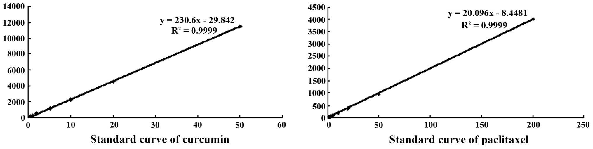
Establishment of analytical
method
The contents of CUR and docetaxel were measured
using the HPLC method; the two standard curves are shown in
Fig. 1. CUR and paclitaxel were
within the linear range of 0–50 and 0–200 µg/ml. The concentration
and peak area showed good correlation.
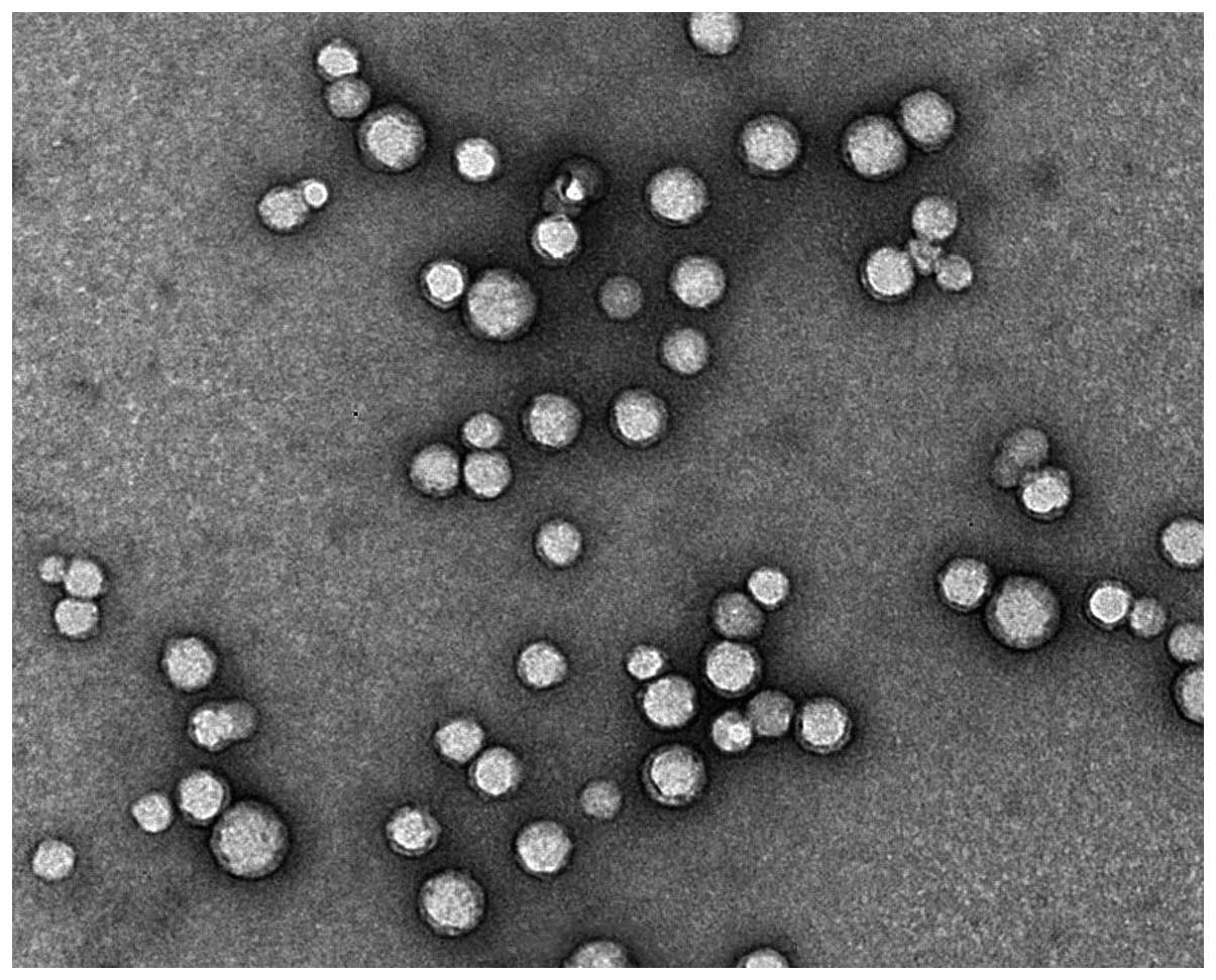
Measurement of the morphology,
particle size and ζ-potential of PLGA-phospholipid-PEG
nanoparticles
The morphology of the nanoparticles was measured
using a transmission electron microscope (Fig. 2). The results showed that the size of
the nanoparticles formed by the self-assembly of PLGA and
phospholipids in solution was uniform and the shape of the
nanoparticles was uniform.
The particle size and ζ-potential of nanoparticles
were determined by the dynamic light scattering method (Table I). The prepared CUR, paclitaxel and
double drug-loaded nanoparticles were <100 nm, and PDI was
<0.2, which showed that the size distribution of the
nanoparticles was small, and suitable for the size of the
nanoparticles. The ultrafiltration centrifugation method was used
to measure the entrapment efficiency of the nanoparticles, which
were all >80%, showing the two drugs can more be completely
encapsulated in nano materials. Additionally, the ζ-potential was
approximately −30 mV.
 | Table I.The particle size, zeta (ζ),
encapsulation efficiency and PDI value of the nanoparticles. |
Table I.
The particle size, zeta (ζ),
encapsulation efficiency and PDI value of the nanoparticles.
| Formulation | Particle size
(nm) | ζ-potential (mV) | Drug encapsulation
(%) | PDI |
|---|
| CUR
nanoparticles | 89.4 | −29.8 | 90.41 | 0.13 |
| Paclitaxel-loaded
nanoparticles | 92.1 | −32.5 | 84.44 | 0.10 |
| Double drug-loaded
nanoparticles | 94.3 | −31.6 | 86.87 | 0.12 |
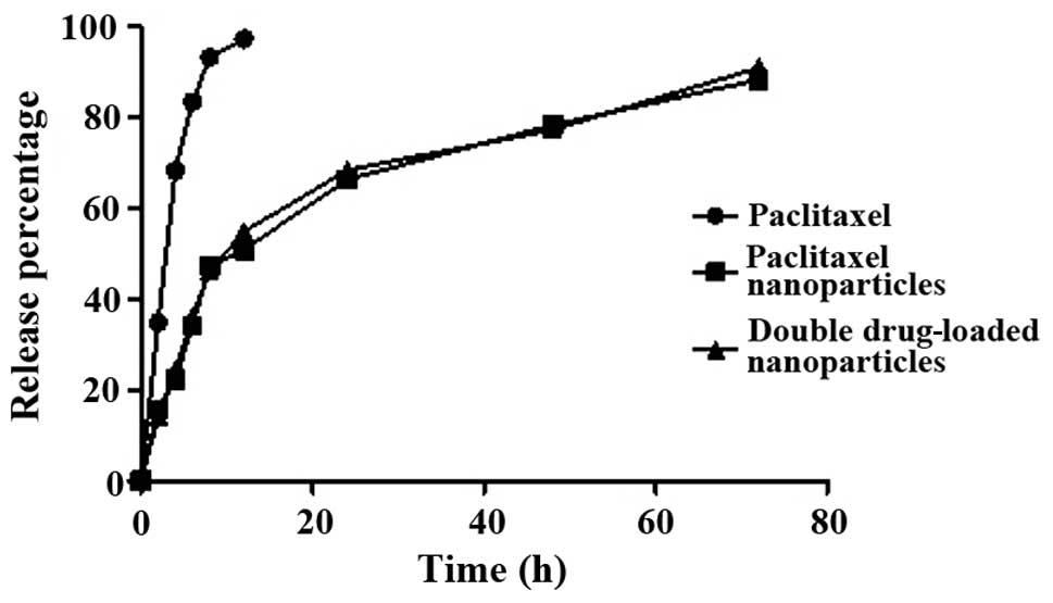
Release of PLGA-phospholipid-PEG
nanoparticles
The release curves of free paclitaxel,
paclitaxel-loaded nanoparticles and double drug-loaded
nanoparticles in the PBS buffer solution of pH 7.4 were studied.
The results are shown in Fig. 3. Free
paclitaxel was released ~70% in 4 h, and almost completely in 8 h.
On the other hand, nanoparticles were released only ~50% in 24 h,
while they were released up to 80% in 48 h and up to 90% in 72 h,
confirming the slow release effect. Furthermore, the release curves
of two types of nanoparticles were without any significant
difference.
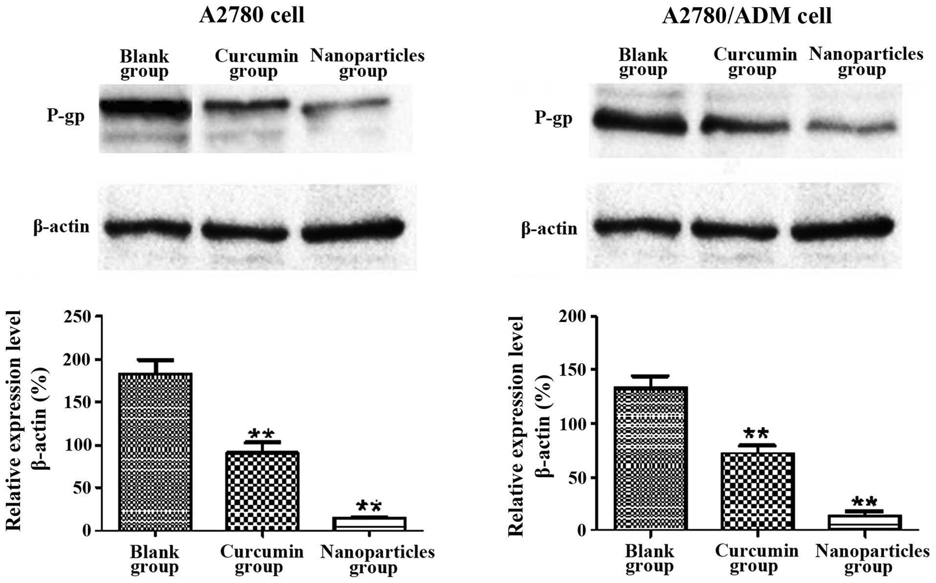
Expression of P-gp protein was
determined by western blot analysis
Western blotting was used to determine the
expression intensity of P-gp in the A2780 and A2780/ADM cells
(Fig. 4). For the two cell lines, the
strips of free CUR and CUR nanoparticles were lighter than the
blank ones, showing CUR was able to reverse the MDR of the two
cells. Compared with free drugs, the downregulation effect of
nanoparticles on P-gp was more obvious. Furthermore, the CUR
nanoparticles had a significant downregulation effect on P-gp cells
of A2780/ADM cells in comparison with A2780 cells. The results
clearly indicated the ability of CUR nanoparticles to reverse the
MDR of tumor cells, in order that the cytotoxic drugs be more
useful for the drug efficacy.
Discussion
New chemotherapy drugs continue to emerge, and
chemotherapy regimens continue to improve. The widespread tumor
MDR, has mainly remained the causative reason responsible for the
failure of cancer therapy. It is a significant problem in chemical
therapy and interventional treatment of tumor (5). For example, for ovarian cancer patients,
more than 70% of patients initially are resistant to the treatment
of drugs from Chinese fir, leading to relapse (6). The protein associated with drug efflux
is the main cause of MDR in tumor cells. This efflux type of
ATP-binding cassette transporter used ATP to produce energy for
decomposition, remove the anticancer drug from inside the cell to
outside to make antitumor drug concentration less than that
required for killing of cells, leading to drug resistance (7). The ATP-binding cassette transporter
family includes P-gp, MDR-associated protein (MRP), breast cancer
resistance protein (BCRP), and the B-cell leukemia/lymphoma gene 2
protein (Bcl-2) (8).
Reverse MDR was used to reduce the probability of
failure of chemotherapy, with a high clinical value. MDR reversal
has become one of the important directions in the research of
chemotherapeutic drugs at home and abroad. Thus, a new type of nano
drug loading system with the ability of coloading MDR modulators
and chemotherapy drugs was selected (9).
The PLGA-phospholipid hybrid nanodrug loading system
is a new type of core shell structure nanoparticle. The main
advantages of the drug loading system in comparison to simple PLGA
nanoparticles as well as liposome are: i) The core is composed of a
biodegradable PLGA material, which is suitable for carrying poor
water insoluble drugs, and can slowly release drugs at a constant
speed; ii) the invisible material forms a hydrophilic outer shell
(phospholipid-PEG), which prevents the nanoparticles from being
swallowed by the immune system, and enhances the half-life of the
nanoparticles in the body circulation; iii) phospholipid monolayer
between the hydrophobic core and hydrophilic shell prevents drug
leaking from the nanoparticles, while reducing the infiltration of
moisture in the nanoparticles, thus, it has a high drug entrapment
efficiency and drug release effect. iv) The surface of the
nanoparticles can improve the active targeting of drugs by
modifying the nucleic acid ligands or antibodies (10–12).
Overcoming MDR through a drug delivery system is an important and
effective reversal strategy in MDR. The drug carriers that have
shown the ability to overcome the MDR of tumor cells included
liposomes, micelles and nanoparticles. Drugs were wrapped in the
carrier, could avoid the identification, binding and efflux of
MDR-related proteins, in order that the drug was able to accumulate
in the cell, without altering the in vivo disposal process
of the drug. In addition, the drug carrier increased the drug
concentration of the tumor tissue through the target function to
overcome the MDR (13–15).
The PLGA phospholipid hybrid nanoparticles carrier
delivery system has been used to successfully encapsulate
anticancer drugs, including paclitaxel, and docetaxel, and examine
the structure of the nanoparticles and in vitro
cytotoxicity. Previous findings have shown that CUR has a variety
of pharmacological effects such as antitumor, anti-inflammatory,
antiviral, and antioxidant effects. Previous studies have indicated
that CUR is effective in inhibiting the three major ATP-binding
cassette transporters, including MDR1, MRP1, and ABCG2 (16). The anticancer activity of CUR occurs
mainly due to its ability to block the transcription factor NF-κB,
a type of regulator of inflammation, cell proliferation, apoptosis
and drug resistance, that can resist apoptosis and regenerate tumor
cells. When CUR was combined with other active drugs, the
pleiotropic effect of CUR was conducive to enhancing the biological
utilization rate of drug activity in tumor tissue, and improving
the level of intracellular drug activity. CUR water solubility was
reduced albeit with a fast metabolism and low bioavailability.
Previous findings have shown that nano preparation is able enhance
the cytotoxicity of different tumor cell lines more than the
prototype drug (17,18). Paclitaxel is increasingly used in the
clinical treatment of breast cancer, non-small cell lung cancer,
pancreatic cancer, soft tissue sarcoma, head and neck cancer,
gastric cancer, ovarian cancer and prostate cancer (19). Paclitaxel has an antitumor effect of
broad spectrum, but almost has no inhibitory activity in MDR tumors
with P-gp overexpression, primarily due to its being a good
substrate for P-gp. Combination therapy was crucial in the
treatment of cancer (20).
Since most of the pharmacological activity of
anticancer drugs have MDR, MDR protein inhibitors with cytotoxic
drugs are loaded in the novel carrier systems. Thus, these novel
systems overcome MDR protein of tumor cells and at the same time,
elevate drug concentrations in tumor cells. Therefore, we selected
this model of the combination of CUR and paclitaxel. The dual
drug-loaded PLGA phospholipid hybrid nano carrier delivery system
overcame MDR and improved the efficacy of chemotherapeutic drugs.
Additionally, the high specificity of nanoparticles was beneficial
in minimizing the toxic and side effects of chemotherapeutic drugs
in normal tissues.
References
|
1
|
Anton-Culver H, Cohen PF, Gildea ME and
Ziogas A: Characteristics of BRCA1 mutations in a population-based
case series of breast and ovarian cancer. Eur J Cancer.
36:1200–1208. 2000. View Article : Google Scholar : PubMed/NCBI
|
|
2
|
Bell DA: Origins and molecular pathology
of ovarian cancer. Mod Pathol. 18(Suppl 2): S19–S32. 2005.
View Article : Google Scholar : PubMed/NCBI
|
|
3
|
Stone GW, Ellis SG, Cox DA, Hermiller J,
O'Shaughnessy C, Mann JT, Turco M, Caputo R, Bergin P, Greenberg J,
et al: TAXUS-IV Investigators: A polymer-based, paclitaxel-eluting
stent in patients with coronary artery disease. N Engl J Med.
350:221–231. 2004. View Article : Google Scholar : PubMed/NCBI
|
|
4
|
Duvoix A, Blasius R, Delhalle S,
Schnekenburger M, Morceau F, Henry E, Dicato M and Diederich M:
Chemopreventive and therapeutic effects of curcumin. Cancer Lett.
223:181–190. 2005. View Article : Google Scholar : PubMed/NCBI
|
|
5
|
Fojo A, Hamilton TC, Young RC and Ozols
RF: Multidrug resistance in ovarian cancer. Cancer. 60(Suppl 8):
2075–2080. 1987. View Article : Google Scholar : PubMed/NCBI
|
|
6
|
Gillet JP, Efferth T and Remacle J:
Chemotherapy-induced resistance by ATP-binding cassette transporter
genes. Biochim Biophys Acta. 1775:237–262. 2007.PubMed/NCBI
|
|
7
|
Shaffer BC, Gillet JP, Patel C, Baer MR,
Bates SE and Gottesman MM: Drug resistance: still a daunting
challenge to the successful treatment of AML. Drug Resist Updat.
15:62–69. 2012. View Article : Google Scholar : PubMed/NCBI
|
|
8
|
Nabekura T, Yamaki T, Hiroi T, Ueno K and
Kitagawa S: Inhibition of anticancer drug efflux transporter
P-glycoprotein by rosemary phytochemicals. Pharmacol Res.
61:259–263. 2010. View Article : Google Scholar : PubMed/NCBI
|
|
9
|
Duncan R: Polymer conjugates as anticancer
nanomedicines. Nat Rev Cancer. 6:688–701. 2006. View Article : Google Scholar : PubMed/NCBI
|
|
10
|
Panyam J, Sahoo SK, Prabha S, Bargar T and
Labhasetwar V: Fluorescence and electron microscopy probes for
cellular and tissue uptake of poly(D, L-lactide-co-glycolide)
nanoparticles. Int J Pharm. 262:1–11. 2003. View Article : Google Scholar : PubMed/NCBI
|
|
11
|
van Vlerken LE and Amiji MM:
Multi-functional polymeric nanoparticles for tumour-targeted drug
delivery. Expert Opin Drug Deliv. 3:205–216. 2006. View Article : Google Scholar : PubMed/NCBI
|
|
12
|
Kumari A, Yadav SK and Yadav SC:
Biodegradable polymeric nanoparticles based drug delivery systems.
Colloids Surf B Biointerfaces. 75:1–18. 2010. View Article : Google Scholar : PubMed/NCBI
|
|
13
|
Alexis F, Pridgen E, Molnar LK and
Farokhzad OC: Factors affecting the clearance and biodistribution
of polymeric nanoparticles. Mol Pharm. 5:505–515. 2008. View Article : Google Scholar : PubMed/NCBI
|
|
14
|
Baratchi S, Kaur RK, Khoshmanesh K, Vasu
P, Ashok C, Hittu M, Paratt A, Krishnakumar S, Sun X, Sahoo SK, et
al: Promises of nanotechnology for drug delivery to brain in
neurodegenerative diseases. Curr Nanosci. 5:15–25. 2009. View Article : Google Scholar
|
|
15
|
Parveen S and Sahoo SK: Nanomedicine:
Clinical applications of polyethylene glycol conjugated proteins
and drugs. Clin Pharmacokinet. 45:965–988. 2006. View Article : Google Scholar : PubMed/NCBI
|
|
16
|
Ong AT, Serruys PW, Aoki J, Hoye A, van
Mieghem CA, Rodriguez-Granillo GA, Valgimigli M, Sonnenschein K,
Regar E, van der Ent M, et al: The unrestricted use of paclitaxel-
versus sirolimus-eluting stents for coronary artery disease in an
unselected population: one-year results of the Taxus-Stent
Evaluated at Rotterdam Cardiology Hospital (T-SEARCH) registry. J
Am Coll Cardiol. 45:1135–1141. 2005. View Article : Google Scholar : PubMed/NCBI
|
|
17
|
Daemen J, Wenaweser P, Tsuchida K, Abrecht
L, Vaina S, Morger C, Kukreja N, Jüni P, Sianos G, Hellige G, et
al: Early and late coronary stent thrombosis of sirolimus-eluting
and paclitaxel-eluting stents in routine clinical practice: data
from a large two-institutional cohort study. Lancet. 369:667–678.
2007. View Article : Google Scholar : PubMed/NCBI
|
|
18
|
Stone GW, Midei M, Newman W, Sanz M,
Hermiller JB, Williams J, Farhat N, Mahaffey KW, Cutlip DE,
Fitzgerald PJ, et al: SPIRIT III Investigators: Comparison of an
everolimus-eluting stent and a paclitaxel-eluting stent in patients
with coronary artery disease: a randomized trial. JAMA.
299:1903–1913. 2008. View Article : Google Scholar : PubMed/NCBI
|
|
19
|
Bierhaus A, Zhang Y, Quehenberger P,
Luther T, Haase M, Müller M, Mackman N, Ziegler R and Nawroth PP:
The dietary pigment curcumin reduces endothelial tissue factor gene
expression by inhibiting binding of AP-1 to the DNA and activation
of NF-kappa B. Thromb Haemost. 77:772–782. 1997.PubMed/NCBI
|
|
20
|
Aggarwal S, Ichikawa H, Takada Y, Sandur
SK, Shishodia S and Aggarwal BB: Curcumin (diferuloylmethane)
down-regulates expression of cell proliferation and antiapoptotic
and metastatic gene products through suppression of IkappaBalpha
kinase and Akt activation. Mol Pharmacol. 69:195–206.
2006.PubMed/NCBI
|