Introduction
Lung cancer is the leading cause of cancer-related
mortality worldwide, with ~1.4 million deaths recorded annually
(1). Approximately 80% of all lung
cancers are classified as non-small-cell lung cancer (NSCLC), which
may be divided phenotypically into two principal subtypes, namely
squamous cell carcinoma (SCC) and adenocarcinoma (AD). SCC and AD
are often treated similarly; however, they differ not only
pathologically, but also in terms of gene expression levels,
clinical manifestation and response to targeted therapeutic agents
(2–4).
Early detection is key to prolonging patient survival; thus, it is
crucial to identify molecular markers that predict prognosis and
design novel treatment strategies for SCC and AD (5).
The B-cell-specific Moloney murine leukemia virus
integration site 1 (BMI1) gene, a member of the polycomb-group
(PcG) proteins, was first isolated as an oncogene that acts
synergistically with c-Myc in the oncogenesis of murine lymphomas
(6,7).
BMI1 functions as a transcriptional repressor of BMI1 and c-Myc
target genes via an epigenetic mechanism (8). In addition, deregulation of BMI1 gene
expression leads to cell proliferation and tumor progression
(8,9).
Aberrant BMI1 expression has been associated with a number of solid
and hematological malignancies (10–18),
including NSCLC (19–28). Although the clinicopathological and
prognostic significance of high BMI1 expression in NSCLC has been
investigated, the results have been inconsistent. For example,
certain studies suggested that BMI1 overexpression is associated
with poor prognosis in SCC (26);
however, Vrzalikova et al (22) found no correlation between BMI1
expression and overall survival (OS) in their cohort, which had a
preponderance of SCCs.
Matrix metalloproteinase-9 (MMP9), a member of the
MMP class of gelatinases, plays a major role in the invasiveness
and metastasis of NSCLC, mainly through specific induction by
vascular endothelial growth factor receptor-1 (VEGFR-1) (29,30). Jiang
et al (31) reported that BMI1
activated nuclear factor-κB (NF-κB) and subsequently upregulated
MMP9 expression, leading to increased migration and invasion of
glioma cells; Li et al (32)
demonstrated that BMI1 contributed to the invasion and metastasis
of hepatocellular carcinoma by increasing MMP9 and VEGF expression
via the phosphoinositide-3-kinase/protein kinase B (PI3K/Akt)
pathway. However, the number of studies on the interaction between
BMI1 and MMP9 in NSCLC progression and metastasis is limited.
In this study, a survival analysis of BMI1, MMP9 and
their combined expression as a marker in lung SCC and AD samples
was performed. The aim was to determine whether the combined high
BMI1 and MMP9 expression levels predict poor survival in SCC. These
results may help elucidate the mechanism underlying tumor
progression and metastasis in patients with NSCLC and ultimately
identify novel diagnostic and/or prognostic markers.
Patients and methods
Patients and histopathology
The study samples consisted of paraffin-embedded
lung tissue samples from 135 patients diagnosed with NSCLC who had
undergone surgery between January, 2008 and January, 2009 at the
Second Affiliated Hospital of Harbin Medical University (Harbin,
China). According to the World Health Organization guidelines
(33), the cases were classified as
SCC or AD. In total, 132 cases were analyzed (AD, n=79; and SCC,
n=53). Of the 132 cases, 10 were well-differentiated, 57 were
moderately and 65 were poorly differentiated. The pathological
tumor stage distribution was I–IV according to the American Joint
Committee on Cancer (34). All the
patients were followed up until October, 2013 or until death. The
median follow-up duration for survivors was 43.15 months (range,
1.30–62.40 months). The patient population comprised 89 men and 43
women (male:female ratio, 2.07:1) and the median age was 61 years
(range, 34–80 years). All the cases representing a spectrum of
NSCLC were retrieved from the records of the Second Affiliated
Hospital of Harbin Medical University. The Ethics Committee of the
hospital granted permission for the study.
Immunohistochemical (IHC)
evaluation
The expression of BMI1 and MMP9 in the 132 patients
with NSCLC was investigated. Paraffin-embedded sections were
processed as previously described (25,27).
Briefly, the sections were deparaffinized in xylene and rehydrated
in a graded series of ethanol. The sections were subsequently
submerged in ethylene diamine tetraacetic acid (pH 8.0) and
autoclaved at 121°C for 5 min to retrieve antigenicity. Endogenous
peroxidase was quenched with 3% H2O2 for 15
min. Following washing with PBS, the sections were incubated with
anti-BMI1 (dilution, 1:80; LS-C98480; LifeSpan Biosciences,
Seattle, WA, USA) and anti-MMP9 (dilution, 1:800; ab38898; Abcam,
MA, USA) antibodies overnight at 4°C. The sections were incubated
with peroxidase-conjugated streptavidin for 30 min and the reaction
products were visualized with the chromogen diaminobenzidine and
counterstained with commercial hematoxylin. The percentage of
positive cells was determined by counting 500 cells in five random
fields per section. Nuclear and cytoplasmic immunostaining was
evaluated using semi-quantitative assessment, in which the
percentage of positive cells was calculated (25). The percentage of positive cells was
determined by counting 500 cells in five random areas per section.
IHC staining of the cells was scored according to the following
criteria: -, no staining; +, 1–10%; ++, 10–50%; +++, 50–75%; and
++++, 75–100% cells stained. The IHC scores for BMI1 and MMP9
expression were classified as low (− and +) and high (++, +++ and
++++).
Statistical analysis
The association between factors was evaluated using
Pearson's χ2 test. Cumulative OS curves were plotted
using the Kaplan-Meier method and log-rank statistics were used to
determine differences between groups. Covariates that remained
significant through the univariate analysis were selected for the
multivariate analysis. Cox regression was used for the multivariate
analysis, with a backward stepwise elimination model. P<0.05 was
considered to indicate statistical significance. Two-sided tests
were used throughout. Statistical calculations were performed using
SPSS 19.0 software (IBM SPSS, Armonk, NY, USA).
Results
BMI1 is overexpressed in NSCLC
tissues
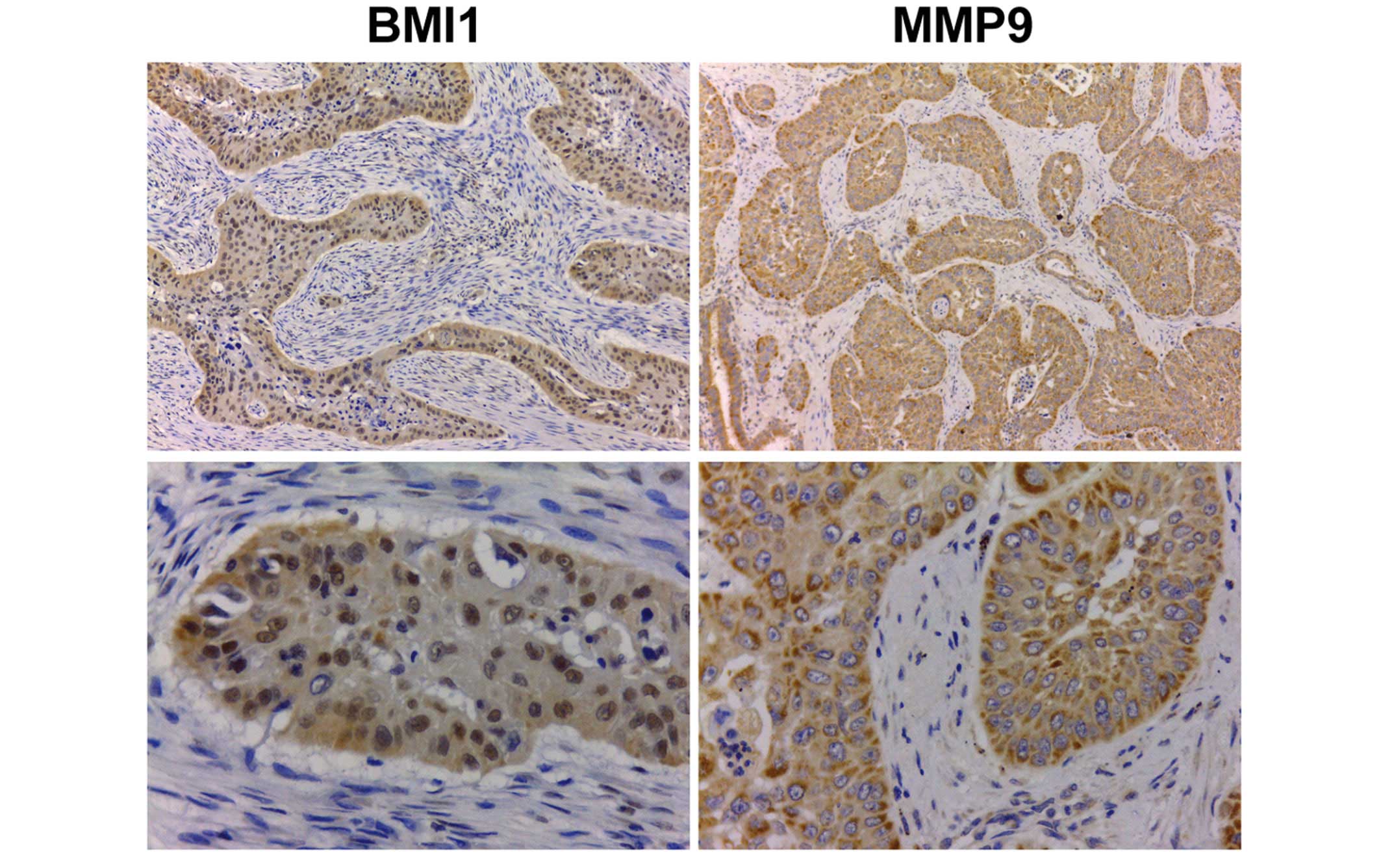
On IHC analysis, the majority of cells in NSCLC
tissues, including SCC and AD cells, were strongly stained,
indicating high expression of BMI1. BMI1 was highly expressed in
95/132 NSCLC cases (72.0%) and 63/79 SCC cases (79.7%) (Fig. 1). The expression of BMI1 in SCC was
higher compared with that in AD (79.7 vs. 60.4%, respectively;
P=0.015). BMI1 was highly expressed in 32/53 (60.4%) AD cases.
Similar to BMI1, MMP9 expression was detected in NSCLC tissues and
its expression level was higher in SCC compared with that in AD
(P=0.012, Fig. 1).
Correlations of BMI1, MMP9 and
combined marker expression with variables in NSCLC samples
BMI1 expression was compared with known
clinicopathological factors and biological markers in the NSCLC
samples. There was a positive correlation between the expression of
BMI1 and that of MMP9 in the SCC samples (χ2=4.241,
P=0.039; Table I). Combined marker
expression in the SCC samples was found to be significantly
correlated with metastasis (χ2=6.392, P=0.014; Table II), but not with other clinical
parameters in all lung cancer or AD cases.
 | Table I.Correlation between BMI1 and MMP9
protein expression in squamous cell carcinoma. |
Table I.
Correlation between BMI1 and MMP9
protein expression in squamous cell carcinoma.
|
| MMP9 expression, n
(%) |
|
|
|---|
|
|
|
|
|
|---|
| BMI1 expression, n
(%) | High | Low | Total | χ2 | P-value |
|---|
| High | 48 (60.8) | 15 (19.0) | 63 (79.8) | 4.241 | 0.039a |
| Low | 8
(10.1) | 8
(10.1) | 16 (20.2) |
|
|
| Total | 56 (70.9) | 23 (29.1) | 79 (100) |
|
|
| Table II.Correlation of BMI1, MMP9 and
combined marker expression levels with clinicopathological
variables in squamous cell carcinoma. |
Table II.
Correlation of BMI1, MMP9 and
combined marker expression levels with clinicopathological
variables in squamous cell carcinoma.
|
|
| BMI1
expression |
| MMP9
expression |
| Combined marker
expression |
|
|---|
|
|
|
|
|
|
|
|
|
|---|
| Variables | All patients
(n=79) | High (n=63) | Low (n=16) | P-value | High (n=56) | Low (n=23) | P-value | High (n=47) | Low (n=32) | P-value |
|---|
| Gender |
|
|
| 0.960 |
|
| 0.275 |
|
| 0.143 |
|
Male | 44 | 35 | 9 |
| 29 | 15 |
| 23 | 21 |
|
|
Female | 35 | 28 | 7 |
| 27 | 8 |
| 24 | 11 |
|
| Age (years) |
|
|
| 0.455 |
|
| 0.760 |
|
| 0.769 |
|
<60 | 46 | 38 | 8 |
| 32 | 14 |
| 28 | 18 |
|
|
≥60 | 33 | 25 | 8 |
| 24 | 9 |
| 19 | 14 |
|
| Smoking |
|
|
| 0.468 |
|
| 0.450 |
|
| 0.116 |
|
Yes | 43 | 33 | 10 |
| 32 | 11 |
| 29 | 14 |
|
| No | 36 | 30 | 6 |
| 24 | 12 |
| 18 | 18 |
|
|
Differentiation |
|
|
| 0.679 |
|
| 0.621 |
|
| 0.465 |
|
Poor | 31 | 24 | 7 |
| 21 | 10 |
| 20 | 11 |
|
|
Moderate/high | 48 | 39 | 9 |
| 35 | 13 |
| 27 | 21 |
|
| Pathological
stage |
|
|
| 0.082a |
|
| 0.312a |
|
| 0.239 |
| I | 41 | 35 | 6 |
| 26 | 15 |
| 23 | 18 |
|
| II | 18 | 11 | 7 |
| 14 | 4 |
| 9 | 9 |
|
|
III/IV | 20 | 17 | 3 |
| 16 | 4 |
| 15 | 5 |
|
| Node/distant
metastasis |
|
|
| 0.248a |
|
| 0.111a |
|
| 0.014b |
|
Yes | 25 | 22 | 3 |
| 21 | 4 |
| 20 | 5 |
|
| No | 54 | 41 | 13 |
| 35 | 19 |
| 27 | 27 |
|
BMI1 protein expression predicts OS in
NSCLC
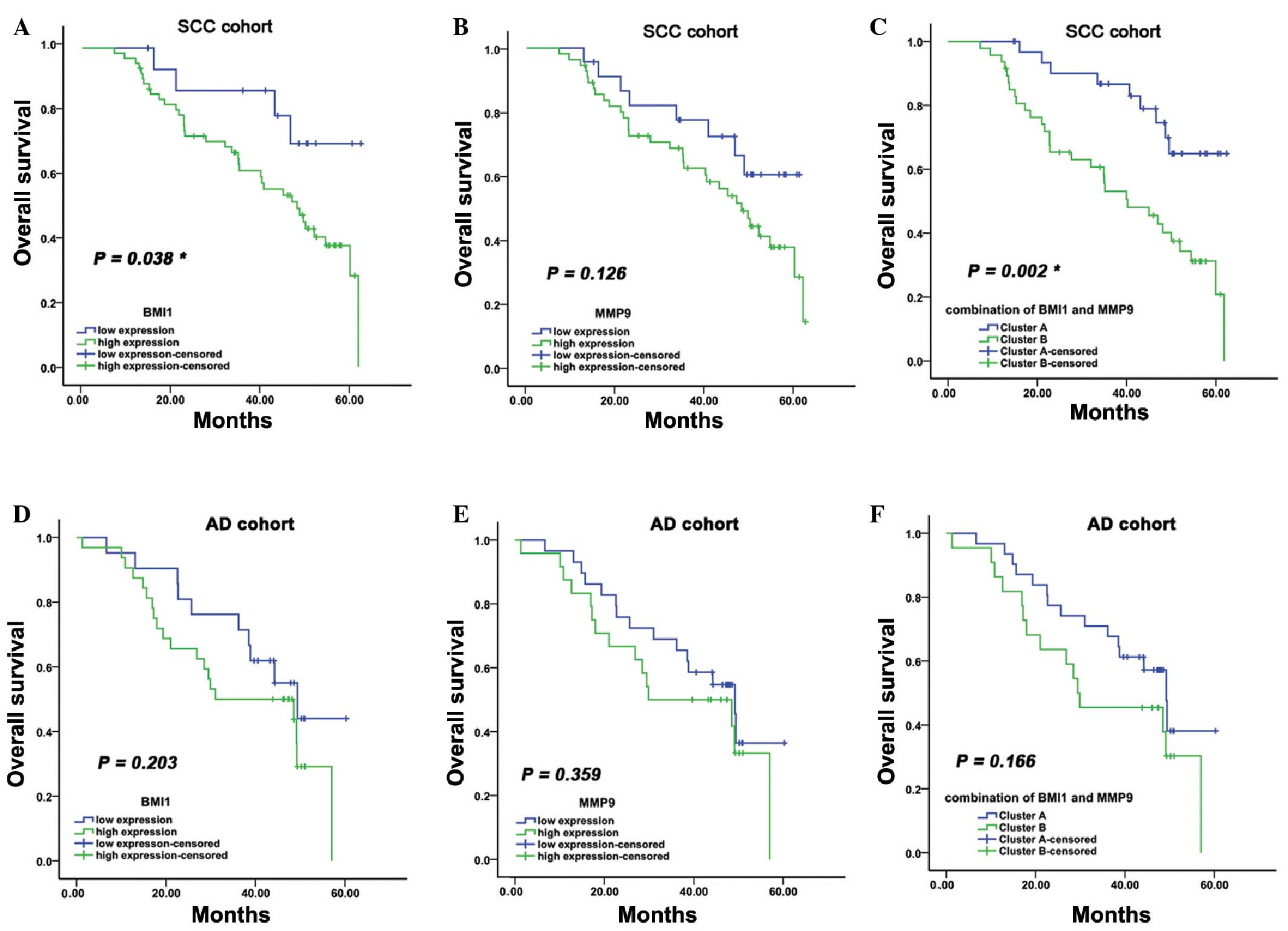
To determine whether BMI1 expression was an
independent prognostic factor for OS in NSCLC, multivariate Cox
regression analyses were performed. Among the immunological
variables analyzed in this study, BMI1 expression was not
associated with clinical outcome in the entire cohort. When
analysis was stratified according to the major histological
subtypes, the results suggested that BMI1 expression was an
unfavorable prognostic factor in SCC (P=0.038; hazard ratio =
2.861; 95% confidence interval: 1.013–8.083; Table III and Fig. 2A). In AD, BMI1 tended to be a negative
prognostic factor for long-term survival, although the difference
was not statistically significant (P=0.203; hazard ratio = 1.633;
95% confidence interval: 0.762–3.501; Fig. 2B).
| Table III.Univariate and multivariate analyses
of overall survival for 79 patients with squamous cell lung
carcinoma. |
Table III.
Univariate and multivariate analyses
of overall survival for 79 patients with squamous cell lung
carcinoma.
|
| Univariate
analysis | Multivariate
analysis |
|---|
|
|
|
|
|---|
| Variables | HR (95% CI) | P-value | HR (95% CI) | P-value |
|---|
| Age (≥60 vs. <60
years) | 1.082
(0.992–1.179) | 0.070 |
|
|
| Gender (male vs.
female) | 0.453
(0.134–1.414) | 0.166 |
|
|
| Smoking status (yes
vs. no) | 1.260
(0.284–5.595) | 0.180 |
|
|
| Differentiation
(poor vs. moderate/high) | 0.233
(0.675–1.287) | 0.233 |
|
|
| Pathological stages
(III/IV vs. II vs. I) | 1.657
(0.889–3.089) | 0.112 |
|
|
| Node/distant
metastasis (yes vs. no) | 2.551
(1.352–4.814) | 0.004a | 17.163
(1.621–5.816) | 0.001a |
| BMI1 (positive vs.
negative) | 2.861
(1.013–8.083) | 0.038a |
|
|
| MMP9 (positive vs.
negative) | 2.157
(0.092–4.888) | 0.066 |
|
|
| Combined markers
(cluster B vs. cluster A) | 3.085
(1.466–6.491) | 0.002a | 2.963
(1.142–7.637) | 0.025a |
BMI1 and MMP9 protein expression
predicts OS in SCC
Although MMP9 protein levels did not predict OS in
NSCLC or its histological subtypes, the combination of high and low
protein expression of BMI1 and MMP9 identified four subgroups with
distinct outcomes in SCC (Fig. 2C).
The combination of high expression of BMI1 and MMP9 (Cluster B) was
associated with shorter OS compared with low BMI1 and high MMP9
expression (Cluster A: High BMI1/low MMP9; low BMI1/high MMP9; and
low BMI1/low MMP9). However, BMI1, MMP9 and combined marker
expression were not significant prognostic factors in AD (Fig. 2D-F).
Discussion
Similar to other cancer types, lung cancer
development is the result of a stepwise progression of malignant
transformation of the normal respiratory epithelium (35). The classification of lung cancer by
Noguchi (35) identifies atypical
adenomatous hyperplasia and cancer in situ as preinvasive
neoplastic lung lesions that serve as precursors of invasive lung
cancer through a progressive transformation into two types,
characterized by survival outcome (35). Well-designed molecular biology
investigations may be used for tumor classification, response
prediction, assigning prognosis or even as therapeutic targets
(36). SCC and AD share a range of
similarities, but there are also differences (2,4,17). Therefore, identification of the roles
of molecular markers in differential prediction of outcome is
important in SCC and AD.
The BMI1 gene, which encodes a PcG protein, was
first isolated as an oncogene that acts synergistically with c-Myc
in the oncogenesis of murine lymphomas (4,6). BMI1
functions as a transcriptional repressor of their target genes via
an epigenetic mechanism (2). A number
of studies have reported that BMI1 is upregulated in a variety of
human malignancies, including lung (19), breast (11), gastric (13), hepatocellular (12) and esophageal (17) cancer, non-Hodgkin lymphoma (10) and cervical (15) cancer, suggesting that BMI1 is a
potential oncogene. As a member of the PcG family, BMI1 plays a
suppressive role, targeting cyclin-dependent kinase inhibitor 2A
(p16INK4A) and cyclin-dependent kinase 2-associated
protein 2 (p14ARF) (21,37,38), and
promotes cell proliferation by suppressing the p16/retinoblastoma
and/or p14ARF/mouse double minute 2/p53 pathways
(39,40). Furthermore, it has been reported that
BMI1 may downregulate transcription of the tumor suppressor
phosphatase and tensin homolog deleted on chromosome 10 (PTEN) via
direct association with the PTEN gene locus (32). BMI1 upregulation also induces
epithelial-to-mesenchymal transition and promotes human SCC
occurrence, aggressiveness, invasion and metastasis by
downregulating E-cadherin and upregulating vimentin expression
(26).
In this study, the expression of BMI1 in NSCLC
samples was investigated. BMI1 was found to be significantly
overexpressed in SCC compared with AD tissues. However, the present
findings are contradictory to those of previous studies (22,25), which
stated that BMI1 expression is not correlated with pathological
type. Vrzalikova et al (22)
reported a trend for higher BMI1 expression in SCC compared with
AD, although the difference was not statistically significant. The
discrepancies across studies may be due to the different
immunohistochemistry protocols used, including antibody dilution,
development time and positivity criteria applied, particularly the
score used to discriminate positivity.
The present study demonstrated that high expression
of BMI1 is associated with shorter OS and indicates that BMI1
expression confers an aggressive phenotype to human SCC. Current
opinions on the significance of BMI1 expression in lung cancer are
diverse, and the effect of BMI1 expression on prognosis in patients
with lung cancer remains controversial (Table IV). The present findings are similar
to those of Huang et al (26),
who reported that BMI1 induced epithelial-to-mesenchymal transition
and enhanced lung SCC cell migration and invasiveness; however,
they did not perform a correlation analysis between BMI1 and OS in
their cohort of 56 SCC cases, but rather between BMI1 and
nodal/distant metastasis. Notably, their cohort comprised a
preponderance of cases with nodal/distant metastasis. The
differences in patient clinicopathological factors between studies
may explain this discrepancy.
| Table IV.Summary of recently published studies
on the correlation between BMI1 expression and clinicopathological
factors or OS. |
Table IV.
Summary of recently published studies
on the correlation between BMI1 expression and clinicopathological
factors or OS.
|
| Correlation |
|---|
|
|
|
|---|
| Study (Refs.) | Year | Cases (n) | Histology | Clinicopathological
factors | OS |
|---|
| Zhang et al
(28) | 2014 | 178 | AD | pStage | Yes |
| Hu et al
(25) | 2012 | 114 | NSCLC | Tumor size,
differentiation, pT classification, pN classification, pStage | Yes |
| Huang et al
(26) | 2012 | 56 | SCC | Tumor size, lymph
node metastasis, distant metastasis | No analysis |
| Zhang et al
(23) | 2010 | 134 | AD | pStage | Yes |
| Vrzalikova et
al (22) | 2008 | 179 | NSCLC | pStage | No |
| Vonlanthen et
al (19) | 2001 | 48 | NSCLC | No | No |
Furthermore, the present study demonstrated that
BMI1 expression is associated with MMP9 expression (P=0.039). Of
note, the combination of high BMI1 and high MMP9 protein levels was
found to be significantly correlated with nodal/distant metastasis
and predicted poor OS in SCC (P=0.002). In human tumors, MMPs play
a critical role in tissue remodeling during development in
pathological processes, including inflammation, tissue repair,
tumor invasion and metastasis (41,42). MMP
expression has been reported to be low in the majority of benign
lesions and is significantly increased in the majority of
malignancies (43,44). Among MMPs, MMP9 is of particular
interest, as it is considered to be a tumor angiogenic factor that
acts through VEGF (45). In NSCLC,
MMP9 promotes the invasion of tumor cells in vitro and
significantly promotes lung metastasis, mainly through specific
induction of VEGFR-1 (29,30). Jiang et al (31) reported that BMI1 activates NF-κB and
subsequently upregulates MMP9 expression, leading to increased
migration and invasion of glioma cells. In hepatocellular
carcinoma, Li et al (32)
reported that BMI1 overexpression contributed to invasion and
metastasis by increasing MMP9 expression via the PTEN/PI3K/Akt
pathway. However, the BMI1-MMP9 interaction in NSCLC invasion and
metastasis is poorly understood. The present results indicate that
the combination of high BMI1 and high MMP9 expression, rather than
high BMI1 expression alone, is an independent prognostic indicator
of OS in SCC. We hypothesize that there is a BMI1-MMP9 signaling
pathway in SCC and that its activity promotes SCC progression and
metastasis, although current models do not fully explain the
complex interactions between BMI1 and MMP9.
In conclusion, BMI1 is upregulated in lung SCC
tissues compared with AD tissues, and BMI1 expression is associated
with adverse prognosis of SCC. BMI1 expression is associated with
that of MMP9, and the combination of high BMI1 and high MMP9
protein levels are significantly correlated with nodal/distant
metastasis. Additionally, the combined marker expression is an
independent prognostic indicator of OS in SCC. The potential
mechanism underlying the involvement of BMI1 and MMP9 in
tissue-specific SCC progression requires further elucidation by
future studies.
Acknowledgements
The authors would like to thank Professor Liu and
Ms. Li for their assistance with collecting patient
information.
References
|
1
|
Siegel R, Naishadham D and Jemal A: Cancer
statistics, 2012. CA Cancer J Clin. 62:10–29. 2012. View Article : Google Scholar : PubMed/NCBI
|
|
2
|
Sy SM, Wong N, Lee TW, et al: Distinct
patterns of genetic alterations in adenocarcinoma and squamous cell
carcinoma of the lung. Eur J Cancer. 40:1082–1094. 2004. View Article : Google Scholar : PubMed/NCBI
|
|
3
|
Arriagada R, Bergman B, Dunant A, Le
Chevalier T, Pignon JP and Vansteenkiste J: International Adjuvant
Lung Cancer Trial Collaborative Group: Cisplatin-based adjuvant
chemotherapy in patients with completely resected non-small-cell
lung cancer. N Engl J Med. 350:351–360. 2004. View Article : Google Scholar : PubMed/NCBI
|
|
4
|
Bruno A, Focaccetti C, Pagani A, et al:
The proangiogenic phenotype of natural killer cells in patients
with non-small cell lung cancer. Neoplasia. 15:133–142. 2013.
View Article : Google Scholar : PubMed/NCBI
|
|
5
|
Scott WJ, Howington J, Feigenberg S,
Movsas B and Pisters K: Treatment of non-small cell lung cancer
stage I and stage II: ACCP evidence-based clinical practice
guidelines (2nd edition). Chest 132 Suppl. 3:234–242. 2007.
View Article : Google Scholar
|
|
6
|
Haupt Y, Alexander WS, Barri G, Klinken SP
and Adams JM: Novel zinc finger gene implicated as myc collaborator
by retrovirally accelerated lymphomagenesis in E mu-myc transgenic
mice. Cell. 65:753–763. 1991. View Article : Google Scholar : PubMed/NCBI
|
|
7
|
van Lohuizen M, Verbeek S, Scheijen B,
Wientjens E, van der Gulden H and Berns A: Identification of
cooperating oncogenes in E mu-myc transgenic mice by provirus
tagging. Cell. 65:737–752. 1991. View Article : Google Scholar : PubMed/NCBI
|
|
8
|
van der Lugt NM, Domen J, Linders K, et
al: Posterior transformation, neurological abnormalities and severe
hematopoietic defects in mice with a targeted deletion of the bmi-1
proto-oncogene. Genes Dev. 8:757–769. 1994. View Article : Google Scholar : PubMed/NCBI
|
|
9
|
Satijn DP and Otte AP: RING1 interacts
with multiple polycomb-group proteins and displays tumorigenic
activity. Mol Cell Biol. 19:57–68. 1999. View Article : Google Scholar : PubMed/NCBI
|
|
10
|
van Kemenade FJ, Raaphorst FM, Blokzijl T,
et al: Coexpression of BMI-1 and EZH2 polycomb-group proteins is
associated with cycling cells and degree of malignancy in B-cell
non-Hodgkin lymphoma. Blood. 97:3896–3901. 2001. View Article : Google Scholar : PubMed/NCBI
|
|
11
|
Kim JH, Yoon SY, Jeong SH, et al:
Overexpression of Bmi-1 oncoprotein correlates with axillary lymph
node metastases in invasive ductal breast cancer. Breast.
13:383–388. 2004. View Article : Google Scholar : PubMed/NCBI
|
|
12
|
Yonemitsu Y, Imazeki F, Chiba T, et al:
Distinct expression of polycomb group proteins EZH2 and BMI1 in
hepatocellular carcinoma. Hum Pathol. 40:1304–1311. 2009.
View Article : Google Scholar : PubMed/NCBI
|
|
13
|
Li W, Li Y, Tan Y, Ma K and Cui J: Bmi-1
is critical for the proliferation and invasiveness of gastric
carcinoma cells. J Gastroenterol Hepatol. 25:568–575. 2010.
View Article : Google Scholar : PubMed/NCBI
|
|
14
|
Li DW, Tang HM, Fan JW, et al: Expression
level of Bmi-1 oncoprotein is associated with progression and
prognosis in colon cancer. J Cancer Res Clin Oncol. 136:997–1006.
2010. View Article : Google Scholar : PubMed/NCBI
|
|
15
|
Chen H, Zhou L, Wan G, Dou T and Tian J:
BMI1 promotes the progression of laryngeal squamous cell carcinoma.
Oral Oncol. 47:472–481. 2011. View Article : Google Scholar : PubMed/NCBI
|
|
16
|
Tong YQ, Liu B, Zheng HY, et al: BMI-1
autoantibody as a new potential biomarker for cervical carcinoma.
PLoS One. 6:e278042011. View Article : Google Scholar : PubMed/NCBI
|
|
17
|
Choy B, Bandla S, Xia Y, et al:
Clinicopathologic characteristics of high expression of Bmi-1 in
esophageal adenocarcinoma and squamous cell carcinoma. BMC
Gastroenterol. 12:1462012. View Article : Google Scholar : PubMed/NCBI
|
|
18
|
Xin T, Zhang FB, Sui GJ and Jin XM: Bmi-1
siRNA inhibited ovarian cancer cell line growth and decreased
telomerase activity. Br J Biomed Sci. 69:62–66. 2012.PubMed/NCBI
|
|
19
|
Vonlanthen S, Heighway J, Altermatt HJ, et
al: The bmi-1 oncoprotein is differentially expressed in non-small
cell lung cancer and correlates with INK4A-ARF locus expression. Br
J Cancer. 84:1372–1376. 2001. View Article : Google Scholar : PubMed/NCBI
|
|
20
|
Breuer RH, Snijders PJ, Smit EF, et al:
Increased expression of the EZH2 polycomb group gene in
BMI-1-positive neoplastic cells during bronchial carcinogenesis.
Neoplasia. 6:736–743. 2004. View Article : Google Scholar : PubMed/NCBI
|
|
21
|
Breuer RH, Snijders PJ, Sutedja GT, et al:
Expression of the p16 (INK4a) gene product, methylation of the p16
(INK4a) promoter region and expression of the polycomb-group gene
BMI-1 in squamous cell lung carcinoma and premalignant
endobronchial lesions. Lung Cancer. 48:299–306. 2005. View Article : Google Scholar : PubMed/NCBI
|
|
22
|
Vrzalikova K, Skarda J, Ehrmann J, et al:
Prognostic value of Bmi-1 oncoprotein expression in NSCLC patients:
a tissue microarray study. J Cancer Res Clin Oncol. 134:1037–1042.
2008. View Article : Google Scholar : PubMed/NCBI
|
|
23
|
Zhang XY, Dong QG, Huang JS, et al: The
expression of stem cell-related indicators as a prognostic factor
in human lung adenocarcinoma. J Surg Oncol. 102:856–862. 2010.
View Article : Google Scholar : PubMed/NCBI
|
|
24
|
Lee MO, Lee HJ, Kim MA, et al: p16Ink4a
suppression of lung adenocarcinoma by Bmi-1 in the presence of p38
activation. J Thorac Oncol. 6:423–431. 2011. View Article : Google Scholar : PubMed/NCBI
|
|
25
|
Hu J, Liu YL, Piao SL, Yang DD, Yang YM
and Cai L: Expression patterns of USP22 and potential targets
BMI-1, PTEN, p-AKT in non-small-cell lung cancer. Lung Cancer.
77:593–599. 2012. View Article : Google Scholar : PubMed/NCBI
|
|
26
|
Huang J, Qiu Y, Chen G, Huang L and He J:
The relationship between Bmi-1 and the epithelial-mesenchymal
transition in lung squamous cell carcinoma. Med Oncol.
29:1606–1613. 2012. View Article : Google Scholar : PubMed/NCBI
|
|
27
|
Meng X, Wang Y, Zheng X, et al:
shRNA-mediated knockdown of Bmi-1 inhibit lung adenocarcinoma cell
migration and metastasis. Lung Cancer. 77:24–30. 2012. View Article : Google Scholar : PubMed/NCBI
|
|
28
|
Zhang X, Sun J, Wang H, et al: IGF-1R and
Bmi-1 expressions in lung adenocarcinoma and their
clinicopathologic and prognostic significance. Tumour Biol.
35:739–745. 2014. View Article : Google Scholar : PubMed/NCBI
|
|
29
|
Hiratsuka S, Nakamura K, Iwai S, et al:
MMP9 induction by vascular endothelial growth factor receptor-1 is
involved in lung-specific metastasis. Cancer Cell. 2:289–300. 2002.
View Article : Google Scholar : PubMed/NCBI
|
|
30
|
Mihaylova Z, Ludovini V, Gregorg V, et al:
Serum level changes of matrix metalloproteinases 2 and 9, vascular
endothelial growth factor and epidermal growth factor receptor
during platinum-based chemotherapy in advanced non-small cell lung
cancer patients. J BUON. 12:105–111. 2007.PubMed/NCBI
|
|
31
|
Jiang L, Wu J, Yang Y, et al: Bmi-1
promotes the aggressiveness of glioma via activating the
NF-kappaB/MMP-9 signaling pathway. BMC Cancer. 12:4062012.
View Article : Google Scholar : PubMed/NCBI
|
|
32
|
Li X, Yang Z, Song W, et al:
Overexpression of Bmi-1 contributes to the invasion and metastasis
of hepatocellular carcinoma by increasing the expression of matrix
metalloproteinase (MMP)2, MMP-9 and vascular endothelial growth
factor via the PTEN/PI3K/Akt pathway. Int J Oncol. 43:793–802.
2013.PubMed/NCBI
|
|
33
|
Wagenaar SS: New WHO-classification of
lung and pleural tumors. Ned Tijdschr Geneeskd. 143:984–990.
1999.(In Dutch). PubMed/NCBI
|
|
34
|
Edge SB and Compton CC: The American Joint
Committee on Cancer: the 7th edition of the AJCC cancer staging
manual and the future of TNM. Ann Surg Oncol. 6:1471–1474. 2010.
View Article : Google Scholar
|
|
35
|
Noguchi M: Stepwise progression of
pulmonary adenocarcinoma-clinical and molecular implications.
Cancer Metastasis Rev. 29:15–21. 2010. View Article : Google Scholar : PubMed/NCBI
|
|
36
|
Hayes DN, Monti S, Parmigiani G, et al:
Gene expression profiling reveals reproducible human lung
adenocarcinoma subtypes in multiple independent patient cohorts. J
Clin Oncol. 24:5079–5090. 2006. View Article : Google Scholar : PubMed/NCBI
|
|
37
|
Silva J, García JM, Peña C, et al:
Implication of polycomb members Bmi-1, Mel-18 and Hpc-2 in the
regulation of p16INK4a, p14ARF, h-TERT and c-Myc expression in
primary breast carcinomas. Clin Cancer Res. 12:6929–6936. 2006.
View Article : Google Scholar : PubMed/NCBI
|
|
38
|
He S, Iwashita T, Buchstaller J, Molofsky
AV, Thomas D and Morrison SJ: Bmi-1 over-expression in neural
stem/progenitor cells increases proliferation and neurogenesis in
culture but has little effect on these functions in vivo. Dev Biol.
328:257–272. 2009. View Article : Google Scholar : PubMed/NCBI
|
|
39
|
Lindström MS, Klangby U and Wiman KG:
p14ARF homozygous deletion or MDM2 overexpression in Burkitt
lymphoma lines carrying wild type p53. Oncogene. 20:2171–2177.
2001. View Article : Google Scholar : PubMed/NCBI
|
|
40
|
Song LB, Li J, Liao WT, et al: The
polycomb group protein Bmi-1 represses the tumor suppressor PTEN
and induces epithelial-mesenchymal transition in human
nasopharyngeal epithelial cells. J Clin Invest. 119:3626–3636.
2009. View
Article : Google Scholar : PubMed/NCBI
|
|
41
|
Stetler-Stevenson WG: Matrix
metalloproteinases in angiogenesis: a moving target for therapeutic
intervention. J Clin Invest. 103:1237–1241. 1999. View Article : Google Scholar : PubMed/NCBI
|
|
42
|
Egeblad M and Werb Z: New functions for
the matrix metalloproteinases in cancer progression. Nat Rev
Cancer. 2:161–174. 2002. View
Article : Google Scholar : PubMed/NCBI
|
|
43
|
Talvensaari-Mattila A, Pääkkö P, Höyhtyä
M, Blanco-Sequeiros G and Turpeenniemi-Hujanen T: Matrix
metalloproteinase-2 immunoreactive protein: a marker of
aggressiveness in breast carcinoma. Cancer. 83:1153–1162. 1998.
View Article : Google Scholar : PubMed/NCBI
|
|
44
|
Wang L, Wang Q, Li HL and Han LY:
Expression of MiR200a, miR93, metastasis-related gene RECK and
MMP2/MMP9 in human cervical carcinoma - relationship with
prognosis. Asian Pac J Cancer Prev. 14:2113–2118. 2013. View Article : Google Scholar : PubMed/NCBI
|
|
45
|
Bergers G, Brekken R, McMahon G, et al:
Matrix metalloproteinase-9 triggers the angiogenic switch during
carcinogenesis. Nat Cell Biol. 2:737–744. 2000. View Article : Google Scholar : PubMed/NCBI
|