Introduction
Colorectal cancer (CRC) is one of the most common
malignant neoplasms worldwide with the third highest morbidity and
mortality rates among all neoplasms (1). In the early stages of disease, numerous
patients diagnosed with CRC may be successfully treated by surgery,
although post-surgery chemotherapy is necessary for patients with
locally advanced cancer and distant metastases. At present,
fluorouracil, leucovorin and oxaliplatin (FOLFOX4) chemotherapy is
one of the more commonly used treatments in patients with locally
advanced CRC.
MicroRNAs (miRNAs) are a class of small non-coding
RNAs containing ~22 nucleotides, which suppress gene expression
post-transcriptionally by base pairing with the 3′-untranslated
regions (UTR) of target mRNA (2).
Over 700 human miRNAs have been indicated to participate in almost
every cellular process investigated, including diverse biological
functions and processes, such as development, differentiation,
metabolism, growth, proliferation and apoptosis (3). The abnormal expressions of miRNAs are
hypothesized to be associated with various human pathologies,
including malignant disease, heart and neurodegenerative diseases
(4). miRNA-196 (miR-196) is one of
the human miRNAs encoded at three paralogous locations in the A, B
and C mammalian homeobox (Hox) clusters and has extensive,
evolutionarily conserved complementarity to messages of HoxB8,
HoxC8 and HoxD8 (5). Previous studies
have shown that miR-196 is overexpressed in several tumor tissue
samples, including CRC, and has an active role in cancer
progression and metastasis, exhibiting tumorigenic (6) or inhibitory effects (7). In addition, increasing numbers of
studies indicate that miR-196 is important in the development of
the immune system through targeted genes, including the Hox gene
family (7–10). The present study aimed to investigate
the association between the expression levels of miR-196 and HoxB8
and the clinicopathological features of CRC. In addition, the
effect of miR-196 on the target gene HoxB8 at the RNA and protein
level was explored. Finally, the difference in the expression
levels of miR-196b and HoxB8 between the chemotherapy-sensitive and
chemotherapy-resistant groups was measured.
Materials and methods
Patients and chemotherapy
The CRC tissue specimens of 80 patients were
collected from patients with CRC at the Department of Colorectal
Surgery in the Union Hospital of Fujian Medical University (Fuzhou,
China) between November 2008 and March 2012. In addition,
normal-appearing mucosae were identified to be directly adjacent to
the focal lesions, 2 samples from which were collected for the
extraction of RNA and protein. The excised tissues were quickly
washed in saline and then immediately placed in liquid nitrogen and
stored at −80°C until use. All patients were diagnosed by
pathological examination, and the clinical and pathological data
are listed in Table I.
 | Table I.Patient characteristics. |
Table I.
Patient characteristics.
|
|
|
| Gender, n | Tumor location,
n | Stage of disease,
n |
|---|
|
|
|
|
|
|
|
|---|
| Group | Total, n | Age range, years
(mean) | Male | Female | Rectum | Colon | II | III | IV |
|---|
| Chemotherapy | 50 | 25–82 (58) | 30 | 20 | 20 | 30 | 14 | 7 | 29 |
|
Sensitive | 25 | 30–82 (59) | 15 | 10 | 11 | 14 | 6 | 3 | 16 |
|
Resistant | 25 | 25–81 (57) | 15 | 10 | 9 | 16 | 8 | 4 | 13 |
| P-value |
|
>0.05a |
>0.05b |
>0.05c |
|
>0.05d |
|
In total, 30 patients did not undergo preoperative
chemotherapy. Among these 30 patients, the median age was 64.5
years (20–90 years) and there were 19 men and 11 women. One-half of
these 30 patients were aged <60 years, and 10 of the patients
had a tumor diameter of >4 cm and 20 patients had tumors <4
cm in diameter. In total, 16 and 14 patients were diagnosed with
colon and rectal cancer, respectively, and 18, 8 and 4 of the
tumors were classified as ulcerative, protrusive and infiltrating,
respectively. Among these 30 tumors, 1 was graded as
well-differentiated, one was identified as poorly-differentiated
and the other 28 were classified as moderately differentiated
tumors. According to the American Joint Committee on Cancer/Union
for International Cancer Control tumor-node-metastasis (TNM)
staging system in CRC (7th edition) (11), 3 cases of stage I, 7 cases of stage
II, 17 cases of stage III and 17 cases of stage IV disease were
identified based on the TNM staging. Also, 3 cases of T1 stage, 5
cases of T2 stage, 21 cases of T3 stage and 1 case of T4 stage
disease were categorized based on the invasion depth. In addition,
18 cases demonstrated lymph node metastasis and 12 cases were
without lymph node metastasis, 3 cases showed distant metastases to
the liver and the remaining 27 did not demonstrate distant
metastasis.
The remaining 50 specimens obtained from patients
that underwent neoadjuvant chemotherapy were quickly placed in 10%
formalin fixative and sent to the Department of Pathology for
paraffin-embedding. All patients signed the informed consent form
and agreed to undergo the FOLFOX4 chemotherapy program. The 50
patients completed at least 3 cycles (2 weeks per cycle) of 85
mg/m2 oxaliplatin (day 1) combined with a 2-h injection
of 200 mg/m2 leucovorin (days 1 and 2), a bolus
injection of 400 mg/m2 and 44-h continuous intravenous
infusion of 1,200 mg/m2 fluorouracil. Routine abdominal
and pelvic computed tomography (SOMATOM Sensation 40; Siemens AG,
Munich, Germany) or magnetic resonance scans (Magnetom Trio 3.0T;
Siemens AG) were performed prior to and subsequent to chemotherapy,
and the Response Evaluation Criteria In Solid Tumors was used to
assess the response to chemotherapy (12). Complete response and partial response
were included in the chemotherapy-sensitive group (25 patients),
and stable disease and progressive disease (PD) were included in
the chemotherapy-resistant group (25 patients). The two groups have
similar demographics, as shown in Table
I.
Methods
Reagents and instruments
The kits for extracting total RNA (miRNeasy Mini
kit), reverse transcription-polymerase chain reaction (RT-PCR;
miScriptReverse Transcription kit) and quantitative fluorescence
PCR (miScript SYBR GreenPCR kit) were purchased from Qiagen GmbH
(Hilden, Germany). The miR-196, HoxB8 and U6 primers were
synthesized and obtained from Shanghai Biotechnology Corporation
(Shanghai, China), as shown in Table
II. The ProteoPrep® Total Extraction Sample kit and
Bicinchoninic Acid (BCA) kit were purchased from GE Healthcare Life
Sciences (Chalfont, UK) and Biyuntian Biotech Co., Ltd., (Nanjing,
China), respectively. Mouse anti-human HoxB8 polyclonal antibody
(#ab55244; dilution, 1:1,000) and mouse anti-β-actin monoclonal
antibody (#ab8226; dilution, 1:2,000) were purchased from Abcam
(Cambridge, UK). The microplate reader (DU-600; Beckman Coulter,
Inc., Brea, CA, USA), real-time PCR instrument (ABI-7500) and image
scanner were purchased from Beckman Coulter, Inc. (Brea, CA, USA),
Ambion (Thermo Fisher Scientific Inc., Waltham, MA, USA) and GE
Healthcare Life Sciences, respectively.
| Table II.Primer sequence of real-time PCR. |
Table II.
Primer sequence of real-time PCR.
| Primer name | Primer
sequence |
|---|
| miR-196 |
TAGGTAGTTTCCTGTTGTTGGG |
| HoxB8-F |
ACGTGCTTCTTTGTAATGACCA |
| HoxB8-R |
TGTAACAATTGCCCACAGCG |
| U6-F |
CTCGCTTCGGCAGCACA |
| U6-R |
AACGCTTCACGAATTTGCGT |
Total RNA extraction
Total RNA, including messenger RNA (mRNA) and miRNA,
was extracted from fresh and pathological paraffin tissues
following the protocol of the miRNeasy Mini kit. The concentration
and purity of RNA were measured at an absorbance (A) value of 280
and 260 nm using a microplate reader (DU-600; Beckman Coulter,
Inc.), and the specimens with A260:A280 ratios of 1:1.8 and 1:2.1
were used for subsequent experiments.
RT-PCR
Prior to RT-PCR, 16 µl DNase Booster Buffer (Qiagen
GmbH) was added to total RNA extracts, followed by adding 10 µl
DNaseI liquid (Qiagen GmbH). Based on the protocol of the miScript
Reverse Transcription kit (Takara Biotechnology Co., Ltd., Dalian,
China), the RT reaction mixture was prepared (2X miRNA RT buffer,
10 µl; 0.1% bovine serum albumin, 2 µl; miRNA reverse transcriptase
mixture, 2 µl; RNA quantification, 0.5 µg; plus RNase-free water up
to a 20-µl total volume of reaction solution) with the reaction
occurring at 37°C for 60 min, then 85°C for 5 sec. The synthesized
complementary DNA (cDNA) were stored at −20°C. RT-PCR was performed
on the Applied Biosystems 7500 Real-Time PCR system (Applied
Biosystems; Thermo Fisher Scientific Inc.). RNAse-free water and
DNase Booster Buffer and DNaseI liquid + U6 served as the negative
and RT controls, respectively.
Quantitative PCR
According to the quantitative PCR kit (Takara
Biotechnology Co., Ltd.), the miR-196 and HoxB8 reaction mixture
was prepared using 10 µl SYBR Premix ExTaq II, 0.8 µl F-Primer and
0.8 µl R-Primer, and correction dye was made with 0.4 µl ROX
Reference Dye II, 6 µl RNase-free water and 2 µl cDNA in the
condition of pre-denaturation at 95°C for 30 sec. Quantitative PCR
was performed on the Applied Biosystems 7500 Real-Time PCR system
(Applied Biosystems; Thermo Fisher Scientific Inc.). The PCR
reaction conditions consisted of 95°C for 3 sec and then at 60°C
for 30 sec. There was a total of 40 cycles of PCR, with U6 as an
internal reference. The average quantification cycle (Cq) was
obtained from triplicate using 2−ΔΔCq (13).
HoxB8 protein expression analysis
Fresh tissue samples, including cancer and normal
mucosa tissue from 30 patients, were analyzed using western blot
analysis. Each sample weighed 50 mg and was ground into powder in
liquid nitrogen. Radio-immunoprecipitation assay lysate buffer (1
ml) was added into the powder and the supernatant was transferred
to a 1.5 ml Eppendorf tube for 30-min lysis on ice. The protein was
obtained from centrifuged supernatant at 4°C for 30 min at 15,000 ×
g and the concentration was determined with a BCA kit (GE
Healthcare Life Sciences). A total of 25 µg of the protein was
separated by sodium dodecyl sulfate polyacrylamide gel
electrophoresis, which was electrotransferred onto hydrophobic
polyvinylidene membranes with a pore size of 0.45 µm (EMD
Millipore, Billerica, MA, USA), and underwent conventional
blocking, primary antibody incubation with mouse monoclonal
anti-HoxB8 (dilution, 1:1,000; catalog no. ab55244; Abcam) and
mouse monoclonal anti-β-actin (dilution, 1:2,000; catalog no.
ab8226; Abcam) antibodies for 30 min at room temperature, secondary
goat anti-mouse IgG horseradish peroxidase-conjugated antibody
(dilution, 1:500; catalog no., SA101-102; Tiangen Biotechnology
Co., Ltd, Beijing, China) incubation for 30 min at room
temperature, enhanced chemiluminescence (ECL) color exposure and
internal reference to β-actin. The film was scanned by the image
scanner (XHCV-3D20; Amersham; GE Healthcare Life Sciences) and
pixel intensity value was obtained by Quantity One software
(version 4.62; Bio-Rad Laboratories, Inc., Hercules, CA, USA). The
expression of HoxB8 protein was shown as the ratio of HoxB8 to
β-actin.
The concentration of HoxB8 protein in the 50 samples
embedded in paraffin wax was measured using the immunohistochemical
streptavidin-biotin complex method (14). Phosphate-buffered saline was used for
the negative control and image of the positive control was from GE
Healthcare Life Sciences. The following standards were used.
Chemiluminescence detection was performed using an
ECL western blotting detection kit (Western Bright ECL kit;
Advansta Inc., Menlo Park, CA, USA) and quantified by scanning
densitometry. β-actin was used as control reference, and the ratio
of HoxB8 to β-actin indicated the HoxB8 expression level. The
following criteria were used: The percentage of positive cells for
each of the sections and colored shades were scored with
semi-quantitative analysis, and the fields were randomly selected
from five directions, consisting of up, center, down, left and
right, under a high magnification (×400). The color was determined
based on intensity score, as follows: 0, no staining; 1, light
yellow staining; 2, brown staining; and 3, deep brown staining. The
percentage of positive cells was also scored, as follows: 0, <5%
stained cells; 1, 5–25% stained cells; 2, 25–50% stained cells; 3,
50–75% stained cells; and 4, >75% stained cells. The mean value
was calculated for each case with the aforementioned scoring
methods and the final score was obtained by multiplying these two
scores. The expression of HoxB8 was qualitatively determined by the
final score: 0, for negative (−); 1–3, for weakly positive (+);
4–7, for positive (++); 8–12, for strongly positive (+++). All
analyses were performed with the double-blind method (15).
Statistical analysis
Statistical analyses were conducted using SPSS 17.0
software (SPSS, Inc., Chicago, IL, USA). The data were expressed as
the mean ± standard deviation. The paired t-test, Kruskal-Wallis
H test and χ2 test were employed to compare the
miR-196 mRNA and HoxB8 mRNA expression level in CRC tissues and
corresponding normal mucosa tissues. The Mann-Whitney U test was
used to analyze the association between the mRNA expression of
miR-196 and HoxB8 and the clinicopathological features of CRC and
sensitivity to chemotherapy. The correlation was examined by
Spearman's rank correlation coefficient analysis. All statistics
were based on two-sample paired tests. P<0.05 was considered to
indicate a statistically significant difference.
Results
Expression levels of miR-196 mRNA and
HoxB8 mRNA
The relative expression levels of miR-196 mRNA and
HoxB8 mRNA were 0.873±1.020 and 1.265±1.040 in CRC tissues,
respectively, and 0.513±0.262 and 0.889±0.548 in the corresponding
normal mucosa tissues, respectively. The expression levels of
miR-196 mRNA and HoxB8 mRNA in CRC tissues were significantly
increased compared with the normal mucosa tissues (P=0.001), with a
negative correlation (r=−0.458; P=0.011). The relative miR-196
expression levels in the chemotherapy-sensitive group and
chemotherapy-resistant group were 0.949±0.691 and 0.345±0.536,
respectively. miR-196 expression in the chemotherapy-sensitive
group was significantly increased compared with the expression in
the chemotherapy-resistant group, and the Mann-Whitney U test
demonstrated a statistically significant difference between the two
groups (Z=−3.172; P=0.002). The relative expression level of HoxB8
mRNA in the chemotherapy-sensitive group (0.490±0.372) was
increased compared with the chemotherapy-resistant group
(0.725±0.438). In addition, there was a significant difference
between the two groups, supported by Mann-Whitney U test (Z=−2.222;
P=0.026). In addition, a negative correlation was identified
(r=−0.595; P=0.001).
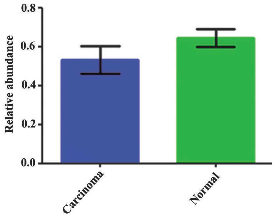
HoxB8 protein expression level
HoxB8 protein was decreased in CRC tissues
(0.532±0.07) and highly expressed in the corresponding normal
mucosa tissues (0.647±0.04). There was a statistically significant
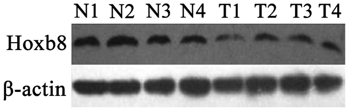
difference between these two groups (P=0.001; Fig. 1). Fig. 2
shows the results of the western blot analysis for the four typical
CRC tissue samples and their corresponding normal mucosa tissue
samples. There was no significant association between HoxB8 protein
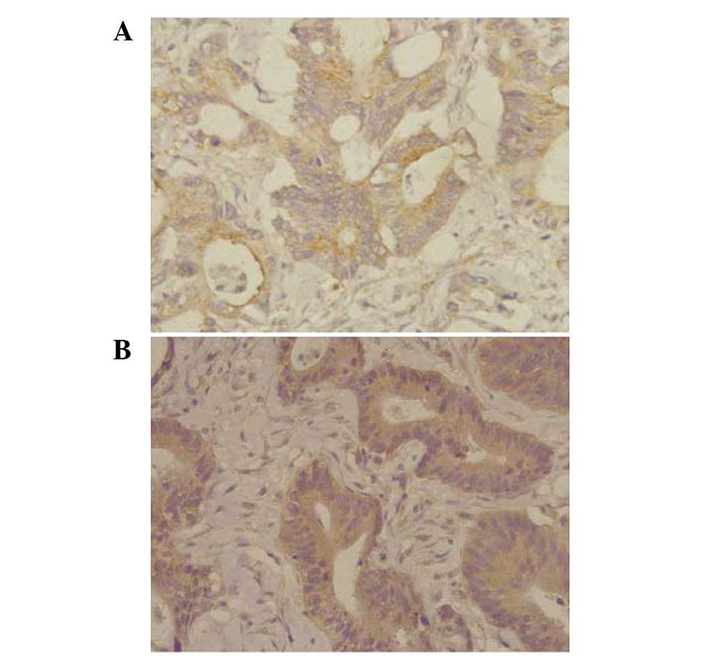
and mRNA expression (r=−0.236; P=0.210). HoxB8 protein in CRC
tissues was mainly expressed in the cytoplasm (Fig. 3). The Mann-Whitney U test demonstrated
that the difference was statistically significant (Z=−2.396;
P=0.017) and the positive rate of HoxB8 expression in the
chemotherapy-sensitive group was decreased compared with the
chemotherapy-resistant group (Table
III).
| Table III.Results of positive rates of HoxB8
protein between the chemotherapy-sensitive and
chemotherapy-resistant groups. |
Table III.
Results of positive rates of HoxB8
protein between the chemotherapy-sensitive and
chemotherapy-resistant groups.
|
|
| HoxB8 expression, n
(%) |
|
|
|---|
|
|
|
|
|
|
|---|
| Group | Cases, n | − | + | ++ | +++ | Z-value | P-value |
|---|
| Sensitive | 25 | 3
(12) | 16 (64) | 4
(16) | 2 (8) | −2.396 | 0.017 |
| Non-sensitive | 25 | 2 (8) | 7
(28) | 13 (52) | 3
(12) |
|
|
Association between miR-196 mRNA and
HoxB8 mRNA expression and the clinical pathological features of
CRC
As shown in Table IV,
miR-196 expression was significantly associated with CRC lymph node
metastasis (P=0.003), tumor stage (I+II and III+IV; P=0.003) and
distant metastasis (P=0.005), but was not associated with the tumor
site (P=0.901), tumor size (P=0.692), gross type (P=0.290), depth
of invasion (P=0.542), tissue differentiation (P=0.615), age
(P=0.178) or gender (P=0.333). HoxB8 mRNA expression was not
associated with any of the aforementioned clinicopathological
features.
| Table IV.Association between the mRNA
expression of miR-196 and HoxB8 and the clinicopathological
features of colorectal cancer. |
Table IV.
Association between the mRNA
expression of miR-196 and HoxB8 and the clinicopathological
features of colorectal cancer.
| Clinicopathological
characteristics | n | Expression of
miR-196 mRNA | Z-value | P-value | Expression of HoxB8
mRNA | Z-value | P-value |
|---|
| Gender |
|
| −0.968 | 0.333 |
| −0.667 | 0.505 |
|
Male | 19 | 1.551±1.100 |
|
| 1.767±1.237 |
|
|
Female | 11 | 2.090±1.311 |
|
| 1.403±0.733 |
|
|
| Age |
|
| −1.348 | 0.178 |
| −1.597 | 0.110 |
| >60
years | 15 | 1.412±1.000 |
|
| 1.879±1.205 |
|
|
| ≤60
years | 15 | 2.085±1.298 |
|
| 1.388±0.914 |
|
|
| Tumor diameter,
cm |
|
| −0.396 | 0.692 |
| −0.352 | 0.725 |
|
>4 | 10 | 1.889±1.195 |
|
| 1.810±1.206 |
|
|
| ≤4 | 20 | 1.678±1.210 |
|
| 1.545±1.033 |
|
|
| Macroscopic type of
tumora |
|
|
2.479b | 0.290 |
|
2.516b | 0.284 |
|
Ulcerative | 18 | 1.658±1.183 |
|
| 1.782±1.175 |
|
|
|
Protrusive | 8 | 1.489±1.151 |
|
| 1.658±1.036 |
|
|
|
Infiltrative | 4 | 2.634±1.167 |
|
| 0.915±0.347 |
|
|
| Tissue
differentiationa |
|
|
0.973b | 0.615 |
|
0.410b | 0.815 |
|
High | 1 | 0.704±0.000 |
|
| 1.202±0.000 |
|
|
|
Moderate | 28 | 1.792±1.214 |
|
| 1.674±1.107 |
|
|
|
Low | 1 | 1.595±0.000 |
|
| 0.942±0.000 |
|
|
| Tumor staging |
|
| −2.948 | 0.003 |
| −1.276 | 0.202 |
|
I+II | 10 | 0.847±0.349 |
|
| 1.854±1.071 |
|
|
III+IV | 20 | 2.199±1.207 |
|
| 1.523±1.095 |
|
|
| Depth of
invasion |
|
| −0.610 | 0.542 |
| −0.516 | 0.606 |
|
T1+T2 | 8 | 1.635±1.391 |
|
| 1.454±0.914 |
|
|
|
T3+T4 | 22 | 1.790±1.140 |
|
| 1.699±1.147 |
|
|
| Tumor location |
|
| −0.125 | 0.901 |
| −0.333 | 0.739 |
|
Rectum | 16 | 1.696±1.133 |
|
| 1.609±0.882 |
|
|
|
Colon | 14 | 1.809±1.290 |
|
| 1.661±1.305 |
|
|
| Lymph node
metastasis |
|
| −2.921 | 0.003 |
| −1.482 | 0.138 |
|
Yes | 18 | 2.245±1.153 |
|
| 1.504±1.132 |
|
|
| No | 12 | 1.005±0.816 |
|
| 1.827±1.012 |
|
|
| Distant
metastasis |
|
| −2.800 | 0.005 |
| −0.726 | 0.468 |
|
Yes | 3 | 4.146±0.758 |
|
| 1.220±0.840 |
|
|
| No | 27 | 1.482±0.892 |
|
| 1.679±1.107 |
|
|
Discussion
miRNA is a class of endogenous gene encoded by
~21–25 nucleotides. miRNAs are small non-coding single stranded RNA
molecules involved in a variety of biological processes, including
early development, apoptosis, proliferation and tumor occurrence
(16,17). For example, miRNA may be combined with
a target gene, through complete or partial binding, by forming the
RNA-induced silencing complex to induce lysis of the target mRNA or
inhibit protein translation (18,19). In
addition, miRNA may be involved in the occurrence and evolution of
malignancies as oncogenes or tumor suppressor genes (20,21).
miR-196 is located in the upstream of the Hox gene
family and has a negative effect on expression of the majority of
Hox gene family members, in which the most common target gene is
HoxB8 (5,22–24).
Previous studies have demonstrated that miR-196 is highly expressed
in gastric cancer (25,26) and malignant glioma (27). Schimanski et al (22) indicated that 29% of 7 CRC tissue
samples overexpressed miR-196a with quantitative RT-PCR compared
with normal colon mucosa. The present study also indicated that
miR-196 was upregulated in the cancer tissue samples, indicating
the possible participation of miR-196 in the occurrence of CRC. In
addition, the expression level of miR-196 was associated with lymph
node metastases, staging and distant metastasis, but was not
associated with the location, size, gross type, depth of invasion,
differentiation, age and gender.
Lymph node metastasis and tumor stage are indicators
of cancer invasion, suggesting that miR-196 is involved in cancer
metastasis and prognosis. According to the 2014 NCCN treatment
guidelines for CRC (28), the
patients with distant metastases are recommended to be administered
with chemotherapy prior to surgery. However, in the present study,
3 patients with distant metastases, all of which were liver
metastases, directly underwent surgery without chemotherapy due to
limited budgets. With these 3 samples, the expression level of
miR-196 was indicated to be significantly correlated with distant
metastasis; this suggests that the overexpression of miR-196 may be
associated with a poor prognosis, and that miR-196 may be a
prognosis marker for CRC. Certain chemotherapy drugs, including
5-fluorouracil and oxaliplatin, may alter the expression level of
miRNAs in CRC. Meng et al (29) indicated that the expression profiles
of miRNA changed with the intervention of chemotherapy drugs, and
proposed to use miRNA as a predictor for evaluating the
chemotherapy drugs. Schimanski et al (22) also identified that the increased
concentration of miR-196 may enhance the sensitivity of CRC cell to
platinum drugs. The results of the present study showed that the
expression levels of miR-196 in patients with CRC decreased
following chemotherapy with FOLFOX4. In addition, the expression
level of miR-196 in the chemotherapy-sensitivity patient group was
increased compared with the chemotherapy-resistant patient group,
suggesting an increased sensitivity to FOLFOX4, which was
consistent with results of the study by Schimanski et al
(22).
HoxB8 is one of the homologous gene family members
encoding the DNA-binding domain of the nucleoprotein, which is
located in human chromosome 17 (30).
HoxB8 is a sequence-specific transcription factor that is important
for cell differentiation, growth and organ formation (31). Rawat et al (32) indicated that the deletion of the
N-terminal domain in the caudal type homeobox 2 led to the
suppression of the Hox gene, which caused abnormal differentiation
of hematopoietic stem cells and the development of acute myeloid
leukemia in mice.
Vider et al (33) supported that HoxB6, HoxB8, HoxC8 and
HoxC9 were abnormally expressed in the various stages of CRC and
that HoxB8 is important in the progression of cancer (34). However, Vider et al (33) did not investigate the association with
clinicopathological features. In the present study, HoxB8 was also
indicated to be highly expressed in the CRC tissues, which is
consistent with the findings of Vider et al (33). Furthermore, the present study explored
the association between HoxB8 and the clinicopathological features
of CRC, and indicated that the expression level of HoxB8 was not
associated with lymph node metastasis, staging, distant metastasis,
tumor gross type, depth of invasion, differentiation, tumor size,
tumor location, age or gender. The preliminary results of the
present study demonstrated that HoxB8 was downregulated in the
FOLFOX4-sensitivity patient group. Using the expression of HoxB8 to
predict the sensitivity to FOLFOX4, the specificity, sensitivity
and accuracy of FOLFOX4 were 76.9, 82.4 and 79.7%, respectively.
HoxB8 may be used to predict the sensitivity to FOLFOX4 in patients
with CRC (35). Furthermore, the
present study indicated that the expression level of HoxB8
decreased with FOLFOX4 chemotherapy and that HoxB8 expression was
decreased in the FOLFOX4-sensitive patient group, suggesting that
an increased level of HoxB8 may decrease the sensitivity to
FOLFOX4.
miRNA lyses target mRNA or inhibits the translation
of target mRNA by complete or partial binding to the 3′-UTR in the
target mRNA, and then downregulates the target mRNA. Kawasaki et
al (36) showed that miR-196
inhibited the expression of HoxB8 in HL60 cells by directly lysing
the target HoxB8 gene. The present study demonstrated that HoxB8
was highly transcripted in CRC but lowly expressed in the CRC
tissues, and that miR-196 was negatively correlated with HoxB8
mRNA. However, miR-196 expression was not associated with the
concentration of HoxB8 protein, which suggests that miR-196
regulates its target gene, HoxB8, in the transcription stage.
Numerous studies have indicated that the expression pattern of
miRNAs has a certain time-sequence and phasic manner, meaning
varied expression levels in various tissues and at various
development stages (20). A
proportional association between miR-196 and HoxB8 may therefore
exist. When the ratio between miR-196 and HoxB8 does not reach the
threshold, miR-196 completely or partially inhibits the expression
of HoxB8 or invokes the development of a tumor (22). Overexpressed miR-196 may cause the
development of a tumor as an oncogene, while downregulated miR-196
may suppress the development of tumor by inhibiting its target
oncogene, HoxB8.
Due to the varied expression of miR-196 and HoxB8
between the chemotherapy-sensitive and chemotherapy-resistant
groups, miR-196 and HoxB8 are hypothesized to aid prediction of the
response of the patient to FOLFOX4 chemotherapy for the CRC. In
addition, miR-196 and HoxB8 may be used to predict the toxicity of
the FOLFOX4 chemotherapy. By measuring the expression of miR-196
and HoxB8, patients that may benefit from FOLFOX4 chemotherapy may
be selected and moved towards to personalized treatment. In
addition, miR-196 and HoxB8 may be used as targets for drugs,
either by inhibiting the expression of HoxB8 to increase the
sensitivity to drugs or by indirectly changing the sensitivity
through the regulation of upstream miR-196. All these findings lay
the foundation of developing novel drugs to treat CRC.
Acknowledgements
The present study was funded by the Natural Science
Foundation of China (grant no., 81472777), the Young and
Middle-aged Talent Training Project of the health ministry of
Fujian Province (grant no., 2013-ZQN-ZD-11), the Ministry of Health
National Clinic Key Special Department Constructive Project (grant
no., 2012 649), the Natural Science Foundation of Fujian Province
(grant no. 2016J01465) and the Youth scientific research fundation
of Fujian Health Department (grant no. 2011-1-9).
References
|
1
|
Jemal A, Siegel R, Xu J and Ward E: Cancer
statistics, 2010. CA Cancer J Clin. 60:277–300. 2010. View Article : Google Scholar : PubMed/NCBI
|
|
2
|
Bartel DP: MicroRNAs: Target recognition
and regulatory functions. Cell. 136:215–233. 2009. View Article : Google Scholar : PubMed/NCBI
|
|
3
|
Volinia S, Calin GA, Liu CG, Ambs S,
Cimmino A, Petrocca F, Visone R, Iorio M, Roldo C, Ferracin M, et
al: A microRNA expression signature of human solid tumors defines
cancer gene targets. Proc Natl Acad Sci USA. 103:2257–2261. 2006.
View Article : Google Scholar : PubMed/NCBI
|
|
4
|
Simpson LJ and Ansel KM: MicroRNA
regulation of lymphocyte tolerance and autoimmunity. J Clin Invest.
125:2242–2249. 2015. View
Article : Google Scholar : PubMed/NCBI
|
|
5
|
Yekta S, Shih IH and Bartel DP:
MicroRNA-directed cleavage of HOXB8 mRNA. Science. 304:594–596.
2004. View Article : Google Scholar : PubMed/NCBI
|
|
6
|
Braig S, Mueller DW, Rothhammer T and
Bosserhoff AK: MicroRNA miR-196a is a central regulator of HOX-B7
and BMP4 expression in malignant melanoma. Cell Mol Life Sci.
67:3535–3548. 2010. View Article : Google Scholar : PubMed/NCBI
|
|
7
|
Li Y, Zhang M, Chen H, Dong Z, Ganapathy
V, Thangaraju M and Huang S: Ratio of miR-196s to HOXC8 messenger
RNA correlates with breast cancer cell migration and metastasis.
Cancer Res. 70:7894–7904. 2010. View Article : Google Scholar : PubMed/NCBI
|
|
8
|
Yang G, Han D, Chen X, et al: MiR-196a
exerts its oncogenic effect in glioblastoma multiforme by
inhibition of IκBα both in vitro and in vivo. Neuro Oncol.
16:652–661. 2014. View Article : Google Scholar : PubMed/NCBI
|
|
9
|
Huang F, Tang J, Zhuang X, Zhuang Y, Cheng
W, Chen W, Yao H and Zhang S: MiR-196a promotes pancreatic cancer
progression by targeting nuclear factor kappa-B-inhibitor alpha.
PLoS One. 9:e878972014. View Article : Google Scholar : PubMed/NCBI
|
|
10
|
Zheng T, Wang J, Chen X and Liu L: Role of
microRNA in anticancer drug resistance. Int J Cancer. 126:2–10.
2010. View Article : Google Scholar : PubMed/NCBI
|
|
11
|
Edge SB and Compton C: The American joint
committee on cancer: The 7th edition of the AJCC cancer staging
manual and the future of TNM. Ann Surg Oncol. 17:143–164. 2010.
View Article : Google Scholar
|
|
12
|
Eisenhauer EA, Therasse P, Bogaerts J,
Schwartz LH, Sargent D, Ford R, Dancey J, Arbuck S, Gwyther S,
Mooney M, et al: New response evaluation criteria in solid tumours:
Revised RECIST guideline (version 1.1). Eur J Cancer. 45:228–247.
2009. View Article : Google Scholar : PubMed/NCBI
|
|
13
|
Kojima A, Uchida I, Sekizaki T, Sasaki Y,
Ogikubo Y and Tamura Y: Rapid detection and identification of
Clostridium chauvoei by PCR based on flagellin gene sequence. Vet
Microbiol. 78:363–371. 2001. View Article : Google Scholar : PubMed/NCBI
|
|
14
|
Qiu JY, Liu P, Shi C and Han B: Low-grade
myofibroblastic sarcomas of the maxilla. Oncol Lett. 9:619–625.
2015.PubMed/NCBI
|
|
15
|
Salmanidis M, Brumatti G, Narayan N, Green
BD, van den Bergen JA, Sandow JJ, Bert AG, Silke N, Sladic R,
Puthalakath H, et al: Hoxb8 regulates expression of microRNAs to
control cell death and differentiation. Cell Death Differ.
20:1370–1380. 2013. View Article : Google Scholar : PubMed/NCBI
|
|
16
|
Blenkiron C and Miska EA: miRNAs in
cancer: Approaches, aetiology, diagnostics and therapy. Hum Mol
Genet. 16:R106–R113. 2007. View Article : Google Scholar : PubMed/NCBI
|
|
17
|
Urbich C, Kuehbacher A and Dimmeler S:
Role of microRNAs in vascular diseases, inflammation and
angiogenesis. Cardiovasc Res. 79:581–588. 2008. View Article : Google Scholar : PubMed/NCBI
|
|
18
|
Stark A, Brennecke J, Bushati N, Russell
RB and Cohen SM: Animal MicroRNAs confer robustness to gene
expression and have a significant impact on 3′UTR evolution. Cell.
123:1133–1146. 2005. View Article : Google Scholar : PubMed/NCBI
|
|
19
|
Carrington JC and Ambros V: Role of
microRNAs in plant and animal development. Science. 301:336–338.
2003. View Article : Google Scholar : PubMed/NCBI
|
|
20
|
Akao Y, Nakagawa Y and Naoe T: Let-7
microRNA functions as a potential growth suppressor in human colon
cancer cells. Biol Pharm Bull. 29:903–906. 2006. View Article : Google Scholar : PubMed/NCBI
|
|
21
|
Hayashita Y, Osada H, Tatematsu Y, Yamada
H, Yanagisawa K, Tomida S, Yatabe Y, Kawahara K, Sekido Y and
Takahashi T: A polycistronic microRNA cluster, miR-17-92, is
overexpressed in human lung cancers and enhances cell
proliferation. Cancer Res. 65:9628–9632. 2005. View Article : Google Scholar : PubMed/NCBI
|
|
22
|
Schimanski CC, Frerichs K, Rahman F,
Berger M, Lang H, Galle PR, Moehler M and Gockel I: High miR-196a
levels promote the oncogenic phenotype of colorectal cancer cells.
World J Gastroenterol. 15:2089–2096. 2009. View Article : Google Scholar : PubMed/NCBI
|
|
23
|
Chopra VS and Mishra RK: ‘Mir’acles in hox
gene regulation. Bioessays. 28:445–448. 2006. View Article : Google Scholar : PubMed/NCBI
|
|
24
|
Zhao Z, Boyle TJ, Liu Z, Murray JI, Wood
WB and Waterston RH: A negative regulatory loop between microRNA
and Hox gene controls posterior identities in Caenorhabditis
elegans. PLoS Genet. 6:e10010892010. View Article : Google Scholar : PubMed/NCBI
|
|
25
|
Tsai KW, Hu LY, Wu CW, Li SC, Lai CH, Kao
HW, Fang WL and Lin WC: Epigenetic regulation of miR-196b
expression in gastric cancer. Genes Chromosomes Cancer. 49:969–980.
2010. View Article : Google Scholar : PubMed/NCBI
|
|
26
|
Liao YL, Hu LY, Tsai KW, Wu CW, Chan WC,
Li SC, Lai CH, Ho MR, Fang WL, Huang KH and Lin WC: Transcriptional
regulation of miR-196b by ETS2 in gastric cancer cells.
Carcinogenesis. 33:760–769. 2012. View Article : Google Scholar : PubMed/NCBI
|
|
27
|
Guan Y, Mizoguchi M, Yoshimoto K, Hata N,
Shono T, Suzuki SO, Araki Y, Kuga D, Nakamizo A, Amano T, et al:
MiRNA-196 is upregulated in glioblastoma but not in anaplastic
astrocytoma and has prognostic significance. Clin Cancer Res.
16:4289–4297. 2010. View Article : Google Scholar : PubMed/NCBI
|
|
28
|
National Comprehensive Cancer Network, .
Clinical Practice Guidelines in Oncology. http://www.nccn.org/professionals/physician_gls/f_guidelines.asp#colonAugust
2–2014
|
|
29
|
Meng F, Henson R, Lang M, Wehbe H,
Maheshwari S, Mendell JT, Jiang J, Schmittgen TD and Patel T:
Involvement of human micro-RNA in growth and response to
chemotherapy in human cholangiocarcinoma cell lines.
Gastroenterology. 130:2113–2129. 2006. View Article : Google Scholar : PubMed/NCBI
|
|
30
|
Yue Y, Farcas R, Thiel G, Bommer C,
Grossmann B, Galetzka D, Kelbova C, Küpferling P, Daser A, Zechner
U and Haaf T: De novo t(12;17) (p13.3;q21.3) translocation with a
breakpoint near the 5′end of the HOXB gene cluster in a patient
with developmental delay and skeletal malformations. Eur J Hum
Genet. 15:570–577. 2007. View Article : Google Scholar : PubMed/NCBI
|
|
31
|
Hornstein E, Mansfield JH, Yekta S, Hu JK,
Harfe BD, McManus MT, Baskerville S, Bartel DP and Tabin CJ: The
microRNA miR-196 acts upstream of Hoxb8 and Shh in limb
development. Nature. 438:671–674. 2005. View Article : Google Scholar : PubMed/NCBI
|
|
32
|
Rawat VP, Thoene S, Naidu VM, Arseni N,
Heilmeier B, Metzeler K, Petropoulos K, Deshpande A,
Quintanilla-Martinez L, Bohlander SK, et al: Overexpression of CDX2
perturbs HOX gene expression in murine progenitors depending on its
N-terminal domain and is closely correlated with deregulated HOX
gene expression in human acute myeloid leukemia. Blood.
111:309–319. 2008. View Article : Google Scholar : PubMed/NCBI
|
|
33
|
Vider BZ, Zimber A, Hirsch D, Estlein D,
Chastre E, Prevot S, Gespach C, Yaniv A and Gazit A: Human
colorectal carcinogenesis is associated with deregulation of
homeobox gene expression. Biochem Biophys Res Commun. 232:742–748.
1997. View Article : Google Scholar : PubMed/NCBI
|
|
34
|
Vider BZ, Zimber A, Chastre E, Gespach C,
Halperin M, Mashiah P, Yaniv A and Gazit A: Deregulated expression
of homeobox-containing genes, HOXB6, B8, C8, C9 and Cdx-1, in human
colon cancer cell lines. Biochem Biophys Res Commun. 272:513–518.
2000. View Article : Google Scholar : PubMed/NCBI
|
|
35
|
Lu X, Pan J, Li S, Shen S, Chi P, Lin H,
Huang Y, Xu Z and Huang S: Establishment of a predictive genetic
model for estimating chemotherapy sensitivity of colorectal cancer
with synchronous liver metastasis. Cancer Biother Radiopharm.
28:552–558. 2013. View Article : Google Scholar : PubMed/NCBI
|
|
36
|
Kawasaki H and Taira K: MicroRNA-196
inhibits HOXB8 expression in myeloid differentiation of HL60 cells.
Nucleic Acids Symposium. 48:211–212. 2004. View Article : Google Scholar
|