Introduction
Colon cancer is the third most common type of
malignant tumor of the gastrointestinal tract, and it has the
highest mortality rate (1). Clinical
data showed that with an improvement in living standards and a
change of diet, the incidence rate of colon cancer showed a rapidly
rising trend year by year and tended to be diagnosed at younger
ages (2,3). The incidence rate of colon cancer rose
from 6.1/100,000 in 1972–1974 to 11.2/100,000 in 1987–1989
worldwide (4). This rise was an 84.6%
increase and the average annual increase was 4.2% (4). The incidence rate of colon cancer ranks
4th in the world, subsequent to stomach, lung and liver cancer,
seriously affecting the social labor force, social production and
quality of life for numerous families (5,6). For the
treatment of colon cancer, radical treatment is surgery, and the
auxiliary treatments consist of radiotherapy and chemotherapy,
reducing tumor burden and the number of tumor cells to achieve a
therapeutic effect. For the majority of colon cancer patients at
the progressive stage it is too late for successful treatment of
the malignancy, and thus, chemotherapy and palliative care may be
the remaining therapeutic strategies. Recurrence and metastasis are
the main causes of treatment failure (7,8).
Therefore, clarification of the pathogenesis of colon cancer may
play an important role in the early diagnosis, early treatment and
prognosis of colon cancer. The Golgi phosphoprotein-3 (GOLPH3) gene
has been identified as an oncogene, localized in the network
structure on the reverse side of the Golgi apparatus (9). GOLPH3 is a phosphorylated Golgi membrane
protein that is involved in the protein sorting of the Golgi
apparatus (10,11). GOLPH3 is involved in the occurrence,
development, apoptosis, proliferation and differentiation of human
cancer, regulating the sensitivity of cancer cells to anti-tumor
drugs (12). There are few studies
that have researched the mechanisms of GOLPH3 in colon cancer
(13). Based on this, the expression
of GOLPH3 in colon cancer and its clinical significance were
analyzed in the present study to provide guidance for the clinical
treatment of colon cancer.
Patients and methods
General data
In total, 98 cases of colon cancer patients randomly
selected for surgery in The First Affiliated Hospital of Henan
University of Science and Technology (Luoyang, China) between June
2011 and June 2013 were taken as the observation group. The
patients did not receive radiotherapy or other associated
anti-tumor treatments prior to the surgery. The colon cancer tissue
was resected and confirmed as cancer tissue by pathology. In total,
61 patients were male and 37 patients were female. The ages ranged
between 30 and 80 years old and the average age was 56.78±10.67
years (mean ± standard deviation). The Dukes' clinical stages
consisted of 42 cases of stage II and 56 cases of stage III colon
cancer (14). The tissue typing,
performed using the World Health Organization histological
classification system, consisted of 53 patients with poorly
differentiated and 45 patients with highly differentiated colon
cancer (15). The infiltration degree
consisted of 40 patients with T1-T2 and 58 patients with T3-T4. In
addition, normal biopsy samples collected from 15 healthy
individuals following routine colonoscopy were used as the control
group. Of these, 9 patients were male and 6 patients were females.
The patient ages ranged between 42 and 78 years and the average age
was 63.26±11.32 years. The present study was conducted in
accordance with the Declaration of Helsinki and was conducted with
approval from the Ethics Committee of the First Affiliated Hospital
of Henan University of Science and Technology (Luoyang, China).
Written informed consent was obtained from all participants.
Reverse transcription-quantitative
polymerase chain reaction (RT-qPCR)
Total RNA was extracted from tumor tissues of the
observation group and biopsy tissues of the control group,
according to the RNA extraction kit protocol (Takara Biotechnology
Co., Ltd., Dalian, China). In total, 25 µl RNase-free water was
added to the precipitate and total RNA was obtained. RNA was
transcribed to complementary DNA (cDNA) using a Moloney Murine
Leukemia Virus Reverse Transcription kit (Takara Biotechnology Co.,
Ltd.), according to the manufacturer's protocol. This cDNA was then
used as the template for PCR following concentration determination.
The primers were designed by Primer Premier 5.0 software (Premier
Biosoft International, Palo Alto, CA, USA) according to the GOLPH3
mRNA sequence provided by GeneBank (ID number, NM_022130.3): GOLPH3
forward, 5′-AAGCCGTTCTTGACAAATGG-3′ and reverse,
5′-ATTGGTGTTGGCCTTCAGAC-3′; and glyceraldehyde 3-phosphate
dehydrogenase (GAPDH) forward, 5′-AGAAGGCTGGGGCTCATTTG-3′ and
reverse, 5′-AGGGGCCATCCACAGTCTTC-3′. The primer was diluted in
double distilled water (10 µmol/l−1). The PCR reaction
system was prepared for RT-qPCR subsequent to the optimization of
primer specificity and annealing temperature. The PCR reaction
system comprised of: 10 µl SYBR Green qPCR Master Mix (Bio-Rad
Laboratories, Inc., Hercules, CA, USA), 1 µl cDNA, 1 µl upstream
and downstream primer, respectively. The PCR reaction conditions
were as follows: Pre-degeneration, 94°C for 2 min; degeneration,
94°C for 30 sec; annealing, 60°C for 30 sec; and extension, 72°C
for 30 sec, for a total of 35 cycles. PCR was performed using DNA
polymerase from Takara Biotechnology Co., Ltd and SYBR green
(Bio-Rad Laboratories, Inc.). To determine the specificity of the
product, a solubility curve was constructed. Finally, the data were
read directly from the fluorescence qPCR instrument (Applied
Biosystems; Thermo Fisher Scientific, Inc., Waltham, MA, USA) using
the 2−ΔΔCq method (16).
Immunohistochemical detection
The tissue specimens in the observation and control
groups were embedded in paraffin, then sectioned into 4-mm slices
and adhered onto poly-lysine acid-treated slides. All slides were
subjected to the same protocol, as follows: Placed in a 67°C oven
and baked for 2 h; immersed in xylene 3 times, 5 min/immersion;
immersed in absolute ethyl alcohol for 5 min; immersed in 95%
ethanol for 5 min; immersed in 75% ethanol for 5 min, washed in
phosphate buffered saline (PBS) 3 times (3 min/wash); and then
placed in 10 mM pH 6.0 citrate sodium buffer solution and heated
for 10 min prior to being cooled for 30 min. The steps were
repeated 2 times. Subsequently, the slides were washed with PBS 3
times (3 min/wash) and incubated in 3% H2O2
solution at room temperature for 10 min to block the activity of
endogenous peroxide hydrogenase. The slides were then washed with
PBS 3 times and incubated in rabbit anti-human GOLPH3 polyclonal
antibody (catalog no., ab82377; dilution, 1:100; Abcam, Cambridge,
UK) at 4°C overnight. Subsequent to incubation the slides were
washed with PBS with Tween 20 (PBST) 3 times (3 min/wash). The
sheep anti-rabbit IgG secondary antibody (catalog no., A0208;
dilution, 1:500; Beyotime Institue of Biotechnology, Shanghai,
China) was then added, the slides were incubated for 1 h at room
temperature and washed with PBST 3 times (3 min/wash). The
substrate 3,3′-diaminobenzidine was used for staining, hematoxylin
was used as a counterstain and 0.1% hydrochloric acid was used for
differentiation, followed by washing in PBS to return the color to
blue. The slides were flushed with unfiltered water, redyed,
dehydrated, hyalinized and sealed.
Observation index
GOLPH3 was located in the network structure on the
opposite side of the Golgi apparatus, so brown yellow or brown
particles in the cytoplasm were considered to be positively stained
cells following immunohistochemical detection. The results were
analyzed according to the cell staining intensity and percentage of
GOLPH3-positive cells. In total, 5 high-power fields were randomly
selected for every section and 100 target cells were counted under
high power fields. The average percentage of 5 high-power fields
was used for result determination. For evaluation the cell staining
criteria were set as: 0% staining was 0; positive cell percentage
≤10% was 1; 11–50% was 2; 51–75% was 3; and >75% was 4. The
intensity of the dye was also classified into numerical criteria:
No dyeing was 0; faint yellow was 1; deep yellow was 2; and brown
yellow was 3. The two scores were multiplied to calculate the final
score. A total score of 4 was the demarcation point. Therefore, a
total score of <4 was defined as GOLPH3-negative and ≥4 was
GOLPH3-positive. The prognosis of the patients was evaluated by the
postoperative recurrence rate and survival rate at 1, 2 and 3
years.
Statistical analysis
All data were analyzed using SPSS 17.0 software
(SPSS Inc., Chicago, IL, USA). The measurement data are shown as
the mean ± standard deviation. The comparison among the multi-group
measurement data was performed using analysis of variance. The
comparison between the two groups was shown using Fisher's least
significant difference test. The cumulative incidence of cancer
development among the groups was performed using Kaplan-Meier
curves. The comparison of the survival rate and recurrence rate
among the groups was performed using Log-rank test. P<0.05 was
considered to indicate a statistically significant difference.
Results
Comparison of GOLPH3 mRNA expression
in 2 groups
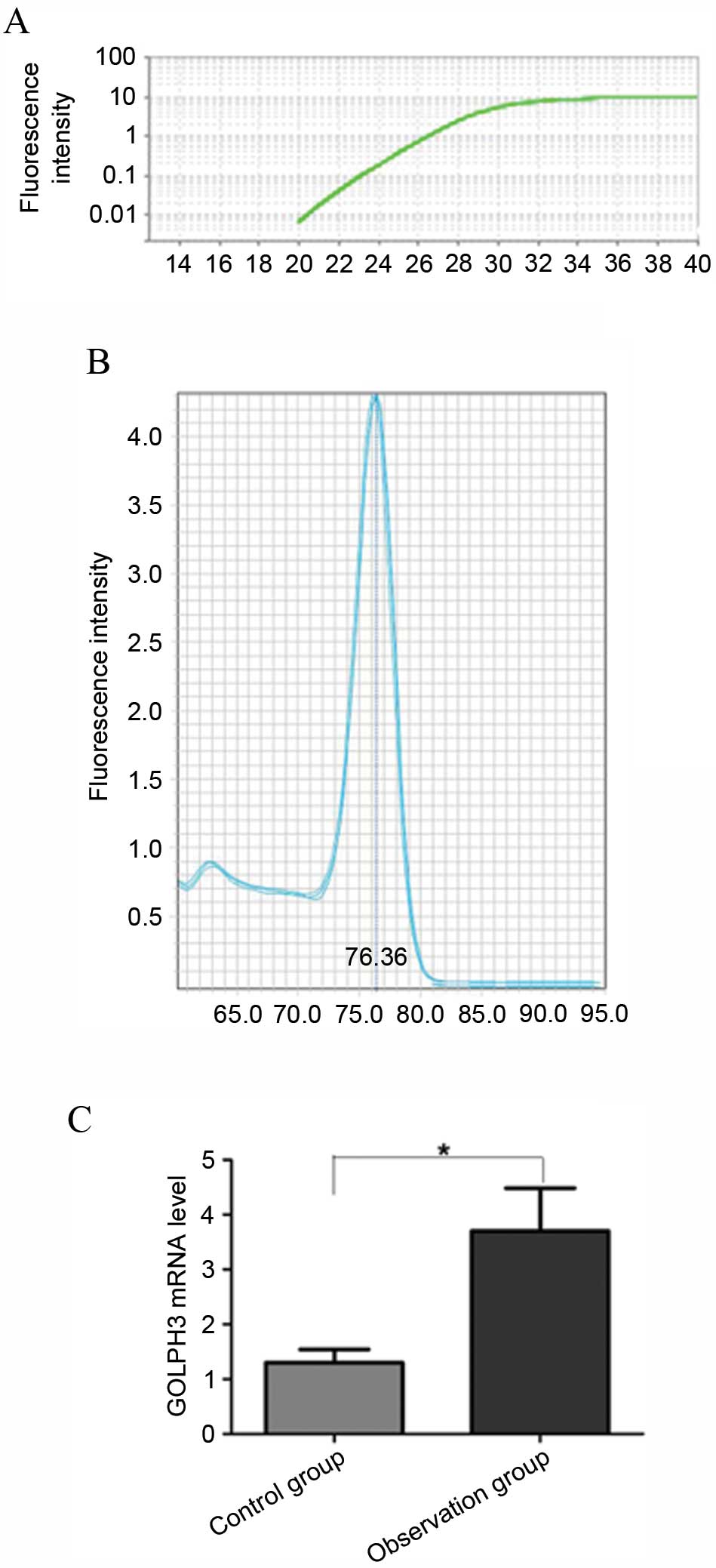
The results of GOLPH3 primer analysis showed that
the primer amplification curve was accurate and the repeatability
was high (Fig. 1A). The primer
specificity was good and there were no miscellaneous peaks
subsequent to amplification (Fig.
1B). GOLPH3 mRNA level in the 2 groups by RT-qPCR method showed
that the average level of GOLPH3 mRNA was significantly higher than
that of the control group (P<0.001) (Fig. 1C).
Comparison of GOLPH3 protein
expression in 2 groups
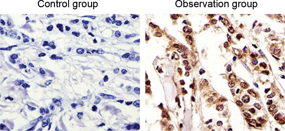
The immunohistochemical results of GOLPH3 protein
expression analysis in the specimens of the observation and control
groups showed that GOLPH3 was mainly expressed in the cytoplasm and
showed low to no expression in normal colon tissue. The expression
of GOLPH3 in colon cancer tissues was increased significantly
(P=0.012; Fig. 2). According to the
expression of GOLPH3 in colon cancer tissues in the observation
group, 29 patients were identified as the GOLPH3-negative group and
69 patients were identified as the GOLPH3-positive group.
Association between GOLPH3 expression
and clinical features
As shown in Table I,
GOLPH3 protein expression was associated with the clinical stage,
histological differentiation degree, lymph node metastasis and
infiltration degree (P<0.05; Table
I), but GOLPH3 expression was unassociated with gender
(P>0.05).
 | Table I.Association between
clinicopathological features and Golgi phosphoprotein-3 expression
level in the observation group. |
Table I.
Association between
clinicopathological features and Golgi phosphoprotein-3 expression
level in the observation group.
|
| Gender | Clinical stages | Tissue types | Lymphatic
metastasis | Infiltration
degree |
|---|
|
|
|
|
|
|
|
|---|
| Pathological
features | Male | Female | II | III | Poorly
differentiated | Highly
differentiated | Yes | No | T1-T2 | T3-T4 |
|---|
| Cases, n | 61 | 37 | 42 | 56 | 53 | 45 | 55 | 43 | 40 | 58 |
| Positive cases,
n | 43 | 26 | 23 | 45 | 42 | 27 | 43 | 26 | 22 | 46 |
| Percentage, % | 70.5 | 70.3 | 54.8 | 80.4 | 79.2 | 60 | 78.2 | 60.5 | 55 | 79.3 |
| χ2 | 0.238 | 7.275 | 4.356 | 5.021 | 6.254 |
| P-value | 0.598 | 0.012 | 0.057 | 0.036 | 0.011 |
Comparison of postoperative recurrence
and survival rate of patients with different levels of GOLPH3
protein
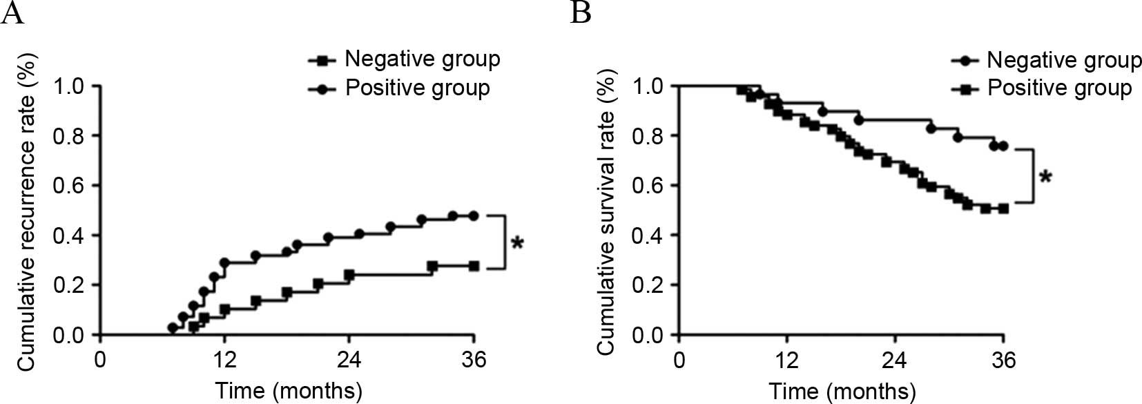
As shown in Fig. 3,
the patients were followed up for >36 months subsequent to being
treated for 3 months. In total, 8 cases in the GOLPH3-negative
group and 34 cases in the GOLPH3-positive group relapsed.
Kaplan-Meier curve showed that the cumulative recurrence rates at
1, 2 and 3 years were 10.34 (3/29 patients), 24.14 (7/29 patients)
and 27.59% (8/29 patients) in the GOLPH3-negative group. The
cumulative recurrence rates at 1, 2 and 3 years were 28.99 (20/69
patients), 39.13 (27/69 patients) and 49.28% (34/69 patients) in
the GOLPH3-positive group. Log-rank test showed that the cumulative
recurrence rate in the GOLPH3-negative group was significantly
lower that of the GOLPH3-positive group (P=0.005). The statistical
results of survival rate showed the cumulative survival rates at 1,
2 and 3 years were 93.10 (27 patients), 86.21 (25 patients) and
72.41% (21 patients) in the GOLPH3-negative patients. The
cumulative survival rates at 1, 2 and 3 years were 88.41 (61
patients), 69.57 (48 patients) and 50.72% (35 patients) in the
GOLPH3-positive patients. The cumulative survival rates at 1, 2 and
3 years in the GOLPH3-negative patients were significantly
increased compared with those of GOLPH3-positive patients
(P=0.012).
Discussion
The preferential site for colon cancer to develop is
at the junction of the rectum and sigmoid colon. Colon cancer is
one of the most common types of malignant tumor of the
gastrointestinal tract, although the incidence and mortality rates
of colon cancer are lower than those of gastric, esophageal and
lung cancers. However, with the development of the economy and
living style, including a change of dietary structure, colon cancer
has become one of the fastest growing types of malignant tumor in
China, which has a detrimental effect upon morbidity and mortality
rates (17,18). In recent years, with the development
of clinical medicine, the treatment success of colon cancer has
improved; however, the 5-year survival rate has not been
significantly improved for advanced stage colon cancer (19). The mortality rate of colon cancer
ranks second for malignant tumors (20). Therefore, additional studies
investigating the prognosis and therapies of colon cancer is
important in order to improve the survival rate.
A previous study showed that GOLPH3 belonged to the
family of Golgi apparatus matrix proteins, and was the first
identified example of an oncogene having powerful trafficking
ability at the reverse side of Golgi network within yeast and
humans (21). Scott et al
(22) conducted a genomic analysis on
a variety of solid tumors; it was found that GOLPH3 was highly
expressed in solid tumors, including melanoma, brain glioma,
prostate cancer and non-small cell cancer. The positive rate of
GOLPH3 was 56% in non-small cell lung cancer. GOLPH3 was confirmed
as an oncogene subsequent to the knockout of RNA interference.
GOLPH3 was attached to the N-terminal of glycosyl-acyl transferase,
and these proteases were fixed on the Golgi apparatus, to regulate
the processing and modification of glycoproteins. GOLPH3 was
identified to move between the Golgi apparatus and cell membrane,
which enables its involvement in a variety of cellular processes.
GOLPH3 overexpression can regulate the signal transduction of
various cellular pathways, particularly the protein glycosylation
pathway, which may eventually lead to tumorigenesis. Studies have
confirmed that the carcinogenic mechanism of GOLPH3 is involved in
tumor occurrence and development via mechanistic target of
rapamycin complex 1 (mTORC1) and mTORC2 signal transductions
(23,24). Therefore, GOLPH3 can be taken as a
critical reference factor of tumor prognosis and may be used in new
drug screening (25,26). Feng et al (27) studied the expression of GOLPH3 in
ovarian carcinoma tissue in 40 cases of ovarian cancer patients.
The results showed that the overall expression of GOLPH3 was
significantly higher than that of para-carcinoma tissue. In
addition, the expression was associated with the patients' surgical
pathological stages, histological differentiation, therapeutic
response and survival. Yu et al (28) detected the expressions of GOLPH3,
caspase-3 and apoptosis index in 62 cases of colon cancer patients.
The results showed the positive expression rate of caspase-3 and
the apoptosis index in colorectal cancer tissues with high
expression of GOLPH3 were significantly decreased compared with
colorectal cancer tissues with low expression of GOLPH3, showing
that the expression of GOLPH3 in colorectal carcinoma was
negatively associated with cell apoptosis.
In the present study, the detection result of GOLPH3
mRNA and protein in colorectal cancer patients showed that the
expressions of GOLPH3 mRNA and protein were significantly increased
in colon tissue of colon cancer patients, indicating that GOLPH3
gene signal transduction pathway in the colorectal cancer tissue
was in an active state, resulting in the increase of GOLPH3
protein. In addition, the current study also revealed that the
expression of GOLPH3 in colorectal carcinoma tissue was positively
associated with: Clinical stages of disease; histological
differentiation degree; lymph node metastasis; and infiltration
degree. The cumulative recurrence rates at 1, 2 and 3 years in
GOLPH3-positive patients were significantly higher than those of
the GOLPH3-negative patients, and the cumulative survival rates
were significantly lower than those of the GOLPH3-negative
patients. Overall, these results confirmed that GOLPH3 was involved
in the occurrence, development, invasion, metastasis and recurrence
of colon cancer.
In conclusion, GOLPH3 was overexpressed in colon
cancer tissue and was closely associated to clinical pathological
features and prognosis, which may be used as a reference for
clinical diagnosis and treatment.
References
|
1
|
Kolligs FT: Diagnostics and Epidemiology
of colorectal cancer. Visc Med. 32:158–164. 2016. View Article : Google Scholar : PubMed/NCBI
|
|
2
|
Slattery ML, Pellatt DF, Wolff RK and
Lundgreen A: Genes, environment and gene expression in colon
tissue: A pathway approach to determining functionality. Int J Mol
Epidemiol Genet. 7:45–57. 2016.PubMed/NCBI
|
|
3
|
Hou TY, Davidson LA, Kim E, Fan YY,
Fuentes NR, Triff K and Chapkin RS: Nutrient-Gene interaction in
colon cancer, from the membrane to cellular physiology. Annu Rev
Nutr. 36:543–570. 2016. View Article : Google Scholar : PubMed/NCBI
|
|
4
|
Jin F, Devesa SS, Chow WH, Zheng W, Ji BT,
Fraumeni JF Jr and Gao YT: Cancer incidence trends in urban
shanghai, 1972–1994: An update. Int J Cancer. 83:435–440. 1999.
View Article : Google Scholar : PubMed/NCBI
|
|
5
|
Aoyagi T, Terracina KP, Raza A and Takabe
K: Current treatment options for colon cancer peritoneal
carcinomatosis. World J Gastroenterol. 20:12493–12500. 2014.
View Article : Google Scholar : PubMed/NCBI
|
|
6
|
Ramalingam S, Subramaniam D and Anant S:
Manipulating miRNA Expression: A novel approach for colon cancer
prevention and chemotherapy. Curr Pharmacol Rep. 1:141–153. 2015.
View Article : Google Scholar : PubMed/NCBI
|
|
7
|
Xia X, Wu W, Zhang K, Cen G, Jiang T, Cao
J, Huang K, Huang C and Qiu Z: Prognostic significance of
complications after laparoscopic colectomy for colon cancer. PLoS
One. 9:e1083482014. View Article : Google Scholar : PubMed/NCBI
|
|
8
|
Kim CW, Kim WR, Kim HY, Kang J, Hur H, Min
BS, Baik SH, Lee KY and Kim NK: Learning curve for single-incision
laparoscopic anterior resection for sigmoid colon cancer. J Am Coll
Surg. 221:397–403. 2015. View Article : Google Scholar : PubMed/NCBI
|
|
9
|
Zhang L, Guo F, Gao X and Wu Y: Golgi
phosphoprotein 3 expression predicts poor prognosis in patients
with prostate cancer undergoing radical prostatectomy. Mol Med Rep.
12:1298–1304. 2015.PubMed/NCBI
|
|
10
|
Ng MM, Dippold HC, Buschman MD, Noakes CJ
and Field SJ: GOLPH3L antagonizes GOLPH3 to determine Golgi
morphology. Mol Biol Cell. 24:796–808. 2013. View Article : Google Scholar : PubMed/NCBI
|
|
11
|
Tu L, Chen L and Banfield DK: A conserved
N-terminal arginine-motif in GOLPH3-family proteins mediates
binding to coatomer. Traffic. 13:1496–1507. 2012. View Article : Google Scholar : PubMed/NCBI
|
|
12
|
Kunigou O, Nagao H, Kawabata N, Ishidou Y,
Nagano S, Maeda S, Komiya S and Setoguchi T: Role of GOLPH3 and
GOLPH3L in the proliferation of human rhabdomyosarcoma. Oncol Rep.
26:1337–1342. 2011.PubMed/NCBI
|
|
13
|
Qiu CZ, Wang MZ, Yu WS, Guo YT, Wang CX
and Yang XF: Correlation of GOLPH3 gene with Wnt signaling pathway
in human colon cancer cells. J Cancer. 7:928–934. 2016. View Article : Google Scholar : PubMed/NCBI
|
|
14
|
Wan DS: Clinical Oncology. Third. Beijing:
Science Press; pp. 2151999, (In Chinese).
|
|
15
|
Ionilă M, Mărgăritescu C, Pirici D and
Mogoantă SS: Mucinous adenocarcinoma of the colon-a histochemical
study. Rom J Morphol Embryol. 52:783–790. 2011.PubMed/NCBI
|
|
16
|
Reuther S, Reiter M, Raabe A and Dikomey
E: Effect of irradiation on the expression of DNA repair genes
studied in human fibroblasts by real-time qPCR using three methods
of reference gene validation. Radiat Environ Biophys. 52:463–469.
2013. View Article : Google Scholar : PubMed/NCBI
|
|
17
|
Lavotshkin S, Jalas JR, Torisu-Itakura H,
Ozao-Choy J, Lee JH, Sim MS, Stojadinovic A, Wainberg Z, Bifulco
CB, Fox BA and Bilchik AJ: Immunoprofiling for prognostic
assessment of colon cancer: A novel complement to ultrastaging. J
Gastrointest Surg. 19:999–1006. 2015. View Article : Google Scholar : PubMed/NCBI
|
|
18
|
Gill S: Adjuvant therapy for resected
high-risk colon cancer: Current standards and controversies. Indian
J Med Paediatr Oncol. 35:197–202. 2014. View Article : Google Scholar : PubMed/NCBI
|
|
19
|
Zakaria F and El-Mashad N: Inadequate
lymph node sampling as a risk factor in stage II colon cancer. Gulf
J Oncolog. 1:46–55. 2014.PubMed/NCBI
|
|
20
|
Uddin S, Bavi PP, Hussain AR, Alsbeih G,
Al-Sanea N, Abduljabbar A, Ashari LH, Alhomoud S, Al-Dayel F, Ahmed
M and Al-Kuraya KS: Leptin receptor expression in Middle Eastern
colorectal cancer and its potential clinical implication.
Carcinogenesis. 30:1832–1840. 2009. View Article : Google Scholar : PubMed/NCBI
|
|
21
|
Abraham RT: GOLPH3 links the Golgi network
to mTOR signaling and human cancer. Pigment Cell Melanoma Res.
22:378–379. 2009. View Article : Google Scholar : PubMed/NCBI
|
|
22
|
Scott KL, Kabbarall O, Liang MC, Ivanova
E, Anagnostou V, Wu J, Dhakal S, Wu M, Chen S, Feinberg T, et al:
GOLPH3 modulates mTOR signalling and rapamycin sensitivity in
cancer. Nature. 459:1085–1090. 2009. View Article : Google Scholar : PubMed/NCBI
|
|
23
|
Lv XL, Tian H, Lu M, Yue WM, Li SH, Gao C
and Si LB: The expression of Golgi phosphoprotein 3 (GOLPH3) in
non-small cell lung cancer and its clinical significance. Chin J
Cancer Biother. 21:554–558. 2014.
|
|
24
|
Isaji T, Im S, Gu W, Wang Y, Hang Q, Lu J,
Fukuda T, Hashii N, Takakura D, Kawasaki N, et al: An oncogenic
protein Golgi phosphoprotein 3 up-regulates cell migration via
sialylation. J Biol Chem. 289:20694–20705. 2014. View Article : Google Scholar : PubMed/NCBI
|
|
25
|
Sechi S, Frappaolo A, Belloni G and
Giansanti MG: The roles of the oncoprotein GOLPH3 in contractile
ring assembly and membrane trafficking during cytokinesis. Biochem
Soc Trans. 43:117–121. 2015. View Article : Google Scholar : PubMed/NCBI
|
|
26
|
Buschman MD, Rahajeng J and Field SJ:
GOLPH3 links the Golgi, DNA damage, and cancer. Cancer Res.
75:624–627. 2015. View Article : Google Scholar : PubMed/NCBI
|
|
27
|
Feng LP and Su WM: Relationship between
Golph3 and prognosis of epithelial ovarian cancer. J Prac Med.
29:243–245. 2013.(In Chinese).
|
|
28
|
Yu WS, Qiu CZ, Wang CX, Wu YY and Huang
ZX: Relationship between Golph3 expression and apoptosis in
colorectal carcinoma tissues. Chin J Clin Oncol. 40:1094–1097.
2013.(In Chinese).
|