Introduction
Inflammatory myofibroblastic tumors (IMT) are
distinctive tumor entities described in almost all anatomical sites
under several definitions, including inflammatory pseudotumor and
plasma cell granuloma. IMT generally shows a benign clinical
outcome and is common in children and young adults, although the
age of occurrence is extremely broad (1). Presenting symptoms are related to the
site of the primary tumor origin, although fever, weight loss,
malaise and night sweats are also reported. Surgical resection is
the mainstay of treatment, while chemoradiotherapy is generally
ineffective (1).
Approximately half of IMT patients carry
rearrangements of the anaplastic lymphoma kinase (ALK) gene
(2–5).
ALK is a tyrosine kinase receptor, which is typically expressed in
the central nervous system (3,4). Fusion of
the ALK gene with different partners may result in the
overexpression of ALK and activation of its kinase domain (6–8).
Identification of ALK fusion genes may support a diagnosis
of IMT, and additionally allow initiation of an effective treatment
regimen with ALK inhibitors (6,7). IMTs
occurring in the head and neck region are extremely rare and are
more commonly located in the larynx, orbit, paranasal sinus,
trachea and parotid gland. The present study describes the
characterization of the 3a/b variants of the echinoderm microtubule
associated protein like 4 (EML4)-ALK gene fusion, which has
not been reported previously in IMT of the hypopharynx.
Materials and methods
Case description
A 74-year-old, non-smoking woman was hospitalized
after presenting with weight loss, progressive dysphagia,
odynophagia and globus sensation. The patient had an unremarkable
medical past and routine laboratory tests, serum tumor markers, a
QuantiFERON® tuberculosis test and antineutrophil
cytoplasmic antibodies test came back negative. A total-body
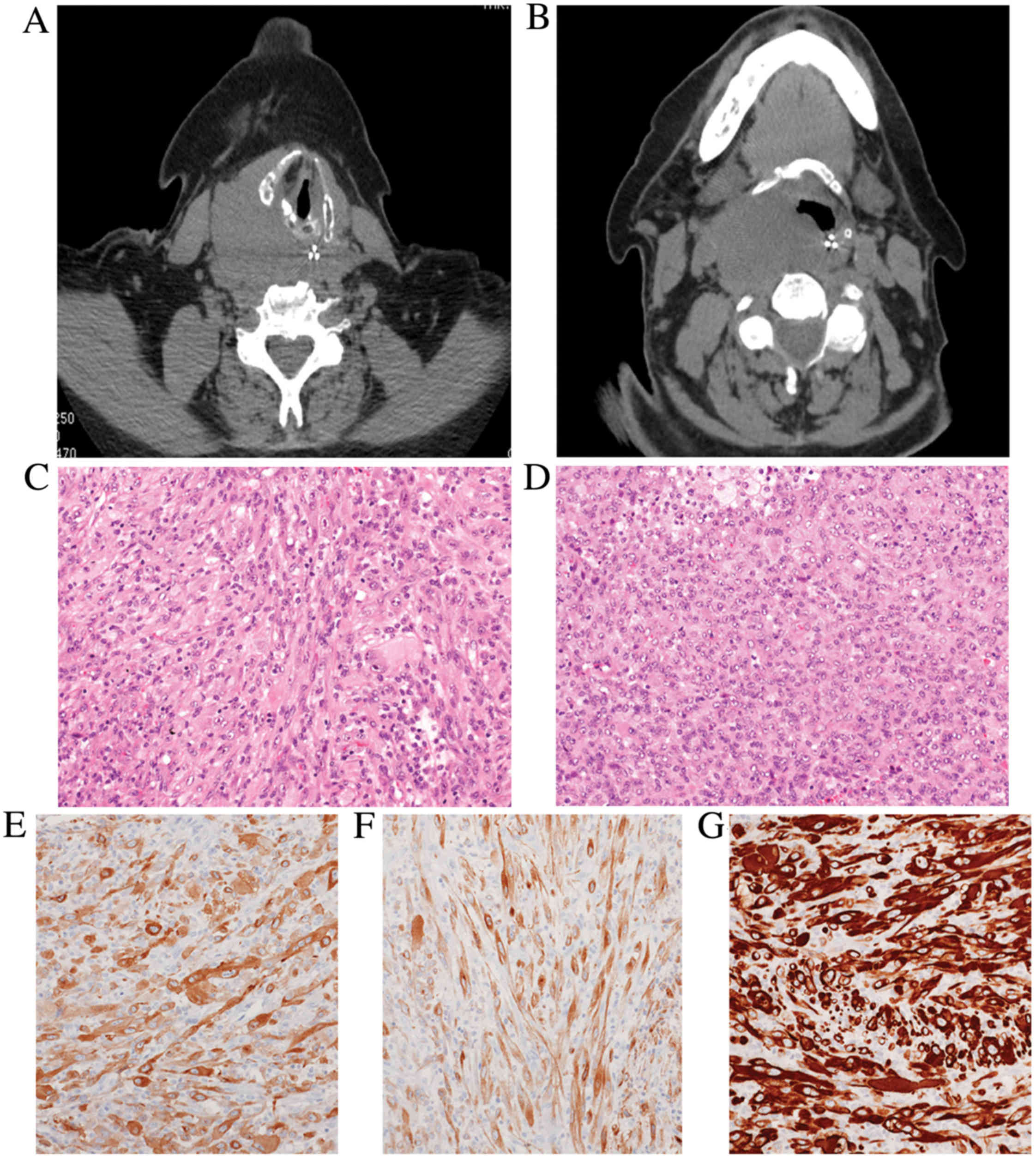
computed tomography scan revealed a 5-cm mass situated at the right
posterior wall of the hypopharynx (Fig.
1A). No other lesions were identified.
Radiological examinations demonstrated the
impossibility to perform a complete surgical resection since the
tumor exhibited infiltrative margins and involved adjacent
structures. However, the patient was symptomatic due to the
progressive occlusion of the pharynx, which could result in a
life-threating condition.
Diagnostic and palliative surgical resection was
performed, and the specimen was fixed in 10% neutralized formalin
for 24 h at room temperature and embedded in paraffin blocks, which
were subsequently cut into 3-µm sections for routine
hematoxylin-eosin staining. Examination under a light microscope
revealed the proliferation of spindle-to-epithelioid cells
intermingled with a mixed inflammatory infiltrate represented by
lymphocytes, plasma cells and foamy histiocytes (Fig. 1B-F). Mitotic rate was extremely high
(>5 mitoses/high-power field) and focal ulceration of the over
lining mucosa was observed. Tumor cells strongly expressed
smooth-muscle actin, desmin and ALK (clones ALK1, 5A4 and D5F3),
while there was no immunoreactivity for pan-cytokeratins, S100
protein, p63, cluster of differentiation (CD) 21, CD35, CD68 and
human herpes virus (HHV)-8. In situ hybridization with an
Epstein-Barr virus-encoded small RNA probe came back negative.
Subsequently, a diagnosis of IMT with epithelioid features was
confirmed. In accordance with the guidelines of the Hospital
Institutional Review Board, consent for anonymous research use of
the tumor specimen and data collection was obtained. The patient
refused enrollment in a clinical trial, which included treatment
with ALK inhibitor, and subsequently succumbed to disease
progression at 11 months post-diagnosis.
Tissue specimen and cell lines
A representative neoplastic formalin-fixed
paraffin-embedded (FFPE) tissue sample from the 74-year-old woman
was used as biological material. NCI-H2228 and MRC5 cell lines were
purchased from the American Type Culture Collection (Manassas, VA,
USA) and Sigma-Aldrich (Merck Millipore, Darmstadt, Germany),
respectively. The cell lines were cultured under the recommended
conditions. Briefly, the NCI-H2228 cell line was cultured in
RPMI-1640 medium containing 1% antibiotics
(penicillin/streptomycin) and 10% fetal bovine serum (FBS) at 37°C
in 5% CO2. The MRC5 cell line was cultured in Eagle's
Minimum Essential Medium supplemented with 2 mM glutamine, 1% non
essential amino acids and 10% FBS at 37°C in 5% CO2. All
media were purchased from Euroclone (EuroClone SpA, Milan, Italy).
RNA and cell blocks from the H2228 and MRC5 cell lines were
utilized as positive and negative controls for fluorescent in
situ hybridization (FISH) analysis and variants 3a/b
EML4-ALK fusion.
Immunohistochemistry
Serial 4-µm-thick sections were obtained from FFPE
blocks representative of tumor tissue for immunohistochemical
analysis. All reactions were performed using a BenchMark XT fully
automated immunostainer (Ventana Medical Systems, Inc., Tucson, AZ,
USA) and antibody incubation was for 8 h at 37°C. The following
antibodies were used: Pan-cytokeratins (prediluted; cat. nos.
760–2521 and 790–4555 for clones AE1 and CAM5.2, respectively;
Ventana Medical Systems, Inc.), smooth-muscle actin (prediluted;
cat. no. 760–2833; Ventana Medical Systems, Inc.), desmin
(prediluted; cat. no. 760–2513; Ventana Medical Systems, Inc.), p63
(prediluted; cat. no. 790–4509; Ventana Medical Systems, Inc.),
CD21 (prediluted; cat. no. 760–4245; Ventana Medical Systems,
Inc.), CD35 (prediluted; cat. no. 760–4439; Ventana Medical
Systems, Inc.), HHV-8 (prediluted; cat. no. 760–4260; Ventana
Medical Systems, Inc.), ALK (prediluted; cat. nos. 790–2918 and
790–4796 for clones ALK1 and D5F3, respectively; Ventana Medical
Systems, Inc.), ALK (1:50 dilution; cat. no. NCL-L-ALK; Novocastra;
Leica Microsystems, Inc., Buffalo Grove, IL, USA), S100
(prediluted; cat. no. 760–2523; Ventana Medical System, Inc.) and
CD68 (prediluted; cat. no. 790–2931; Ventana Medical Systems,
Inc.).
Cytogenetics and ALK break apart FISH
assay
FISH assay was performed on 4-µm FFPE sections using
Vysis ALK Break Apart FISH Probe kit (Abbott Molecular, Abbott
Park, IL, USA). The hybridized slides were reviewed on an Olympus
IX-50 microscope (Olympus Corporation, Tokyo, Japan) at ×100
magnification.
RNA extraction and reverse
transcription-polymerase chain reaction (RT-PCR)
Total RNA extraction from tumor tissue was performed
with the RecoverAll™ Total Nucleic Acid Isolation kit (Ambion;
Thermo Fisher Scientific, Inc., Waltham, MA, USA) according to the
manufacturer's protocol. A total of 500 ng RNA was reverse
transcribed using the SuperScript® III First-Strand
Synthesis system (Invitrogen; Thermo Fisher Scientific, Inc.) for
RT-PCR reactions. To analyze variants 1 and 2, two specific
synthetic DNA double strands of the EML4-ALK fusion region
were synthesized. Amplified products were characterized by cloning
PCR products into the StrataClone™ PCR Cloning Vector pSC-A
(Agilent Technologies, Inc., Santa Clara, CA, USA), straight
amplifying from crude lysates of single bacterial colonies was
performed using the following primers: EML4_exon6_F,
5′-CTGCAGACAAGCATAAAGATG-3′ and ALK_exon20_R,
5′-GCTTGCTCAGCTTGTACTC-3′. Direct sequencing was performed using
the BigDye® Terminator v3.1 Cycle Sequencing kit
(Applied Biosystems; Thermo Fisher Scientific, Inc.) and an ABI
PRISM® 3130 Sequence Detection system (Applied
Biosystems; Thermo Fisher Scientific, Inc.).
Fluorescent fragment analysis
Fluorescent PCR amplification was performed using a
reverse fluorescein amidite-labeled primer specific to exon 20 of
ALK and forward primers specific to exons 13–20 and 6 of
EML4 to amplify variants 1, 2 and 3a/b, respectively
(8). As a control for RNA quality,
primers specific to β-2-microglobulin were employed. RT-PCR
products were size-fractionated by capillary electrophoresis in an
ABI 3130 Genetic Analyzer (Applied Biosystems; Thermo Fisher
Scientific, Inc.) and results were analyzed with
GeneMapper® software v.4 (Applied Biosystems; Thermo
Fisher Scientific, Inc.).
To confirm the results of fluorescent analysis, DNA
fragments were analyzed by microchip electrophoresis using an
Agilent 2100 Bioanalyzer with the DNA 1000 assay sizing reagent kit
(Agilent Technologies, Inc.).
Results
Detection of ALK involvement in
IMT
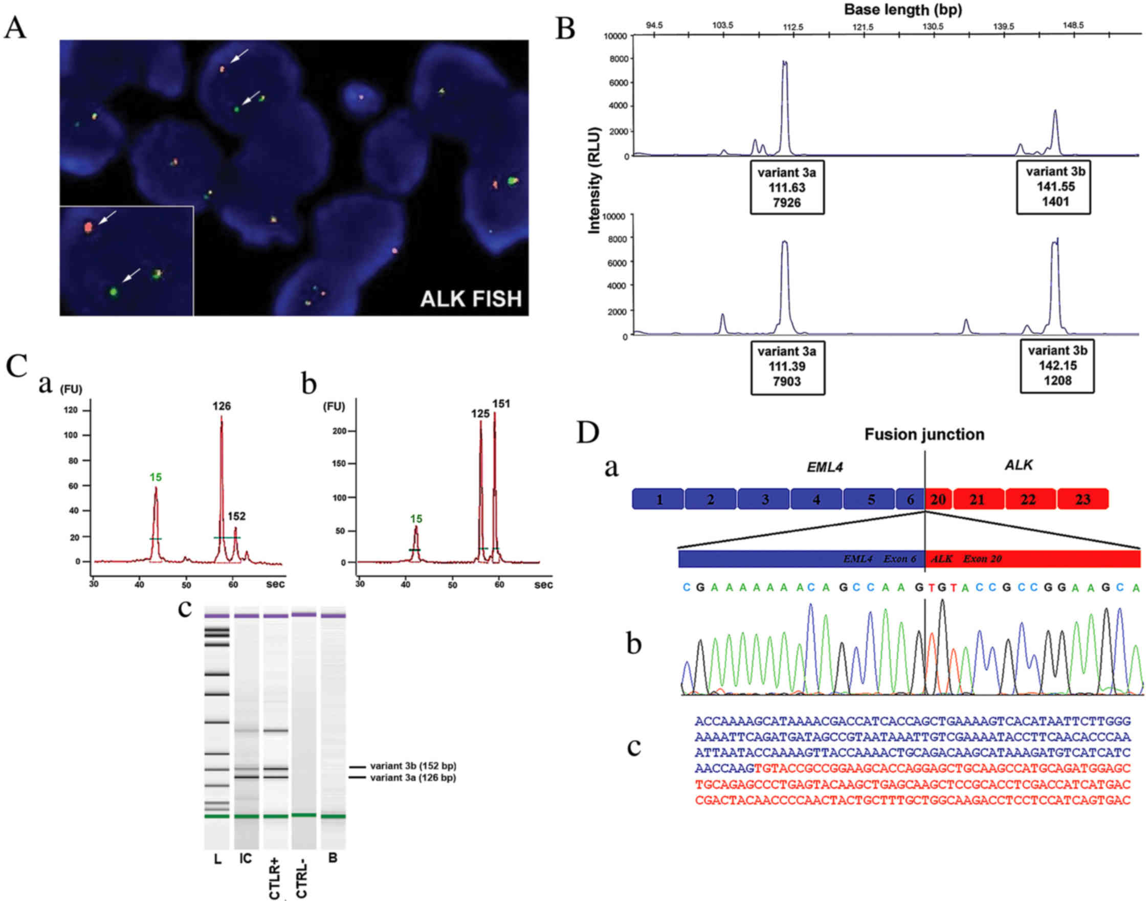
ALK rearrangement was assessed by FISH
analysis. Single and multiple copies of the intact ALK
fusion together with the abnormal split pattern were observed in
the tumor cells of the tissues specimen with a frequency of 65%
ALK rearranged gene (combined EML4-ALK fusion and
5′end-ALK deletion), (Fig.
2A).
Identification of the EML4-ALK 3a/b
fusion variants
Two fragments of the expected length (112 and 142
bp) corresponding to the fusion 3a/b variants were detected during
the first round of RT-PCR, which is designed to detect the most
frequent 1,2 and 3a/b types of transcript. Using specific primers
for the amplification of the EML4-ALK 3a/b variants fusion
cDNA, the same two pair of PCR products were detected in the
NCI-H2228 cell line and in the IMT patient sample. These molecular
findings were confirmed using two different fluorescent fragment
analysis methods; the first was previously described by Sanders
et al (8), (Fig. 2B) and the second uses novel, fast and
efficient microchip electrophoresis to carefully detect ALK
variation through the use of capillary microelectrophoresis and
detection (Fig. 2C). Molecular
cloning and direct sequencing of each PCR product confirmed the
presence of the variant 3a fusion (linking exon 6 of EML4 to
exon 20 of ALK) and the 3b fusion, containing an insertion
of 33 bp mapped to intron 6 of EML4 that was located between
the same exons of EML4 and ALK (Fig. 2D).
Discussion
The World health organization (WHO) classification
of soft tissue tumors defines IMT as the neoplastic counterpart
among the wastebasket group of lesions previously known as
inflammatory pseudotumors (9). IMT
represents a proliferation of myofibroblastic cells, often
intermingled with lymphocytes, plasma cells and histiocytes, which
eventually forms a highly aggressive lesion (9). IMT is ubiquitous and margin-free
surgical resection is considered the only curative treatment. By
contrast, aggressive or metastatic lesions have no standard therapy
and chemotherapy is usually ineffective.
In the head and neck region, the larynx is the most
common location of IMT, while a few cases in the hypopharynx have
been reported (10,11). Clinical and radiological differential
diagnosis is broad and includes infections (tuberculosis and
mycoses), Wegener's granulomatosis, amyloidosis and other
malignancies, such as carcinoma, melanoma and salivary gland tumors
(10,11).
Up to 70% of IMTs harbour ALK gene
rearrangements resulting in the formation of a chimeric fusion
protein, which is detectable by FISH or immunohistochemistry
(2–5).
ALK is a receptor-type protein tyrosine kinase that is currently
being analysed by oncologists as an essential growth driver, which
defines its potential susceptibility to ALK inhibitors (7,12–15). The oncogenic function of ALK occurs as
a result of it forming a fusion protein with various gene partners
through chromosomal translocations, most commonly TPM3 or
TPM4 in IMT, resulting in oncogenic activation of ALK
(3). Alternatively, a small inversion
[inv2(2)(p21p23)] on the short arm of
chromosome 2 leads to a functional EML4-ALK fusion-type
tyrosine kinase (3–22) (Table I).
Initially described in non-small cell lung carcinoma (15), this abnormality was also identified in
~20% of ALK-rearranged pulmonary IMT cases (5). To date, a total of 10 cases of
EML4-ALK fusions in IMT have been reported (3,5,9). The present study described a
EML4-ALK rearranged IMT located in the hypopharynx. By using
two different fluorescent fragmentation analysis methods, the
specific 3a/b variants were identified within the RNA of the tumor
specimen and confirmed by standard Sanger sequencing. This
description of the ALK fusion 3a/b variants in an IMT case,
together with the few cases of ALK-EML4 fusions reported in
literature, corroborates the existing hypothesis that identical
ALK fusions detected in different tumor types may drive an
inappropriate activation of the same kinase signalling pathway,
which could be oncogenic in disparate cellular lineages. In
addition, the present case also noted the epithelioid appearance of
the tumor cells, which could potentially lead to a dismal clinical
course. Notably, Mariño-Enríquez et al (23) described ALK-positive intra-abdominal
IMT composed predominantly of sheets of round-to-epithelioid cells
significantly associated with aggressive course with rapid local
recurrences.
 | Table I.ALK fusion partners in IMT. |
Table I.
ALK fusion partners in IMT.
| Authors, year | IMT location | Patient
age/gender | ALK fusion | Refs. |
|---|
| Lawrence et
al, 2000 | Abdomen | 23 years/F | TMP3-ALK | (3) |
|
| Lung | 30 years/F | TMP3_ALK | (3) |
|
| Abdomen | 1 year/M | TMP4-ALK | (3) |
| Bridge et al,
2001 | Neck | 3 years/F | CLTC-ALK | (16) |
|
| Pelvis | 37 years/M | CLTC-ALK | (16) |
| Cools et al,
2002 | Abdomen | 4 months/M | CARS-ALK | (17) |
| Ma et al,
2003 | Abdomen | 1 year/M |
RANBP2-ALK | (18) |
|
| Abdomen | 7 months/M |
RANBP2-ALK | (18) |
| Debiec-Rychter
et al, 2003 | Bladder | 46 years/M |
ATIC-ALK | (19) |
| Panagopoulos et
al, 2006 | Abdomen | 23 years/M |
SEC31L1-ALK | (20) |
| Takeuchi et
al, 2011 | Lung | 45 years/M |
PPFIBP1-ALK | (21) |
|
| Lung | 34 years/F |
PPFIBP1-ALK | (21) |
| Wang et al,
2012 | Neck | 7 years/F |
DCTN1-ALK | (22) |
| Lovly et al,
2014 | Lung | 38 years/F |
EML4-ALK | (12) |
|
| Mesentery | 11 years/F |
LMNA-ALK | (12) |
|
| Shoulder | 1 year/F |
PRAKAR1A-ALK | (12) |
|
| Bladder | 26 years/F | FN1-ALK | (12) |
|
| Pelvis | 14 years/M | TFG-ALK | (12) |
In conclusion, to the best of our knowledge, the
present study describes for the first time the presence of the
EML4-ALK 3a/b variant in a case of malignant IMT of the
hypopharynx. This was achieved by integrating different
methodologies, which is considered the most suitable molecular
approach for gene fusion characterization. Furthermore, the
identification of ALK rearrangement in cases of malignant
IMTs may offer a rationale to adopt selective targeted therapies in
this ‘orphan’ tumor.
Acknowledgements
The present study was supported by the Italian
Ministry of Health (grant nos. RC1503LO51, 2010-2316264 and
RC1502AP18), the ‘5×1000′ voluntary contributions and Associazione
Italiana per la Ricerca sul Cancro (grant no. 12983).
References
|
1
|
Surabhi VR, Chua S, Patel RP, Takahashi N,
Lalwani N and Prasad SR: Inflammatory myofibroblastic tumors:
Current update. Radiol Clin North Am. 54:553–563. 2016. View Article : Google Scholar : PubMed/NCBI
|
|
2
|
Griffin CA, Hawkins AL, Dvorak C, Henkle
C, Ellingham T and Perlman EJ: Recurrent involvement of 2p23 in
inflammatory myofibroblastic tumors. Cancer Res. 59:2776–2780.
1999.PubMed/NCBI
|
|
3
|
Lawrence B, PerezAtayde A, Hibbard MK,
Rubin BP, Dal Cin P, Pinkus JL, Pinkus GS, Xiao S, Yi ES, Fletcher
CD and Fletcher JA: TPM3-ALK and TPM4-ALK oncogenes in inflammatory
myofibroblastic tumors. Am J Pathol. 157:377–384. 2000. View Article : Google Scholar : PubMed/NCBI
|
|
4
|
Coffin CM, Patel A, Perkins S,
ElenitobaJohnson KS, Perlman E and Griffin CA: ALK1 and p80
expression and chromosomal rearrangements involving 2p23 in
inflammatory myofibroblastic tumor. Mod Pathol. 14:569–576. 2001.
View Article : Google Scholar : PubMed/NCBI
|
|
5
|
Antonescu CR, Suurmeijer AJ, Zhang L, Sung
YS, Jungbluth AA, Travis WD, AlAhmadie H, Fletcher CD and Alaggio
R: Molecular characterization of inflammatory myofibroblastic
tumors with frequent ALK and ROS1 fusions and rare novel RET
rearrangement. Am J Surg Pathol. 39:957–967. 2015. View Article : Google Scholar : PubMed/NCBI
|
|
6
|
Butrynski JE, D'Adamo DR, Hornick JL, Dal
Cin P, Antonescu CR, Jhanwar SC, Ladanyi M, Capelletti M, Rodig SJ,
Ramaiya N, et al: Crizotinib in ALK-rearranged inflammatory
myofibroblastic tumor. N Engl J Med. 363:1727–1733. 2010.
View Article : Google Scholar : PubMed/NCBI
|
|
7
|
Mano H: ALKoma: A cancer subtype with a
shared target. Cancer Discov. 2:495–502. 2012. View Article : Google Scholar : PubMed/NCBI
|
|
8
|
Sanders H, Li H, Bruey JM, Scheerle JA,
MeloniEhrig AM, Kelly JC, Novick C and Albitar M: Exon scanning by
reverse transcriptase polymerase chain reaction for detection of
known and novel EML4-ALK fusion variants in non-small cell lung
cancer. Cancer Genet. 204:45–52. 2011. View Article : Google Scholar : PubMed/NCBI
|
|
9
|
Fletcher C, Bridge JA, Hogendoorn PC, et
al: WHO Classification of Tumours of Soft Tissue and Bone. IARC;
Lyon: 2013
|
|
10
|
Graefe H, Stellmacher F, Sotlar K,
Wollenberg B and Gehrking E: Inflammatory pseudotumor of the
hypopharynx: Clinical diagnosis, immunohistochemical findings and
treatment of this rare disease. In Vivo. 22:817–820.
2008.PubMed/NCBI
|
|
11
|
Nakayama K, Inoue Y, Aiba T, Kono K,
Wakasa K and Yamada R: Unusual CT and MR findings of inflammatory
pseudotumor in the parapharyngeal space: Case report. AJNR Am J
Neuroradiol. 22:1394–1397. 2001.PubMed/NCBI
|
|
12
|
Lovly CM, Gupta A, Lipson D, Otto G,
Brennan T, Chung CT, Borinstein SC, Ross JS, Stephens PJ, Miller VA
and Coffin CM: Inflammatory myofibroblastic tumors harbor multiple
potentially actionable kinase fusions. Cancer Discov. 4:889–895.
2014. View Article : Google Scholar : PubMed/NCBI
|
|
13
|
Cook JR, Dehner LP, Collins MH, Ma Z,
Morris SW, Coffin CM and Hill DA: Anaplastic lymphoma kinase (ALK)
expression in the inflammatory myofibroblastic tumor: A comparative
immunohistochemical study. Am J Surg Pathol. 25:1364–1371. 2001.
View Article : Google Scholar : PubMed/NCBI
|
|
14
|
Ni C, Xu YY, Zhou SH and Wang SQ:
Differential diagnosis of inflammatory myofibroblastic tumour and
low-grade myofibroblastic sarcoma: Two case reports with a
literature review. J Intern Med Res. 39:311–320. 2011. View Article : Google Scholar
|
|
15
|
Soda M, Choi YL, Enomoto M, Takada S,
Yamashita Y, Ishikawa S, Fujiwara S, Watanabe H, Kurashina K,
Hatanaka H, et al: Identification of the transforming EML4-ALK
fusion gene in non-small-cell lung cancer. Nature. 448:561–566.
2007. View Article : Google Scholar : PubMed/NCBI
|
|
16
|
Bridge JA, Kanamori M, Ma Z, Pickering D,
Hill DA, Lydiatt W, Lui MY, Colleoni GW, Antonescu CR, Ladanyi M
and Morris SW: Fusion of the ALK gene to the clathrin heavy chain
gene, CLTC, in inflammatory myofibroblastic tumor. Am J Pathol.
159:411–415. 2001. View Article : Google Scholar : PubMed/NCBI
|
|
17
|
Cools J, Wlodarska I, Somers R, Mentens N,
Pedeutour F, Maes B, De Wolf-Peeters C, Pauwels P, Hagemeijer A and
Marynen P: Identification of novel fusion partners of ALK, the
anaplastic lymphoma kinase, in anaplastic large-cell lymphoma and
inflammatory myofibroblastic tumor. Genes Chromosomes Cancer.
34:354–362. 2002. View Article : Google Scholar : PubMed/NCBI
|
|
18
|
Ma Z, Hill DA, Collins MH, Morris SW,
Sumegi J, Zhou M, Zuppan C and Bridge JA: Fusion of ALK to the
Ran-binding protein 2 (RANBP2) gene in inflammatory myofibroblastic
tumor. Genes Chromosomes Cancer. 37:98–1053. 2003. View Article : Google Scholar : PubMed/NCBI
|
|
19
|
Debiec-Rychter M, Marynen P, Hagemeijer A
and Pauwels P: ALK-ATIC fusion in urinary bladder inflammatory
myofibroblastic tumor. Genes Chromosomes Cancer. 38:187–190. 2003.
View Article : Google Scholar : PubMed/NCBI
|
|
20
|
Panagopoulos I, Nilsson T, Domanski HA,
Isaksson M, Lindblom P, Mertens F and Mandahl N: Fusion of the
SEC31L1 and ALK genes in an inflammatory myofibroblastic tumor. Int
J Cancer. 118:1181–1161. 2006. View Article : Google Scholar : PubMed/NCBI
|
|
21
|
Takeuchi K, Soda M, Togashi Y, Sugawara E,
Hatano S, Asaka R, Okumura S, Nakagawa K, Mano H and Ishikawa Y:
Pulmonary inflammatory myofibroblastic tumor expressing a novel
fusion, PPFIBP1-ALK: Reappraisal of anti-ALK immunohistochemistry
as a tool for novel ALK fusion identification. Clin Cancer Res.
17:3341–3381. 2011. View Article : Google Scholar : PubMed/NCBI
|
|
22
|
Wang X, Krishnan C, Nguyen EP, Meyer KJ,
Oliveira JL, Yang P, Yi ES, EricksonJohnson MR, Yaszemski MJ, Maran
A and Oliveira AM: Fusion of dynactin 1 to anaplastic lymphoma
kinase in inflammatory myofibroblastic tumor. Hum Pathol.
43:2047–2052. 2012. View Article : Google Scholar : PubMed/NCBI
|
|
23
|
Mariño-Enríquez A, Wang WL, Roy A,
LopezTerrada D, Lazar AJ, Fletcher CD, Coffin CM and Hornick JL:
Inflammatory myofibroblastic sarcoma: An aggressive intra-abdominal
variant of inflammatory myofibroblastic tumor with nuclear membrane
or perinuclear ALK. Am J Surg Pathol. 35:135–144. 2011. View Article : Google Scholar : PubMed/NCBI
|