Introduction
Esophageal cancer is the fourth leading cause of
cancer mortality worldwide, and is one of the most aggressive types
of digestive tract tumor (1). The
poor prognosis of esophageal cancer is due to the high incidence
rate of lymph node metastasis and the invasion of neighboring
organs within a 5-year period following surgery, despite
improvements in treatment (2).
Investigations into the mechanisms underlying invasion and
metastasis in esophageal cancer may contribute to an improved
patient prognosis.
Loss of epithelial-cadherin (E-cad) and simultaneous
acquisition of vimentin (Vim) in epithelial cells is an alteration
indicative of the epithelial-mesenchymal transition (EMT), which,
in tumors, is a crucial step for cancer cells to obtain malignant
properties (3). The epithelial
transmembrane glycoprotein E-cad, a cell adhesion molecule, can
regulate cell motility in a calcium-dependent manner (4). Dysregulated E-cad expression is able to
induce impairments in adhesive ability and abnormalities in
differentiation due to genetic mutation, promoter region
hypermethylation and suppression of transcription, allowing tumor
cells to dissociate from the primary lesion (5).
Transcriptional repression is considered one of the
most critical mechanisms underlying the downregulation of E-cad
expression. The type III intermediate filament protein Vim is
normally expressed in cells of mesenchymal origin. Vim is a
component of the cytoskeleton, is involved in cell adhesion and
migration, and also regulates apoptosis and signal transduction in
tumor tissues (6–8). EMT, as a target involved in invasion and
metastasis mechanisms, has been revealed to result in the poor
prognosis of patients with triple negative breast cancer and
pancreatic cancer (9,10). EMT may be directly or indirectly
mediated by various factors including snail, slug and twist.
Signaling of transforming growth factor-β is considered to exert a
pivotal function in triggering and promoting EMT (11). Periostin (POSTN) is an important
downstream target in this process, and has been implicated to
mediate the initiation of EMT triggered by twist in prostate cancer
(11). As such, POSTN is considered
as a key proteins involved in EMT.
POSTN, also termed osteoblast-specific factor 2, is
an evolutionarily conserved extracellular matrix (ECM) protein and
a member of the fasciclin family (12), and has been reported to be
overexpressed in various types of human cancer (13,14). POSTN
is able to interact with other ECM proteins, specific cell surface
receptors and integrins through multiple signal pathways affecting
metastasis, invasion and angiogenesis in cancer development
(15,16). Previous experimental results have
revealed that POSTN has a role in mediating the promotion of
esophageal cancer (16,17). However, reports have yet to elucidate
the association between POSTN and EMT in esophageal squamous cell
carcinoma (ESCC). The present study hypothesized that POSTN is
involved in the EMT of ESCC cells, and consequently facilitates
cancer invasion and metastasis.
In the present study, the expression levels of
POSTN, E-cad and Vim in ESCC cells and corresponding
paracarcinomatous normal epithelium were examined using
immunohistochemistry. The associations of POSTN, E-cad and Vim with
clinicopathological features and the patients' survival status were
investigated using univariate and multivariate analyses to verify
whether POSTN is useful for predicting tumor malignant properties
and poor prognosis in patients with ESCC.
Materials and methods
Patients and tumor biopsies
A total of 58 tumor specimens were obtained from
patients with ESCC who underwent curative esophagectomy at the
Department of Thoracic Surgery (Anhui Medical University, Hefei,
China), between January 2007 and January 2008. All cases were
diagnosed on a clinical basis with pathological confirmation and no
patient received additional treatment prior to surgery. Of the 58
patients, there were 50 males and 8 females, with a median age of
61 years (range, 40–82). The obtained patient characteristics
included tumor node metastasis (TNM) stage I/II (n=16), TNM stage
III/IV (n=42), poor differentiation (n=33), moderate and well
differentiation (n=25), lymphatic metastasis (n=42) and vascular
invasion (n=44). Clinical status was determined in accordance with
the National Comprehensive Cancer Network Clinical Practice
Guidelines in Oncology Version 2009 (18). All patients’ tissue specimens were
fixed in 10% formalin, paraffin-embedded and stained with
hematoxylin and eosin for immunohistochemical analysis. Clinical
and pathological data of all patients was obtained from medical
records, and the follow-up deadline was 17th March, 2015. The
present study was approved by the Institutional Review Board of the
Anhui Provincial Hospital (Anhui Medical University), and written
informed consent was obtained from all participants.
Immunohistochemistry and
evaluation
Immunohistochemical staining of POSTN, E-cad, and
Vim in ESCC and adjacent normal tissues was performed by the
two-step method (Beijing Zhongshan Jinqiao Biotechnology Co., Ltd.,
Beijing, China). Specimen sections (thickness, 4 µm) were dewaxed
by dimethylbenzene, rinsed in phosphate-buffered saline (PBS),
boiled under high pressure in citrate-buffer (10 mmol/l; pH=6.0)
for 2 min for antigen retrieval, and treated with 3% hydrogen
peroxide to block endogenous peroxidase activity. Each section was
incubated with the primary mouse anti-E-cad monoclonal antibody
(cat. no. ZM-0092; 1:200), mouse anti-Vim monoclonal antibody (cat.
no. ZM-0260) (both 1:200; Beijing Zhongshan Jinqiao Biotechnology
Co., Ltd.), and mouse anti-POSTN polyclonal antibody (cat. no.
TA804575; 1:150; Abcam, Cambridge, UK) at 4°C overnight. Subsequent
to washing with PBS, the sections were incubated with the
appropriate HRP-labeled secondary antibody (goat antimouse IgG;
pre-diluted; from PV-6000 Polink-1 HRP DAB Detection System
One-step polymer detection system for Mouse and Rabbit antibodies,
Beijing Zhongshan Jinqiao Biotechnology Co., Ltd.) at room
temperature for 30 min. The reaction products were developed using
a 3,3-diaminobenzidine (DAB; Beijing Zhongshan Jinqiao
Biotechnology Co., Ltd.) solution for 10 min, and dehydrated
following hematoxylin counterstaining. Negative control slides were
processed with PBS and a confirmed positive tissue section was used
as a positive control.
Semi-quantitative integration was used to assess the
results of immunohistochemistry. Yellow-brown granules located in
cytoplasm and cytoplasmic membrane were defined as positive
staining. Staining intensity was scored as follows: 0, negative; 1,
yellow; 2, brown; and 3, sepia. An image of the background was
taken for reference. Scores for the average percentage of
immunopositive cells were based on the number of stained cells per
100 cells in 10 high-power fields (magnification, ×400) from the
hot spots in low power fields (magnification, ×100), and rated as
follows: 0, <10% positive cells; 1, 10–25%; 2, >25–50%; 3,
>50–75%; and 4, >75% positive cells. The final assessment
based on their product was as follows: 0, negative (−); 1–4, weak
positive (+); 5–8, moderate positive (++); and 9–12, strong
positive (+++). The slices were independently evaluated by two
pathologists who were blind to the clinical diagnosis.
Statistical analysis
Statistical analyses were completed with SPSS 13.0
(SPSS, Inc., Chicago, IL, USA). Experiments were performed in
triplicate. All data were presented as the mean ± standard
deviation. The χ2 test was used as appropriate for the
comparison of variables. Correlations between POSTN and EMT markers
were performed using the Spearman's rank correlation test. Overall
survival (OS) and disease-free survival (DFS) rates were determined
using a Kaplan-Meier analysis and a log-rank test. The Cox
regression model was used for multivariate survival analysis.
P<0.05 was considered to indicate a statistically significant
difference.
Results
Expression levels of E-cad, Vim, and
POSTN in the intratumoral and paracarcinomatous tissues of
ESCC
E-cad was observed to be expressed on the squamous
cell membranes, and attenuated notably in the ESCC tissues compared
with the paracarcinomatous epithelium (32.8 vs. 74.1%;
χ2=19.957; P<0.001; Fig.
1). Vim was located in the cytoplasm of fibroblasts,
endothelial cells and squamous cells; the focus of the present
study was just squamous cells. Vim expression was positive in 24/58
intratumoral cases (41.4%, χ2=26.973, P<0.001),
significantly more than 1/58 paracarcinomatous cases (1.7%;
Fig. 1). POSTN expression was
detected in the cytoplasm of fibroblasts, endothelial cells, and
squamous cells. Similar to Vim, the present study also analyzed
POSTN expression in squamous cells. The positive rate of POSTN
expressed in the tumor squamous cells was 41/58 (70.7%,
χ2=11.293, P=0.001), notably higher than 23/58 (39.7%)
in the paracarcinomatous epithelium (Fig.
1). Data above are presented in Table
I.
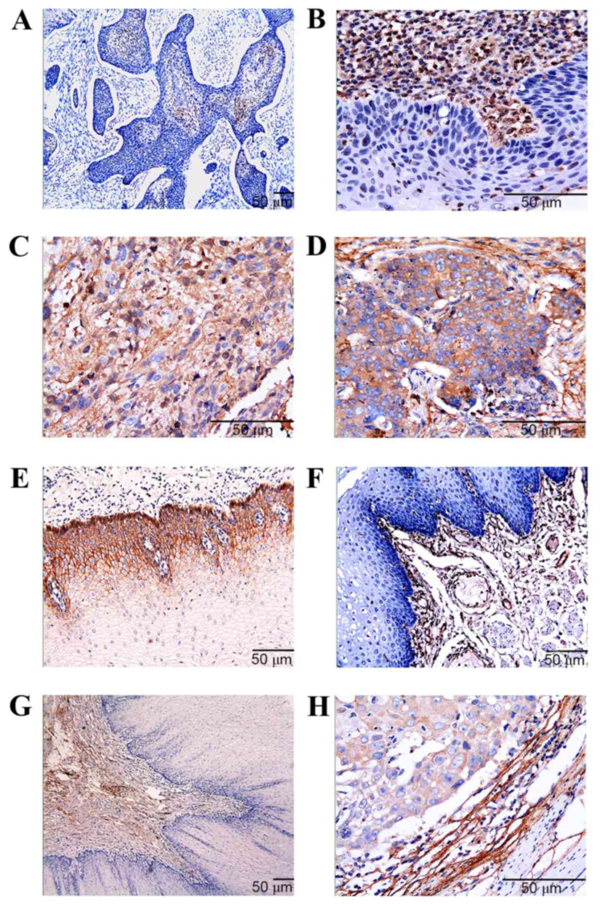 | Figure 1.Immunohistochemistry demonstrates
expression diversity of E-cad, Vim, and POSTN in the ESCC tissues.
(A) E-cad expression in the tumor cells (magnification, ×100)
receded significantly comparing with the strong staining in (E) the
paracarcinomatous squamous cells (magnification, ×200). (B) Vim
expression in the tumor cells (magnification, ×400) was stronger
than that in (F) the paracarcinomatous squamous cells
(magnification, ×200). (C) POSTN was observed to be overexpressed
in the cancer cells (magnification, ×400), whereas it demonstrated
low positive rate in (G) the paracarcinomatous squamous cells
(magnification, ×100). (D) POSTN expression in the tumor tissue
with poor differentiation (magnification, ×400) was denser than
that in (H) the well-differentiated tumor tissue (magnification,
×400). Scale bar=50 µm. E-cad, epithelial cadherin; Vim, vimentin;
POSTN, periostin; ESCC, esophageal squamous cell carcinoma. |
 | Table I.Positive rate of E-cad, Vim, and POSTN
expression in the squamous cells of tumor and paracarcinomatous
tissues in esophageal squamous cell carcinoma. |
Table I.
Positive rate of E-cad, Vim, and POSTN
expression in the squamous cells of tumor and paracarcinomatous
tissues in esophageal squamous cell carcinoma.
|
| Tumor tissues | Paracarcinomatous
tissues |
|
|---|
|
|
|
|
|
|---|
| Variable | Positive rate
(%) | Positive rate
(%) | χ2 | P-value |
|---|
| E-cad | 19/58 (32.8) | 43/58 (74.1) | 19.957 | <0.001 |
| Vim | 24/58 (41.4) | 1/58 (1.7) | 26.973 | <0.001 |
| POSTN | 41/58 (70.7) | 23/58 (39.7) | 11.293 |
0.001 |
Correlations between POSTN and the EMT
hallmark proteins in ESCC squamous cells
The present study analyzed the correlations between
POSTN and the EMT hallmark proteins E-cad and Vim in the ESCC
squamous cells according to the immunohistochemical score. There
was an inverse correlation between expression levels of E-cad and
POSTN (r=−0.339; P<0.001; Table
II). No case expressing E-cad was observed in the samples that
had a POSTN score >6. By contrast, Vim expression was found to
have a positive correlation with POSTN expression (r=0.130;
P<0.001; Table II). Intensity of
Vim expression was weak to moderate/high, and Vim was observed to
be expressed positively in the presence of POSTN. This trend in our
experiment revealed that POSTN overexpression was associated with
E-cad attenuation as well as Vim enrichment (Fig. 2).
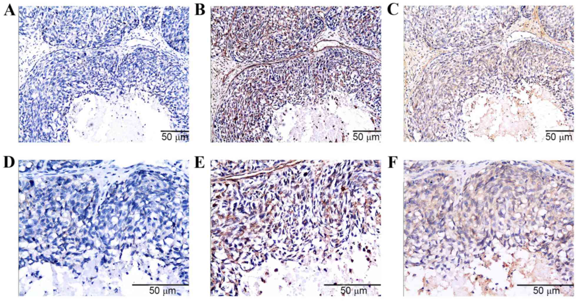 | Figure 2.Consecutive slides from the same
specimen of ESCC tissue exhibited the reciprocal association
between the expression levels of POSTN and the EMT hallmark
proteins. (A) Negative staining of E-cad in the cancer cells under
low power lens (magnification, ×200) and (D) high power lens
(magnification, ×400). (B) Positive staining of Vim in the cancer
cells from the same location of the same specimen under low power
lens (magnification, ×200) and (E) high power lens (magnification,
×400). (C) Positive staining of POSTN in the cancer cells from the
same location of the same specimen under low power lens
(magnification, ×200) and (F) high power lens (magnification,
×400). Scale bar=50 µm. ESCC, esophageal squamous cell carcinoma;
POSTN, periostin; EMT, epithelial-mesenchymal transition; E-cad,
epithelial cadherin; Vim, vimentin. |
| Table II.Associations between POSTN and the EMT
hallmark proteins in the esophageal squamous cell carcinoma
squamous cells. |
Table II.
Associations between POSTN and the EMT
hallmark proteins in the esophageal squamous cell carcinoma
squamous cells.
|
| E-cad (SC) | Vim (SC) |
|---|
|
|
|
|
|---|
| POSTN (SC) | r-value | P-value | r-value | P-value |
|---|
|
| −0.339 | <0.001 | 0.130 | <0.001 |
Associations between
clinicopathological characteristics and expression levels of E-cad,
Vim, and POSTN in the ESCC squamous cells
The associations between expression levels of the
proteins in the ESCC squamous cells and clinicopathological factors
were analyzed by the χ2 test. E-cad expression exhibited
a higher positive rate in the tumor tissues with advanced TNM stage
(χ2=12.994; P<0.001), lymphatic metastasis
(χ2=12.994; P<0.001) and venous invasion
(χ2=4.981; P=0.026). Conversely, there was no
statistical association between E-cad status and tumor
differentiation (P=0.647). The positive rate of Vim expression was
elevated significantly in the cases with a terminal TNM stage
(χ2=4.665; P=0.039) and lymphatic metastasis
(χ2=4.665; P=0.039), but did not exhibit an association
with the degree of tumor differentiation (P=0.072) or venous
invasion (P=0.121). Notably, the overexpression of POSTN was
validated to be significantly associated with all
clinicopathological factors in this study, including TNM stage
(χ2=7.739; P=0.009), tumor differentiation degree
(χ2=15.106; P<0.001; Fig.
1), lymphatic metastasis (χ2=7.739; P=0.009) and
venous invasion (χ2=10.896; P=0.002). These results are
presented in detail in Table
III.
| Table III.Associations between
clinicopathological characteristics and positive rate of E-cad, Vim
and POSTN in ESCC squamous cells. |
Table III.
Associations between
clinicopathological characteristics and positive rate of E-cad, Vim
and POSTN in ESCC squamous cells.
|
|
| E-cad | Vim | POSTN |
|---|
|
|
|
|
|
|
|---|
| Variable | n | Positive rate,
% | χ2 | P-value | Positive rate,
% | χ2 | P-value | Positive rate,
% | χ2 | P-value |
|---|
| TNM stage |
|
|
|
|
|
|
|
|
|
|
|
I/II | 16 | 68.8 | 12.994 | <0.001 | 18.8 | 4.665 | 0.039 | 43.8 | 7.739 | 0.009 |
|
III/IV | 42 | 19.0 |
|
| 50.0 |
|
| 81.0 |
|
|
| Differentiation
degree |
|
|
|
|
|
|
|
|
|
|
|
Low | 33 | 30.3 | 0.210 | 0.647 | 51.5 | 3.243 | 0.072 | 90.9 | 15.106 | <0.001 |
|
Moderate/high | 25 | 36.0 |
|
| 28.0 |
|
| 44.0 |
|
|
| Lymphatic
metastasis |
|
|
|
|
|
|
|
|
|
|
|
Positive | 42 | 19.0 | 12.994 | <0.001 | 50.0 | 4.665 | 0.039 | 81.0 | 7.739 | 0.009 |
|
Negative | 16 | 68.8 |
|
| 18.8 |
|
| 43.8 |
|
|
| Venous
invasion |
|
|
|
|
|
|
|
|
|
|
|
Positive | 44 | 25.0 | 4.981 | 0.026 | 47.7 | 3.028 | 0.121 | 81.8 | 10.896 | 0.002 |
|
Negative | 14 | 57.1 |
|
| 21.4 |
|
| 35.7 |
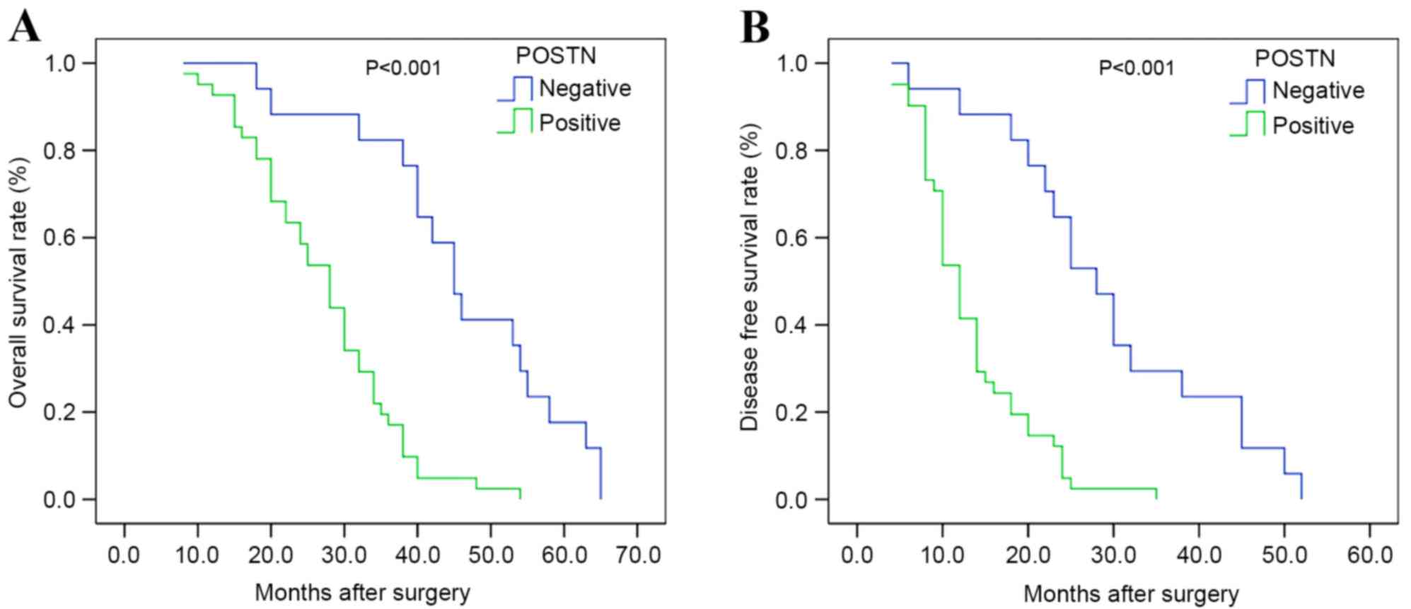
Survival analysis
Survival analysis according to the Kaplan-Meier
method revealed that the OS (27.171±1.584 months; Fig. 3A) and DFS (13.341±1.039 months;
Fig. 3B) of patients with positive
POSTN expression was markedly shorter than that of patients with
negative POSTN expression (OS, 45.824±3.408 months,
χ2=22.252, P<0.001; DFS, 29.471±3.155 months,
χ2=22.696, P<0.001). Similarly, patients with
positive Vim expression tended to have a poorer prognosis for OS
(27.833±2.235 vs. 36.029±2.628 months; χ2=5.936;
P=0.015) and DFS (14.125±1.809 vs. 20.853±2.145 months;
χ2=5.750; P=0.016). However, the OS (40.895±3.327
months) and DFS (25.263±3.100 months) of patients with positive
E-cad expression were markedly longer than those of patients with
negative E-cad expression (OS, 28.615±1.968 months,
χ2=9.255, P=0.002; DFS, 14.564±1.389 months,
χ2=9.667, P=0.002).
Multivariate analysis using the Cox regression model
indicated that POSTN positive expression (P=0.022) and TNM stage
(P=0.004) were independent prognostic factors for the OS of
patients with ESCC. POSTN positive expression (P=0.019) remained
one of the independent prognostic factors for the DFS of patients
with ESCC, along with venous invasion (P=0.004) and TNM stage
(P=0.008; Table IV).
| Table IV.Multivariate analysis of factors
associated with OS and DFS in the ESCC patients. |
Table IV.
Multivariate analysis of factors
associated with OS and DFS in the ESCC patients.
| Factors | OS RR | RR 95% CI | P-value | DFS RR | RR 95% CI | P-value |
|---|
| POSTN |
|
|
|
|
|
|
|
Positive vs. negative | 3.126 | 1.177–8.300 | 0.022 | 2.721 | 1.180–6.277 | 0.019 |
| E-cad |
|
|
|
|
|
|
|
Positive vs. negative | 0.852 | 0.378–1.920 | 0.699 | 0.687 | 0.305–1.547 | 0.365 |
| Vim |
|
|
|
|
|
|
|
Positive vs. negative | 1.067 | 0.524–2.170 | 0.859 | 1.122 | 0.569–2.212 | 0.739 |
| Gender |
|
|
|
|
|
|
| Male
vs. female | 1.918 | 0.812–4.526 | 0.137 | 2.085 | 0.877–4.957 | 0.096 |
| Age |
|
|
|
|
|
|
| ≥60 vs.
<60 | 1.345 | 0.590–3.066 | 0.481 | 1.823 | 0.810–4.104 | 0.147 |
| TNM stage |
|
|
|
|
|
|
| III/IV
vs. I/II | 4.600 | 1.649–12.834 | 0.004 | 3.788 | 1.422–10.090 | 0.008 |
| Differentiation
degree |
|
|
|
|
|
|
|
Moderate/high vs. low | 0.581 | 0.255–1.320 | 0.195 | 0.723 | 0.359–1.455 | 0.363 |
| Venous
invasion |
|
|
|
|
|
|
|
Positive vs. negative | 2.250 | 0.871–5.813 | 0.094 | 4.174 | 1.567–11.118 | 0.004 |
Discussion
EMT has been demonstrated to be involved in several
underlying mechanisms leading to tumor invasion, migration and the
induction of cancer stem-like cells in esophageal cancer (19,20). In
the present study, expression levels of E-cad and Vim in ESCC
squamous cells changed abnormally and inversely. Reduction of the
E-cad positive rate and elevation of the Vim positive rate in ESCC
tissues indicated that EMT occurred in the ESCC tissues. In
addition, attenuation of E-cad and enrichment of Vim in cancer
cells was associated with the invasion and metastasis of ESCC, thus
potentially affecting the prognosis of patients with ESCC.
The association between POSTN and EMT has been
investigated in several types of tumors. However, conclusions vary
depending on the type of cancer. POSTN is reported to be capable of
enhancing EMT of nicotine-mediated tumor cells in lung and gastric
cancer (21,22), whereas exhibits biphasic effects on
EMT in pancreatic cancer cells and even has a suppressive role in
the regulation of EMT in bladder cancer cells (23,24). The
present study aimed to elucidate the reciprocal correlation between
POSTN and EMT in ESCC, and revealed that POSTN overexpression was
associated with E-cad attenuation as well as Vim enrichment, via
immunohistochemistry.
Additionally, POSTN has been reported to be able to
upregulate snail and twist, which are regarded as negative
regulators of E-cad in transcription, and enhance protein kinase B
(Akt) phosphorylation to decrease E-cad expression (24). POSTN was deemed to be involved in the
crosstalk between epidermal growth factor receptor and integrins at
the plasma membranes, activating the Akt and the focal adhesion
kinase (FAK) signaling pathways consecutively to promote EMT in
non-small cell lung, ovarian and colon cancer (25–27).
Consequently, the present data showing the reciprocal correlations
between POSTN and the EMT markers E-cad and Vim in ESCC are
consistent with previous studies.
Considering the role of POSTN as a promotional
mediator of EMT in ESCC cells, upregulated POSTN with concomitant
repression of E-cad and overexpression of Vim may result in
advanced tumor characteristics. The present study therefore
demonstrated that POSTN expression was elevated in the ESCC tissues
with poor differentiation, lymphatic metastasis, venous invasion
and thus a severe TNM stage. In terms of the mechanism, POSTN was
previously reported to be able to combine to multiple integrin
receptors including αvβ3, αvβ5, and α6β4, partially regulating the
intracellular signaling pathways linked to phosphatidylinositol 3
kinase (PI3K) /AKT as well as FAK, and sequentially induce the
proliferation, metastasis and invasion of cancer cells, and alter
tumor microenvironment (11,16,28). The
results of the present study validate the negative influences on
the ESCC tissue due to the increased POSTN expression in cancer
cells. In addition, previous studies of POSTN in esophageal cancer
accentuated the significance of microenvironment invasion mediated
by POSTN, suggesting that POSTN may induce vascular endothelial
growth factor expression and cooperate with mutant P53 to promote
tumor angiogenesis and invasion in microenvironment (16,29,30). These
findings suggest that POSTN may be responsible for malignant
clinicopathological properties, and could therefore be considered
as a predictive marker for the presence of lymph node and blood
vessel invasion as well as histologically poor differentiation.
Since the functions of POSTN lead to the aggressive
properties in ESCC cells, the survival period could be affected in
the patients with these features induced by upregulated POSTN
expression. The OS and DFS of POSTN positive patients with ESCC
were both markedly shorter than that of the POSTN negative patients
in the univariate analysis. Additionally, multivariate analysis
revealed that overexpression of POSTN in the ESCC cells can predict
poor prognosis independently in OS and DFS. These data are in
agreement with the negative impact of POSTN on the patients'
survival in non-small cell lung cancer, breast, colorectal,
prostate, and ovarian cancers (31–35). The
present results therefore indicated the pivotal role of POSTN in
the progression of invasion and metastasis in ESCC tissues, as well
as the prognostic value in the survival of patients with ESCC.
There were several limitations to the present study.
Immunohistochemistry could only reflect the correlations between
POSTN and EMT. Therefore, transfection of target genes, cytological
analyses and xenografts on animal models are required to further
the understanding of these correlations. In addition, a bias may
have occurred due to the insufficient sample amount. The above
limitations made the present conclusions preliminary.
In conclusion, the present study revealed that POSTN
could be regarded as an upregulated factor in ESCC cells, promoting
EMT, facilitating the invasion and metastasis of tumor cells, thus
leading to a poor prognosis. These results indicated that
suppression of the regulation between POSTN and EMT may provide a
valid means to control tumor metastasis and the relapse of patients
with ESCC.
Acknowledgements
The authors would like to thank Dr Ke Chen and Dr
Xiao-Qiu Wang (Department of Pathology, Anhui Provincial Hospital,
Hefei, China) for their assistance with pathology. The present
study was partly supported by the National Natural Science
Foundation of China (grant nos. 81472329 and 81201906).
References
|
1
|
Lin Y, Totsuka Y, He Y, Kikuchi S, Qiao Y,
Ueda J, Wei W, Inoue M and Tanaka H: Epidemiology of esophageal
cancer in Japan and China. J Epidemiol. 23:233–242. 2013.
View Article : Google Scholar : PubMed/NCBI
|
|
2
|
Tu CC, Hsu PK, Chien LI, Liu WC, Huang CS,
Hsieh CC, Hsu HS and Wu YC: Prognostic histological factors in
patients with esophageal squamous cell carcinoma after preoperative
chemoradiation followed by surgery. BMC Cancer. 17:622017.
View Article : Google Scholar : PubMed/NCBI
|
|
3
|
Marcucci F, Stassi G and De Maria R:
Epithelial-mesenchymal transition: A new target in anticancer drug
discovery. Nat Rev Drug Discov. 15:311–325. 2016. View Article : Google Scholar : PubMed/NCBI
|
|
4
|
Miyoshi A, Kitajima Y, Sumi K, Sato K,
Hagiwara A, Koga Y and Miyazaki K: Snail and SIP1 increase cancer
invasion by upregulating MMP family in hepatocellular carcinoma
cells. Br J Cancer. 90:1265–1273. 2014. View Article : Google Scholar
|
|
5
|
Jang SY, Park SY, Lee HW, Choi YK, Park
KG, Yoon GS, Tak WY, Kweon YO, Hur K and Lee WK: The combination of
periostin overexpression and microvascular invasion is related to a
poor prognosis for hepatocellular carcinoma. Gut Liver. 10:948–954.
2016. View
Article : Google Scholar : PubMed/NCBI
|
|
6
|
Vardaki I, Ceder S, Rutishauser D,
Baltatzis G, Foukakis T and Panaretakis T: Periostin is identified
as a putative metastatic marker in breast cancer-derived exosomes.
Oncotarget. 7:74966–74978. 2016.PubMed/NCBI
|
|
7
|
Molena D, Mungo B, Stem M, Poupore AK,
Chen SY and Lidor AO: Does quality of care matter? A study of
adherence to national comprehensive cancer network guidelines for
patients with locally advanced esophageal cancer. J Gastrointest
Surg. 19:1739–1747. 2015. View Article : Google Scholar : PubMed/NCBI
|
|
8
|
Jiang Y, Liao L, Shrestha C, Ji S, Chen Y,
Peng J, Wang L, Liao E and Xie Z: Reduced expression of E-cadherin
and p120-catenin and elevated expression of PLC-γ1 and PIKE are
associated with aggressiveness of oral squamous cell carcinoma. Int
J Clin Exp Pathol. 8:9042–9051. 2015.PubMed/NCBI
|
|
9
|
Lanier MH, Kim T and Cooper JA: CARMIL2 is
a novel molecular connection between vimentin and actin essential
for cell migration and invadopodia formation. Mol Biol Cell.
26:4577–4588. 2015. View Article : Google Scholar : PubMed/NCBI
|
|
10
|
Paccione RJ, Miyazaki H, Patel V, Waseem
A, Gutkind JS, Zehner ZE and Yeudall WA: Keratin down-regulation in
vimentin-positive cancer cells is reversible by vimentin RNA
interference, which inhibits growth and motility. Mol Cancer Ther.
7:2894–2903. 2008. View Article : Google Scholar : PubMed/NCBI
|
|
11
|
Bayo P, Jou A, Stenzinger A, Shao C, Gross
M, Jensen A, Grabe N, Mende CH, Rados PV, Debus J, et al: Loss of
SOX2 expression induces cell motility via vimentin up-regulation
and is an unfavorable risk factor for survival of head and neck
squamous cell carcinoma. Mol Oncol. 9:1704–1719. 2015. View Article : Google Scholar : PubMed/NCBI
|
|
12
|
Jin MS, Hyun CL, Park IA, Kim JY, Chung
YR, Im SA, Lee KH, Moon HG and Ryu HS: SIRT1 induces tumor invasion
by targeting epithelial mesenchymal transition-related pathway and
is a prognostic marker in triple negative breast cancer. Tumour
Biol. 37:4743–4753. 2016. View Article : Google Scholar : PubMed/NCBI
|
|
13
|
Yan TT, Fu XL, Li J, Bian YN, Liu DJ, Hua
R, Ren LL, Li CT, Sun YW, Chen HY, et al: Downregulation of RPL15
may predict poor survival and associate with tumor progression in
pancreatic ductal adenocarcinoma. Oncotarget. 6:37028–37042.
2015.PubMed/NCBI
|
|
14
|
Hu Q, Tong S, Zhao X, Ding W, Gou Y, Xu K,
Sun C and Xia G: Periostin mediates TGF-β-induced epithelial
mesenchymal transition in prostate cancer cells. Cell Physiol
Biochem. 36:799–809. 2015. View Article : Google Scholar : PubMed/NCBI
|
|
15
|
Litvin J, Selim AH, Montgomery MO, Lehmann
K, Rico MC, Devlin H, Bednarik DP and Safadi FF: Expression and
function of periostin-isoforms in bone. J Cell Biochem.
92:1044–1061. 2004. View Article : Google Scholar : PubMed/NCBI
|
|
16
|
Contié S, Voorzanger-Rousselot N, Litvin
J, Clézardin P and Garnero P: Increased expression and serum levels
of the stromal cell-secreted protein periostin in breast cancer
bone metastases. Int J Cancer. 128:352–360. 2011. View Article : Google Scholar : PubMed/NCBI
|
|
17
|
Wang W, Sun QK, He YF, Ma DC, Xie MR, Ji
CS and Hu B: Overexpression of periostin is significantly
correlated to the tumor angiogenesis and poor prognosis in patients
with esophageal squamous cell carcinoma. Int J Clin Exp Pathol.
7:593–601. 2014.PubMed/NCBI
|
|
18
|
Underwood TJ, Hayden AL, Derouet M, Garcia
E, Noble F, White MJ, Thirdborough S, Mead A, Clemons N, Mellone M,
et al: Cancer-associated fibroblasts predict poor outcome and
promote periostin-dependent invasion in oesophageal adenocarcinoma.
J Pathol. 235:466–477. 2015. View Article : Google Scholar : PubMed/NCBI
|
|
19
|
Li S, Qin X, Li Y, Zhang X, Niu R, Zhang
H, Cui A, An W and Wang X: MiR-133a suppresses the migration and
invasion of esophageal cancer cells by targeting the EMT regulator
SOX4. Am J Transl Res. 7:1390–1403. 2015.PubMed/NCBI
|
|
20
|
Sato F, Kubota Y, Natsuizaka M, Maehara O,
Hatanaka Y, Marukawa K, Terashita K, Suda G, Ohnishi S, Shimizu Y,
et al: EGFR inhibitors prevent induction of cancer stem-like cells
in esophageal squamous cell carcinoma by suppressing
epithelial-mesenchymal transition. Cancer Biol Ther. 16:933–940.
2015. View Article : Google Scholar : PubMed/NCBI
|
|
21
|
Wu SQ, Lv YE, Lin BH, Luo LM, Lv SL, Bi AH
and Jia YS: Silencing of periostin inhibits nicotine-mediated tumor
cell growth and epithelial-mesenchymal transition in lung cancer
cells. Mol Med Rep. 7:875–880. 2013.PubMed/NCBI
|
|
22
|
Liu Y and Liu BA: Enhanced proliferation,
invasion, and epithelial-mesenchymal transition of
nicotine-promoted gastric cancer by periostin. World J
Gastroenterol. 17:2674–2680. 2011. View Article : Google Scholar : PubMed/NCBI
|
|
23
|
Kanno A, Satoh K, Masamune A, Hirota M,
Kimura K, Umino J, Hamada S, Satoh A, Egawa S, Motoi F, et al:
Periostin, secreted from stromal cells, has biphasic effect on cell
migration and correlates with the epithelial to mesenchymal
transition of human pancreatic cancer cells. Int J Cancer.
122:2707–2718. 2008. View Article : Google Scholar : PubMed/NCBI
|
|
24
|
Kim CJ, Sakamoto K, Tambe Y and Inoue H:
Opposite regulation of epithelial-to-mesenchymal transition and
cell invasiveness by periostin between prostate and bladder cancer
cells. Int J Oncol. 38:1759–1766. 2011.PubMed/NCBI
|
|
25
|
Soltermann A, Tischler V, Arbogast S,
Braun J, Probst-Hensch N, Weder W, Moch H and Kristiansen G:
Prognostic significance of epithelial-mesenchymal and
mesenchymal-epithelial transition protein expression in non-small
cell lung cancer. Clin Cancer Res. 14:7430–7437. 2008. View Article : Google Scholar : PubMed/NCBI
|
|
26
|
Gillan L, Matei D, Fishman DA, Gerbin CS,
Karlan BY and Chang DD: Periostin secreted by epithelial ovarian
carcinoma is a ligand for alpha (V)beta(3) and alpha(V)beta(5)
integrins and promotes cell motility. Cancer Res. 62:5358–5364.
2002.PubMed/NCBI
|
|
27
|
Bao S, Ouyang G, Bai X, Huang Z, Ma C, Liu
M, Shao R, Anderson RM, Rich JN and Wang XF: Periostin potently
promotes metastatic growth of colon cancer by augmenting cell
survival via the Akt/PKB pathway. Cancer Cell. 5:329–339. 2004.
View Article : Google Scholar : PubMed/NCBI
|
|
28
|
Mosher DF, Johansson MW, Gillis ME and
Annis DS: Periostin and TGF-β-induced protein: Two peas in a pod?
Crit Rev Biochem Mol Biol. 50:427–439. 2015.PubMed/NCBI
|
|
29
|
Heidari P, Esfahani SA, Turker NS, Wong G,
Wang TC, Rustgi AK and Mahmood U: Imaging of secreted extracellular
periostin, an important marker of invasion in the tumor
microenvironment in esophageal cancer. J Nucl Med. 56:1246–1251.
2015. View Article : Google Scholar : PubMed/NCBI
|
|
30
|
Wong GS, Lee JS, Park YY, Klein-Szanto AJ,
Waldron TJ, Cukierman E, Herlyn M, Gimotty P, Nakagawa H and Rustgi
AK: Periostin cooperates with mutant p53 to mediate invasion
through the induction of STAT1 signaling in the esophageal tumor
microenvironment. Oncogenesis. 2:e592013. View Article : Google Scholar : PubMed/NCBI
|
|
31
|
Hong LZ, Wei XW, Chen JF and Shi Y:
Overexpression of periostin predicts poor prognosis in non-small
cell lung cancer. Oncol Lett. 6:1595–1603. 2013.PubMed/NCBI
|
|
32
|
Nuzzo PV, Rubagotti A, Argellati F, Di
Meglio A, Zanardi E, Zinoli L, Comite P, Mussap M and Boccardo F:
Prognostic value of preoperative serum levels of periostin (PN) in
early breast cancer (BCa). Int J Mol Sci. 16:17181–17192. 2015.
View Article : Google Scholar : PubMed/NCBI
|
|
33
|
Li Z, Zhang X, Yang Y, Yang S, Dong Z, Du
L, Wang L and Wang C: Periostin expression and its prognostic value
for colorectal cancer. Int J Mol Sci. 16:12108–12118. 2015.
View Article : Google Scholar : PubMed/NCBI
|
|
34
|
Nuzzo PV, Rubagotti A, Zinoli L, Ricci F,
Salvi S, Boccardo S and Boccardo F: Prognostic value of stromal and
epithelial periostin expression in human prostate cancer:
Correlation with clinical pathological features and the risk of
biochemical relapse or death. BMC Cancer. 12:6252012. View Article : Google Scholar : PubMed/NCBI
|
|
35
|
Ryner L, Guan Y, Firestein R, Xiao Y, Choi
Y, Rabe C, Lu S, Fuentes E, Huw LY, Lackner MR, et al: Upregulation
of periostin and reactive stroma is associated with primary
chemoresistance and predicts clinical outcomes in epithelial
ovarian cancer. Clin Cancer Res. 21:2941–2951. 2015. View Article : Google Scholar : PubMed/NCBI
|