Spandidos Publications Logo
  • About
    • About Spandidos
    • Aims and Scopes
    • Abstracting and Indexing
    • Editorial Policies
    • Reprints and Permissions
    • Job Opportunities
    • Terms and Conditions
    • Contact
  • Journals
    • All Journals
    • Oncology Letters
      • Oncology Letters
      • Information for Authors
      • Editorial Policies
      • Editorial Board
      • Aims and Scope
      • Abstracting and Indexing
      • Bibliographic Information
      • Archive
    • International Journal of Oncology
      • International Journal of Oncology
      • Information for Authors
      • Editorial Policies
      • Editorial Board
      • Aims and Scope
      • Abstracting and Indexing
      • Bibliographic Information
      • Archive
    • Molecular and Clinical Oncology
      • Molecular and Clinical Oncology
      • Information for Authors
      • Editorial Policies
      • Editorial Board
      • Aims and Scope
      • Abstracting and Indexing
      • Bibliographic Information
      • Archive
    • Experimental and Therapeutic Medicine
      • Experimental and Therapeutic Medicine
      • Information for Authors
      • Editorial Policies
      • Editorial Board
      • Aims and Scope
      • Abstracting and Indexing
      • Bibliographic Information
      • Archive
    • International Journal of Molecular Medicine
      • International Journal of Molecular Medicine
      • Information for Authors
      • Editorial Policies
      • Editorial Board
      • Aims and Scope
      • Abstracting and Indexing
      • Bibliographic Information
      • Archive
    • Biomedical Reports
      • Biomedical Reports
      • Information for Authors
      • Editorial Policies
      • Editorial Board
      • Aims and Scope
      • Abstracting and Indexing
      • Bibliographic Information
      • Archive
    • Oncology Reports
      • Oncology Reports
      • Information for Authors
      • Editorial Policies
      • Editorial Board
      • Aims and Scope
      • Abstracting and Indexing
      • Bibliographic Information
      • Archive
    • Molecular Medicine Reports
      • Molecular Medicine Reports
      • Information for Authors
      • Editorial Policies
      • Editorial Board
      • Aims and Scope
      • Abstracting and Indexing
      • Bibliographic Information
      • Archive
    • World Academy of Sciences Journal
      • World Academy of Sciences Journal
      • Information for Authors
      • Editorial Policies
      • Editorial Board
      • Aims and Scope
      • Abstracting and Indexing
      • Bibliographic Information
      • Archive
    • International Journal of Functional Nutrition
      • International Journal of Functional Nutrition
      • Information for Authors
      • Editorial Policies
      • Editorial Board
      • Aims and Scope
      • Abstracting and Indexing
      • Bibliographic Information
      • Archive
    • International Journal of Epigenetics
      • International Journal of Epigenetics
      • Information for Authors
      • Editorial Policies
      • Editorial Board
      • Aims and Scope
      • Abstracting and Indexing
      • Bibliographic Information
      • Archive
    • Medicine International
      • Medicine International
      • Information for Authors
      • Editorial Policies
      • Editorial Board
      • Aims and Scope
      • Abstracting and Indexing
      • Bibliographic Information
      • Archive
  • Articles
  • Information
    • Information for Authors
    • Information for Reviewers
    • Information for Librarians
    • Information for Advertisers
    • Conferences
  • Language Editing
Spandidos Publications Logo
  • About
    • About Spandidos
    • Aims and Scopes
    • Abstracting and Indexing
    • Editorial Policies
    • Reprints and Permissions
    • Job Opportunities
    • Terms and Conditions
    • Contact
  • Journals
    • All Journals
    • Biomedical Reports
      • Information for Authors
      • Editorial Policies
      • Editorial Board
      • Aims and Scope
      • Abstracting and Indexing
      • Bibliographic Information
      • Archive
    • Experimental and Therapeutic Medicine
      • Information for Authors
      • Editorial Policies
      • Editorial Board
      • Aims and Scope
      • Abstracting and Indexing
      • Bibliographic Information
      • Archive
    • International Journal of Epigenetics
      • Information for Authors
      • Editorial Policies
      • Editorial Board
      • Aims and Scope
      • Abstracting and Indexing
      • Bibliographic Information
      • Archive
    • International Journal of Functional Nutrition
      • Information for Authors
      • Editorial Policies
      • Editorial Board
      • Aims and Scope
      • Abstracting and Indexing
      • Bibliographic Information
      • Archive
    • International Journal of Molecular Medicine
      • Information for Authors
      • Editorial Policies
      • Editorial Board
      • Aims and Scope
      • Abstracting and Indexing
      • Bibliographic Information
      • Archive
    • International Journal of Oncology
      • Information for Authors
      • Editorial Policies
      • Editorial Board
      • Aims and Scope
      • Abstracting and Indexing
      • Bibliographic Information
      • Archive
    • Medicine International
      • Information for Authors
      • Editorial Policies
      • Editorial Board
      • Aims and Scope
      • Abstracting and Indexing
      • Bibliographic Information
      • Archive
    • Molecular and Clinical Oncology
      • Information for Authors
      • Editorial Policies
      • Editorial Board
      • Aims and Scope
      • Abstracting and Indexing
      • Bibliographic Information
      • Archive
    • Molecular Medicine Reports
      • Information for Authors
      • Editorial Policies
      • Editorial Board
      • Aims and Scope
      • Abstracting and Indexing
      • Bibliographic Information
      • Archive
    • Oncology Letters
      • Information for Authors
      • Editorial Policies
      • Editorial Board
      • Aims and Scope
      • Abstracting and Indexing
      • Bibliographic Information
      • Archive
    • Oncology Reports
      • Information for Authors
      • Editorial Policies
      • Editorial Board
      • Aims and Scope
      • Abstracting and Indexing
      • Bibliographic Information
      • Archive
    • World Academy of Sciences Journal
      • Information for Authors
      • Editorial Policies
      • Editorial Board
      • Aims and Scope
      • Abstracting and Indexing
      • Bibliographic Information
      • Archive
  • Articles
  • Information
    • For Authors
    • For Reviewers
    • For Librarians
    • For Advertisers
    • Conferences
  • Language Editing
Login Register Submit
  • This site uses cookies
  • You can change your cookie settings at any time by following the instructions in our Cookie Policy. To find out more, you may read our Privacy Policy.

    I agree
Search articles by DOI, keyword, author or affiliation
Search
Advanced Search
presentation
Oncology Letters
Join Editorial Board Propose a Special Issue
Print ISSN: 1792-1074 Online ISSN: 1792-1082
Journal Cover
July-2017 Volume 14 Issue 1

Full Size Image

Sign up for eToc alerts
Recommend to Library

Journals

International Journal of Molecular Medicine

International Journal of Molecular Medicine

International Journal of Molecular Medicine is an international journal devoted to molecular mechanisms of human disease.

International Journal of Oncology

International Journal of Oncology

International Journal of Oncology is an international journal devoted to oncology research and cancer treatment.

Molecular Medicine Reports

Molecular Medicine Reports

Covers molecular medicine topics such as pharmacology, pathology, genetics, neuroscience, infectious diseases, molecular cardiology, and molecular surgery.

Oncology Reports

Oncology Reports

Oncology Reports is an international journal devoted to fundamental and applied research in Oncology.

Experimental and Therapeutic Medicine

Experimental and Therapeutic Medicine

Experimental and Therapeutic Medicine is an international journal devoted to laboratory and clinical medicine.

Oncology Letters

Oncology Letters

Oncology Letters is an international journal devoted to Experimental and Clinical Oncology.

Biomedical Reports

Biomedical Reports

Explores a wide range of biological and medical fields, including pharmacology, genetics, microbiology, neuroscience, and molecular cardiology.

Molecular and Clinical Oncology

Molecular and Clinical Oncology

International journal addressing all aspects of oncology research, from tumorigenesis and oncogenes to chemotherapy and metastasis.

World Academy of Sciences Journal

World Academy of Sciences Journal

Multidisciplinary open-access journal spanning biochemistry, genetics, neuroscience, environmental health, and synthetic biology.

International Journal of Functional Nutrition

International Journal of Functional Nutrition

Open-access journal combining biochemistry, pharmacology, immunology, and genetics to advance health through functional nutrition.

International Journal of Epigenetics

International Journal of Epigenetics

Publishes open-access research on using epigenetics to advance understanding and treatment of human disease.

Medicine International

Medicine International

An International Open Access Journal Devoted to General Medicine.

Journal Cover
July-2017 Volume 14 Issue 1

Full Size Image

Sign up for eToc alerts
Recommend to Library

  • Article
  • Citations
    • Cite This Article
    • Download Citation
    • Create Citation Alert
    • Remove Citation Alert
    • Cited By
  • Similar Articles
    • Related Articles (in Spandidos Publications)
    • Similar Articles (Google Scholar)
    • Similar Articles (PubMed)
  • Download PDF
  • Download XML
  • View XML
Article

Location of the meso-pancreatoduodenum as a regional lymphatic basin for pancreatic head carcinoma

  • Authors:
    • Hirofumi Terakawa
    • Hirohisa Kitagawa
    • Isamu Makino
    • Hironori Hayashi
    • Katsunobu Oyama
    • Hisatoshi Nakagawara
    • Tomoharu Miyashita
    • Hidehiro Tajima
    • Hiroyuki Takamura
    • Sachio Fushida
    • Noriyuki Ozaki
    • Tetsuo Ohta
  • View Affiliations / Copyright

    Affiliations: Department of Gastroenterologic Surgery, Graduate School of Medical Science, Kanazawa University, Kanazawa 920‑8641, Japan, Department of Functional Anatomy, Graduate School of Medical Science, Kanazawa University, Kanazawa 920‑8641, Japan
  • Pages: 397-403
    |
    Published online on: May 9, 2017
       https://doi.org/10.3892/ol.2017.6138
  • Expand metrics +
Metrics: Total Views: 0 (Spandidos Publications: | PMC Statistics: )
Metrics: Total PDF Downloads: 0 (Spandidos Publications: | PMC Statistics: )
Cited By (CrossRef): 0 citations Loading Articles...

This article is mentioned in:


Abstract

The meso-pancreatoduodenum is the primary site of tumor infiltration in patients with pancreatic head cancer, with numerous patients exhibiting lymph node metastases. Effective dissection of the regional lymphatic basin requires knowledge of the patterns of the arterial branches. The present study examined the patterns of the arteries feeding the pancreatic head and the distribution of the meso‑pancreatoduodenum. The present study included 123 patients with pancreatic cancer who underwent contrast‑enhanced preoperative 64‑multidetector‑computed tomography to determine the routes of the inferior pancreaticoduodenal and first jejunal arteries. Surgical specimens and cadavers were also evaluated histologically to clarify the distribution of the meso‑pancreatoduodenum. The feeding arteries were divided into three types, with 64.2% of patients having type A, 28.4% having type B and 7.3% having type C branches. The branches emerged from the back or left side of the superior mesenteric artery and ran to the far side of the pancreatic head in an arc. Consequently, the meso‑pancreatoduodenum had a roll‑shaped appearance, surrounding the trunk arteries and extending to the left side of the superior mesenteric artery. Dissecting the right and left sides of the superior mesenteric artery during lymphadenectomy could improve the effectiveness of resection.

Introduction

The prognosis of patients with pancreatic head cancer (PHC) is poor. Curative surgical resection is the main treatment modality contributing to good prognosis (1,2). Even following curative pancreaticoduodenectomy, however, numerous patients experience local recurrence around the superior mesenteric artery (SMA) margin (3,4), adversely affecting prognosis. Curative surgical resection requires control of tumor infiltration and lymph node metastasis. Standard lymphadenectomy instead of extended lymphadenectomy has been recommended as the cure for PHC (5). Lymphatic vessels form networks, allowing lymph node metastases to spread. Standard lymphadenectomy should therefore include identification of the primary site of lymph node metastasis and its regional lymphatic basin.

The mesopancreas, is defined as the soft connective tissue located between the SMA and the uncinate process, or the structure situated to the right side of the SMA, and is regarded as the primary site of cancer cell infiltration (3,6,7). Excision of the entire mesopancreas can result in complete clearance of peripancreatic retroperitoneal tissue and improve the prognosis of patients with PHC. However, numerous patients with PHC possess lymph node metastases on the left side of the SMA (8,9), and lymphadenectomy involving the left side of the SMA does not include the mesopancreas.

The meso-pancreatoduodenum (meso-pd), consisting of a cluster of soft connective tissue situated along the inferior pancreaticoduodenal artery (IPDA) and the first jejunal artery (FJA), is thought to be a site of lymphatic spread, and total excision of the meso-pancreatoduodenum (tMPDe) is regarded as necessary for pathological cure (10). Although tMPDe includes the left side of the SMA, arterial branches from the SMA to the head of the pancreas exhibit various patterns. The present study examined the patterns of the arteries feeding the pancreatic head and the distribution of the meso-pd.

Patients and methods

Between January 2003 and December 2013, 123 patients with pancreatic cancer underwent preoperative 64-multidetector-computed tomography (CT). Written informed consent was obtained from each patient for the use of their data in the present study. Iodinated contrast material (350 mg/ml) was delivered at a dose of 1.8 ml/kg over 30 sec. Early and late arterial phase images were captured at 25 and 40 sec, respectively. These images were examined to determine the routes of the IPDA and the FJA.

The branches of the pancreatic head were categorized into three types as described (11) (Fig. 1). In type A, the IPDA formed a common vessel with the FJA; in type B, the IPDA branched directly from the SMA; and in type C, the anterior inferior pancreaticoduodenal artery and the posterior inferior pancreaticoduodenal artery (PIPDA) branched out separately from the SMA. The angles formed at the point of emergence from the SMA of these branches were measured using horizontal CT; using the SMA as the axis, the ventral side was considered 0°, the left side 90° and the dorsal side 180°.

Figure 1.

Illustration of the three types of IPDA branching. (A) Type A: The IPDA forms a common vessel with the FJA. (B) Type B: The IPDA branches directly from the SMA. (C) Type C: The AIPDA and PIPDA branch from separate parts of the SMA. IPDA, inferior pancreaticoduodenal artery; FJA, first jejunal artery; SMA, superior mesenteric artery; AIPDA, anterior inferior pancreaticoduodenal artery; PIPDA, posterior inferior pancreaticoduodenal artery.

The surgical specimens and cadavers were assessed histologically to determine the distribution of meso-pd. The cadaveric study was conducted at the Department of Functional Anatomy, Kanazawa University (Kanazawa, Japan). All cadavers were donated to the university for medical education and study purposes. Cadavers with any intra-abdominal injury or gross intra-abdominal pathology were excluded. Dissection was started with a midline vertical abdominal incision. All the tissues between the pancreas and aorta, including the SMA, inferior vena cava, superior mesenteric vein and posterior abdominal wall, were preserved. The aorta and inferior vena cava were ligated and divided from the level of the celiac trunk to the level of the inferior mesenteric artery. The SMA was preserved at the level of the third duodenal portion. Surgical and cadaveric specimens were fixed in 10% formalin, cut horizontally into 5-mm tissue blocks corresponding with the CT scan images, dehydrated and embedded in paraffin. Sections 5 µm in thickness were cut and stained with hematoxylin and eosin, elastica van Gieson and D2-40, the latter for detection of lymphatic vessels.

Immunohistochemical assays were performed on formalin-fixed paraffin-embedded tissues. Sections were cut 5 µm thick, deparaffinized in xylene, rehydrated in graded alcohols (100%) and washed in PBS. Slides were boiled in citrate buffer (pH 6.0) at 120°C for 5 min and allowed to cool for 20 min. Endogenous peroxidases were blocked by incubation in 0.3% hydrogen peroxide in distilled water, followed by washing in PBS three times. Sections were incubated with D2-40 monoclonal antibody (cat. no. M3619; dilution, 1:50, Dako; Agilent Technologies, Inc., Santa Clara, CA, USA) for 90 min at room temperature, with immunohistochemical staining performed using an EnVision + horseradish peroxidase DAB system (Dako; Agilent Technologies, Inc.). The tissue sections were subsequently counterstained with hematoxylin and dehydrated through a graded ethanol series (100%) and xylene three times.

Results

The characteristics of patients enrolled in this study are summarized in Table I. All three types of branching of the IPDA and FJA in the pancreatic head were observed in our patients, with 79 (64.2%) having type A, 35 
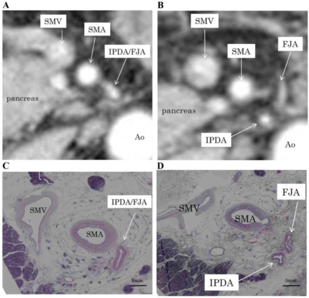
(28.5%) having type B and 9 (7.3%) having type C branches (Table II). In type A, the vessel common to the IPDA and FJA emerged between the left and dorsal sides of the SMA (148±40.0°). In types B and C, the IPDA and PIPDA emerged from the dorsal side of the SMA, at 187±47.0° and 182±37.4°, respectively (Fig. 2; Table II). The routes of the IPDA, IPDA/FJA common vessel and PIPDA exhibited bending.

Figure 2.

Illustration of the branching patterns of the IPDA, FJA, and PIPDA. (A) In type A branching, the IPDA/FJA common vessel arises between the left and dorsal sides of the SMA. (B) In type B branching, the IPDA arises from the dorsal side of the SMA. (C) In type C branching, the PIPDA arises from the dorsal side of the SMA. IPDA, inferior pancreaticoduodenal artery; FJA, first jejunal artery; PIPDA, posterior inferior pancreaticoduodenal artery; SMA, superior mesenteric artery.

Table I.

Location of pancreatic cancers by age and sex.

Table I.

Location of pancreatic cancers by age and sex.

Location of the pancreatic cancerPancreatic headPancreatic body and tail
Sex, male/female51/3125/16
Age, years, mean (range)63.5 (34–81)66.5 (45–84)

Table II.

IPDA branching types of pancreatic cancers.

Table II.

IPDA branching types of pancreatic cancers.

IPDA branching typeNumber of patients, n (%)Angle formed by the IPDA or PIPDA
Type A79 (64.2)148±40.0°
Type B35 (28.4)187±47.0°
Type C9 (7.3)182±37.4°

[i] IPDA, inferior pancreaticoduodenal artery; PIPDA, posterior inferior pancreaticoduodenal artery. Type A, Type B and Type C. Type A: The IPDA forms a common vessel with the FJA. Type B: The IPDA branches directly from the SMA. Type C: The AIPDA and PIPDA branch from separate parts of the SMA.

The distribution of the meso-pd was determined by evaluating the histological continuity of tissue in surgical specimens and cadavers. Immunohistochemistry with antibodies to D2-40 revealed lymphatic vessels beside the collagen fibers in this 
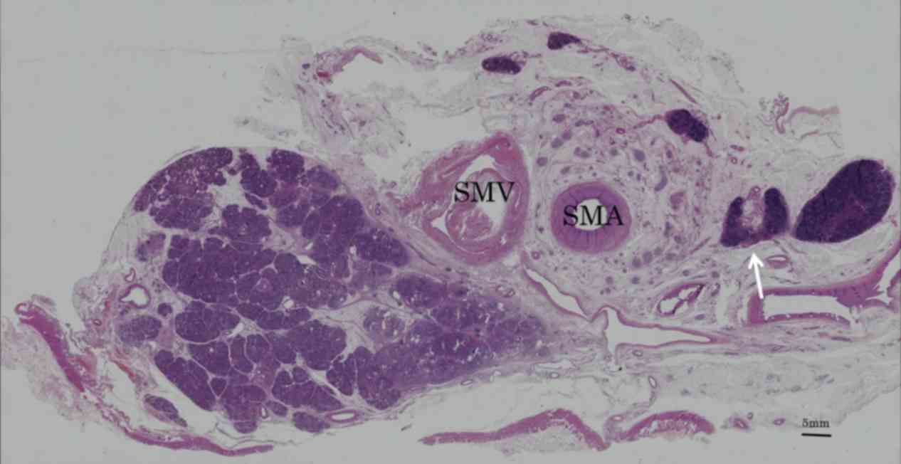
area (Fig. 3). Collagen fibers, forming two fibrous bunches, were observed regularly in the soft tissue. Around the SMA, which was covered grossly by fibrous tissue, collagen fibers formed concentric circles ~3 mm thick, a structure called the superior mesenteric arterial plexus (PLsma) (12). By contrast, collagen fibers near the uncinated process ran vertically from the uncinated process of the pancreas to the PLsma, with continuity through the posterior and left side of the SMA (Fig. 4). These collagen fibers, which were also bent, gradually fused and joined with the PLsma, making the PLsma thicker as it proceeded towards the origin of the SMA (Fig. 5).

Figure 3.

Patterns of D2-40 staining demonstrating (A) a lymph duct (arrow) adjacent to (B) collagen fibers. Magnification, ×40.

Figure 4.

Histological assessment of collagen fibers in tissue sections. Hematoxylin and eosin staining revealed that (A) at the level of the third portion of the duodenum, the collagen fibers (arrows) that surround the SMA are not continuous with those surrounding the pancreatic head (asterisk) but gradually develop continuity at the (B) upper level. (C and D) Elastica van Gieson stained tissue sections. SMA, superior mesenteric artery.

Figure 5.

(A) CT scan showing the IPDA/FJA common vessel emerging between the left and dorsal sides of the SMA. (B) CT scan showing the IPDA branching from the FJA. (C) Surgical specimen showing the IPDA/FJA common vessel emerging between the left and dorsal sides of the SMA. (D) Surgical specimen showing the IPDA branching from the FJA. Magnification, ×1. CT, computed tomography; IPDA, inferior pancreaticoduodenal artery; FJA, first jejunal artery; SMA, superior mesenteric artery; SMV, superior mesenteric vein; Ao, aorta.

Discussion

We previously determined that the manner of lymphatic extension and nerve plexus infiltration of the PHC were dependent on whether the tumor originated from the embryonic dorsal or ventral pancreatic bud (12,13). Tumors confined to the ventral pancreas extend toward the SMA, whereas tumors confined to the dorsal pancreas extend towards the common HA or hepatoduodenal ligament. If the tumor infiltrates deeply into the two areas, the cancer is likely to extend in the two directions. Therefore, the SMA margin is important for the carcinoma of the ventral pancreas.

The present study examined the distribution of the meso-pd, which was considered to be the mesentery of the embryonic ventral pancreas. The SMA margin is the most frequent site of PHC recurrence (3), despite the soft tissue on the right side of the SMA being regularly resected by pancreaticoduodenectomy. Lymphatic vessel involvement is common in PHC. Regional lymphatic basin resection is required to avoid local recurrence, since this area is thought to remain the regional lymphatic basin of PHC subsequent to soft tissue resection of the right side of the SMA, resulting in local recurrence. The mesopancreas (3,6,7), defined as the structure located on the right side of the SMA, is the primary site of cancer cell infiltration. However, lymph node metastasis is often observed on the left side of the SMA in patients with PHC (8,9). Resection based on the mesopancreas may therefore be insufficient for curative resection of the regional lymphatic basin.

The meso-pd, consisting of a cluster of soft connective tissue situated along the IPDA and the FJA, is regarded as the site of lymphatic spread, with tMPDe regarded as necessary for pathological cure (10). Patterns of arterial branches differ in the pancreatic head. Dissection of the lymphatic basin in PHC patients requires assessment of these patterns by multi detector CT, thus clarifying the direction of the SMA. At emergence, the arteries feeding the pancreatic head can be classified into three types (11). All of these branches emerged from the back or left side of the SMA and ran to the far side of the pancreatic head in an arc. Therefore, the meso-pd was located in a roll shape around the trunk arteries, extending to the left side of the SMA.

By investigating the continuity of the meso-pd histologically in this area of soft tissue, it was revealed by immunohistochemistry that the lymphatic vessels were alongside the collagen fibers. The present study therefore examined the distribution of the meso-pd relative to the continuity of the collagen fibers. The meso-pd originates from the uncinated process of the pancreas and connects to the PLsma, defined as the left back side of the soft tissue around the SMA (14). The distribution of the meso-pd is the same as the route of the IPDA. Its lower limit was vertically above the third duodenal portion. This soft tissue is also the mesentery of the jejunum, which is the route of the lymphatics and the nerves 
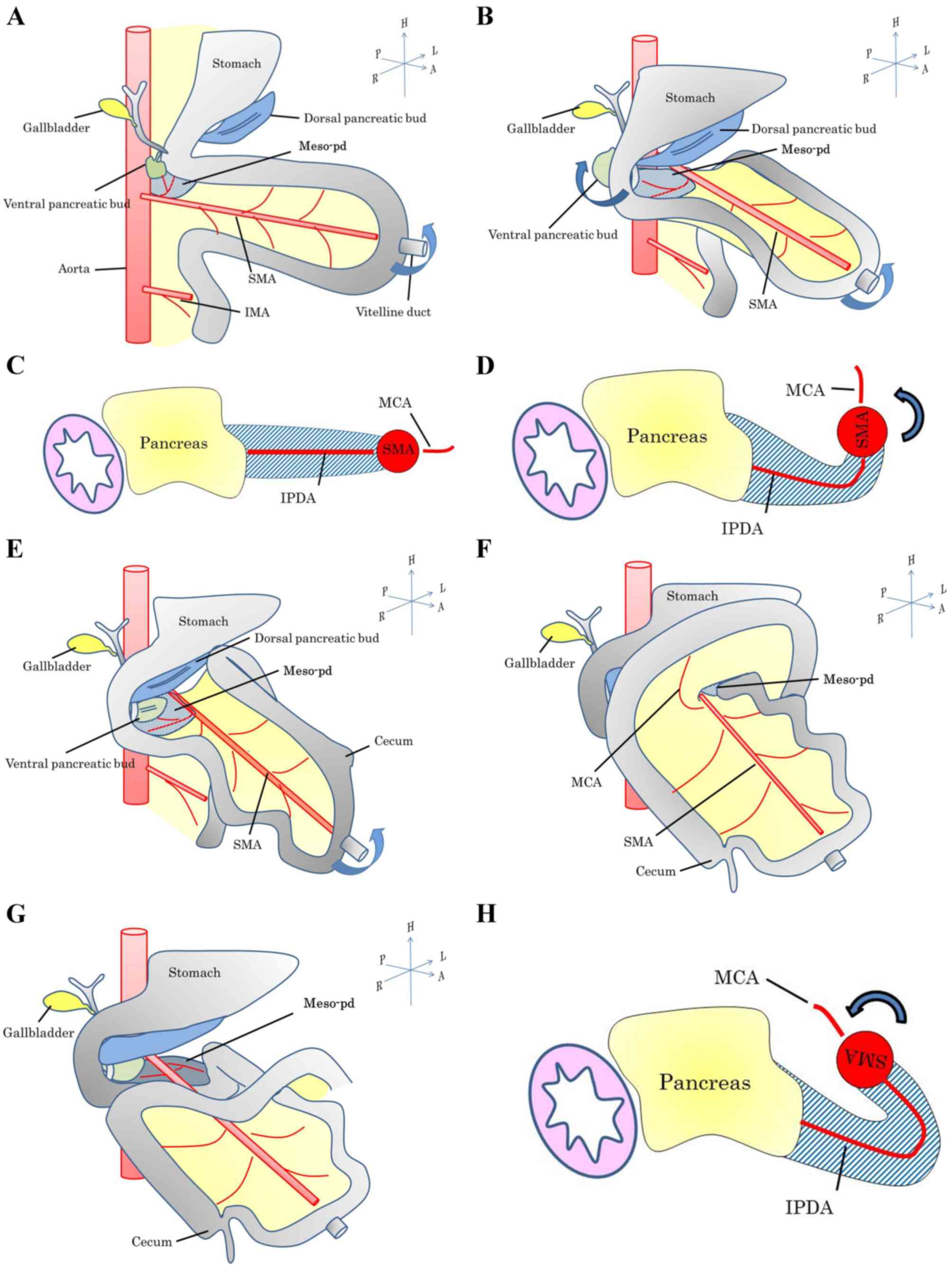
connecting the jejunum. These findings suggest that the regional lymphatic basin of the pancreatic head spreads towards the left side of the SMA, with an appearance resembling a Swiss roll. This appearance was likely caused by intestinal rotation in the fetus (Figs. 6 and 7) (15). The mid-gut rotated 270° anticlockwise around the SMA, which acted as the axis. This resulted in a bend in the meso-pd, which subsequently extended to the left of the SMA.

Figure 6.

Diagram showing the mesopancreas situated to the right of the SMA (shaded area). SMA, superior mesenteric artery; SMV, superior mesenteric vein; Ao, aorta; IVC, inferior vena cava.

Figure 7.

Diagrams showing (A) the position of the mid-gut prior to rotation during the prenatal period. (B) The position of the mid-gut following an anticlockwise rotation of 90° around the SMA as the axis. (C) Horizontal slice of B. The meso-pd is linear. (E) Position of the mid-gut following anticlockwise rotation for another 90°. (D) Horizontal slice of c-1. A bend occurs in the meso-pd. (F) Position of the mid-gut following an anticlockwise rotation for another 90°; such a rotation is noted perinatally. (G) The transverse colon is pulled down until the third portion of the duodenum is contained within the meso-pd. (H) A bend recurring in the meso-pd, extending to the left of the SMA. SMA, superior mesenteric artery; IMA, inferior mesenteric artery; IPDA, inferior pancreaticoduodenal artery; MCA, middle colic artery; FJA, first jejunal artery.

The artery first approached during pancreatoduodenectomy has been revealed to contribute to the determination of resectability and reduction of bleeding; several procedures have been described (16). Early shutdown of arterial blood feeding the head of the pancreas is therefore important. The present evaluation of the location of the branches of the pancreatic head from the SMA supports the artery first approach. Among the various surgical methods, the mesenteric approach is considered optimal for en bloc resection since the meso-pd, which contains the nerve plexus and lymphatics associated with the head of the pancreas, extends from the right to the back side of the SMA. Thus, tMPDe has theoretical advantages for en bloc and curative resection of carcinomas of the ventral pancreas. By contrast, the mesentery corresponding to the embryonic dorsal pancreas is currently unclear, although it has been associated with the HA. Survival rate following HA resection was poor in the present study and procedure, which focuses on the meso-pd, and was shown to be insufficient for treatment of carcinomas of the dorsal pancreas (17).

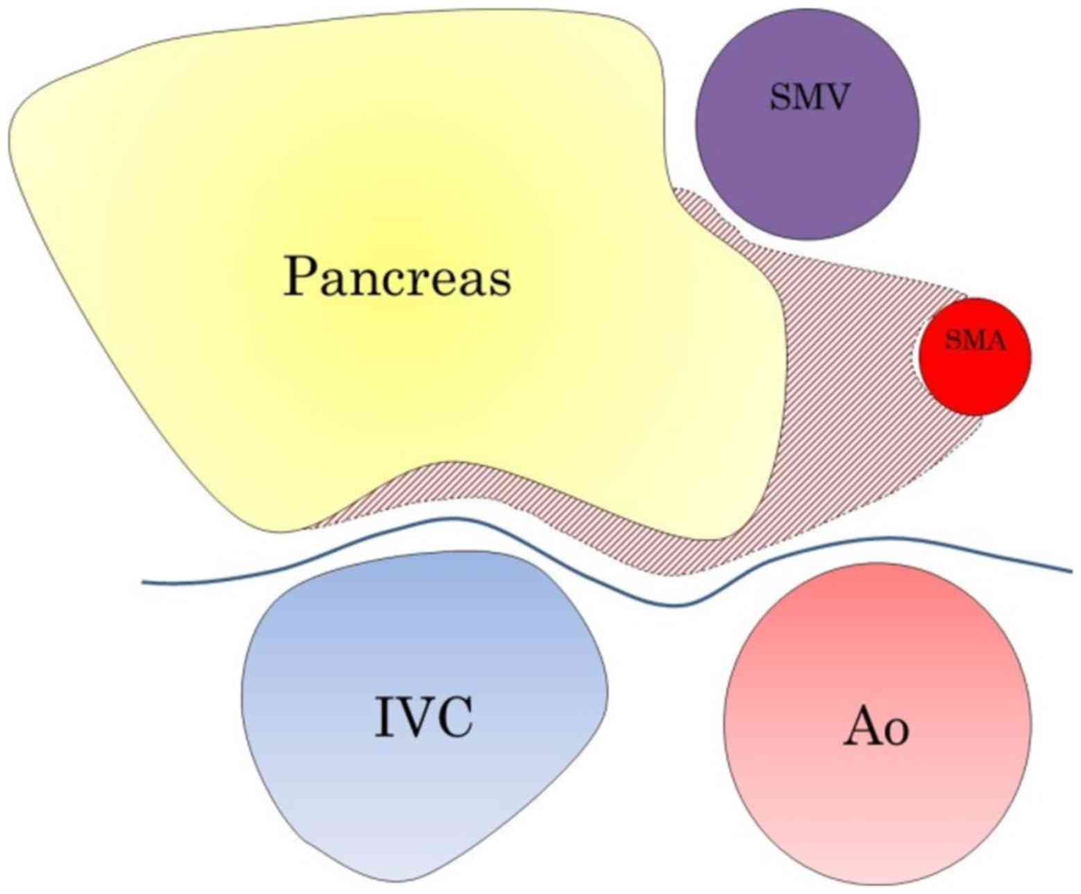
The present study concluded that the meso-pd, which spans the dorsal and left sides of the SMA (Fig. 8), is the proper mesentery of the pancreas and duodenum. Therefore, in patients with PHC, dissecting the right and left sides of the SMA during lymphadenectomy may be advantageous.

Figure 8.

Photomicrograph of stained tissue section showing lymph node metastasis (white arrow) on the left side of the SMA. SMA, superior mesenteric artery; SMV, superior mesenteric vein.

References

1 

Wagner M, Redaelli C, Lietz M, Seiler CA, Friess H and Büchler MW: Curative resection is the single most important factor determining outcome in patients with pancreatic adenocarcinoma. Br J Surg. 91:586–594. 2004. View Article : Google Scholar : PubMed/NCBI

2 

Stojadinovic A, Brooks A, Hoos A, Jaques DP, Conlon KC and Brennan MF: An evidence-based approach to the surgical management of resectable pancreatic adenocarcinoma. J Am Coll Surg. 196:954–964. 2003. View Article : Google Scholar : PubMed/NCBI

3 

Gaedcke J, Gunawan B, Grade M, Szöke R, Liersch T, Becker H and Ghadimi BM: The mesopancreas is the primary site for R1 resection in pancreatic head cancer: Relevance for clinical trials. Langenbecks Arch Surg. 395:451–458. 2010. View Article : Google Scholar : PubMed/NCBI

4 

Heye T, Zausig N, Klauss M, Singer R, Werner J, Richter GM, Kauczor HU and Grenacher L: CT diagnosis of recurrence after pancreatic cancer: Is there a pattern? World J Gastroenterol. 17:1126–1134. 2011. View Article : Google Scholar : PubMed/NCBI

5 

Pedrazzoli S, DiCarlo V, Dionigi R, Mosca F, Pederzoli P, Pasquali C, Klöppel G, Dhaene K and Michelassi F: Standard versus extended lymphadenectomy associated with pancreaticoduodenectomy in the surgical treatment of adenocarcinoma of the head of the pancreas: A multicenter, prospective, randomized study. Lymphadenectomy Study Group. Ann Surg. 228:508–517. 1998. View Article : Google Scholar : PubMed/NCBI

6 

Gockel I, Domeyer M, Wolloscheck T, Konerding MA and Junginger T: Resection of the mesopancreas (RMP): A new surgical classification of a known anatomical space. World J Surg Oncol. 5:442007. View Article : Google Scholar : PubMed/NCBI

7 

Adham M and Singhirunnusorn J: Surgical technique and results of total mesopancreas excision (TMpE) in pancreatic tumors. Eur J Surg Oncol. 38:340–345. 2012. View Article : Google Scholar : PubMed/NCBI

8 

Kayahara M, Nagakawa T, Ueno K, Ohta T, Tsukioka Y and Miyazaki I: Surgical strategy for carcinoma of the pancreas head area based on clinicopathologic analysis of nodal involvement and plexus invasion. Surgery. 117:616–623. 1995. View Article : Google Scholar : PubMed/NCBI

9 

Noto M, Miwa K, Kitagawa H, Kayahara M, Takamura H, Shimizu K and Ohta T: Pancreas head carcinoma: Frequency of invasion to soft tissue adherent to the superior mesenteric artery. Am J Surg Pathol. 29:1056–1061. 2005.PubMed/NCBI

10 

Kawabata Y, Tanaka T, Nishi T, Monma H, Yano S and Tajima Y: Appraisal of a total meso-pancreatoduodenum excision with pancreaticoduodenectomy for pancreatic head carcinoma. Eur J Surg Oncol. 38:574–579. 2012. View Article : Google Scholar : PubMed/NCBI

11 

Horiguchi A, Ishihara S, Ito M, Asano Y, Yamamoto T and Miyakawa S: Three-dimensional models of arteries constructed using multidetector-row CT images to perform pancreatoduodenectomy safely following dissection of the inferior pancreaticoduodenal artery. J Hepatobiliary Pancreat Sci. 17:523–526. 2010. View Article : Google Scholar : PubMed/NCBI

12 

Kitagawa H, Ohta T, Makino I, Tani T, Tajima H, Nakagawara H, Ohnishi I, Takamura H, Kayahara M, Watanabe H, et al: Carcinomas of the ventral and dorsal pancreas exhibit different patterns of lymphatic spread. Front Biosci. 13:2728–2735. 2008. View Article : Google Scholar : PubMed/NCBI

13 

Makino I, Kitagawa H, Ohta T, Nakagawara H, Tajima H, Ohnishi I, Takamura H, Tani T and Kayahara M: Nerve plexus invasion in pancreatic cancer: Spread patterns on histopathologic and embryological analyses. Pancreas. 37:358–365. 2008. View Article : Google Scholar : PubMed/NCBI

14 

Japan Pancreas Society, . Classification of Pancreatic Carcinoma. 3rd. Kanehara & Co., Ltd.; Tokyo: 2011

15 

Shinohara H, Kurahashi Y, Kanaya S, Haruta S, Ueno M, Udagawa H and Sakai Y: Topographic anatomy and laparoscopic technique for dissection of no. 6 infrapyloric lymph nodes in gastric cancer surgery. Gastric Cancer. 16:615–620. 2013. View Article : Google Scholar : PubMed/NCBI

16 

Inoue Y, Saiura A, Yoshioka R, Ono Y, Takahashi M, Arita J, Takahashi Y and Koga R: Pancreatoduodenectomy with systematic mesopancreas dissection using a supracolic anterior artery-first approach. Ann Surg. 262:1092–1101. 2015. View Article : Google Scholar : PubMed/NCBI

17 

Kitagawa H, Tajima H, Nakagawara H, Makino I, Miyashita T, Shoji M, Nakanuma S, Hayashi N, Takamura H, Ohta T and Ohtake H: En bloc vascular resection for the treatment of borderline resectable pancreatic head carcinoma. Mol Clin Oncol. 2:369–374. 2014.PubMed/NCBI

Related Articles

  • Abstract
  • View
  • Download
  • Twitter
Copy and paste a formatted citation
Spandidos Publications style
Terakawa H, Kitagawa H, Makino I, Hayashi H, Oyama K, Nakagawara H, Miyashita T, Tajima H, Takamura H, Fushida S, Fushida S, et al: Location of the meso-pancreatoduodenum as a regional lymphatic basin for pancreatic head carcinoma. Oncol Lett 14: 397-403, 2017.
APA
Terakawa, H., Kitagawa, H., Makino, I., Hayashi, H., Oyama, K., Nakagawara, H. ... Ohta, T. (2017). Location of the meso-pancreatoduodenum as a regional lymphatic basin for pancreatic head carcinoma. Oncology Letters, 14, 397-403. https://doi.org/10.3892/ol.2017.6138
MLA
Terakawa, H., Kitagawa, H., Makino, I., Hayashi, H., Oyama, K., Nakagawara, H., Miyashita, T., Tajima, H., Takamura, H., Fushida, S., Ozaki, N., Ohta, T."Location of the meso-pancreatoduodenum as a regional lymphatic basin for pancreatic head carcinoma". Oncology Letters 14.1 (2017): 397-403.
Chicago
Terakawa, H., Kitagawa, H., Makino, I., Hayashi, H., Oyama, K., Nakagawara, H., Miyashita, T., Tajima, H., Takamura, H., Fushida, S., Ozaki, N., Ohta, T."Location of the meso-pancreatoduodenum as a regional lymphatic basin for pancreatic head carcinoma". Oncology Letters 14, no. 1 (2017): 397-403. https://doi.org/10.3892/ol.2017.6138
Copy and paste a formatted citation
x
Spandidos Publications style
Terakawa H, Kitagawa H, Makino I, Hayashi H, Oyama K, Nakagawara H, Miyashita T, Tajima H, Takamura H, Fushida S, Fushida S, et al: Location of the meso-pancreatoduodenum as a regional lymphatic basin for pancreatic head carcinoma. Oncol Lett 14: 397-403, 2017.
APA
Terakawa, H., Kitagawa, H., Makino, I., Hayashi, H., Oyama, K., Nakagawara, H. ... Ohta, T. (2017). Location of the meso-pancreatoduodenum as a regional lymphatic basin for pancreatic head carcinoma. Oncology Letters, 14, 397-403. https://doi.org/10.3892/ol.2017.6138
MLA
Terakawa, H., Kitagawa, H., Makino, I., Hayashi, H., Oyama, K., Nakagawara, H., Miyashita, T., Tajima, H., Takamura, H., Fushida, S., Ozaki, N., Ohta, T."Location of the meso-pancreatoduodenum as a regional lymphatic basin for pancreatic head carcinoma". Oncology Letters 14.1 (2017): 397-403.
Chicago
Terakawa, H., Kitagawa, H., Makino, I., Hayashi, H., Oyama, K., Nakagawara, H., Miyashita, T., Tajima, H., Takamura, H., Fushida, S., Ozaki, N., Ohta, T."Location of the meso-pancreatoduodenum as a regional lymphatic basin for pancreatic head carcinoma". Oncology Letters 14, no. 1 (2017): 397-403. https://doi.org/10.3892/ol.2017.6138
Follow us
  • Twitter
  • LinkedIn
  • Facebook
About
  • Spandidos Publications
  • Careers
  • Cookie Policy
  • Privacy Policy
How can we help?
  • Help
  • Live Chat
  • Contact
  • Email to our Support Team