Introduction
Preoperative 5-fluorouracil-based chemoradiotherapy
(CRT) followed by mesorectal excision is the standard treatment for
patients with locally advanced rectal cancer (LARC) (1–3). Several
previous studies have demonstrated that CRT significantly
downstages the disease and reduces the risk of local tumor
recurrence (4–6). The response rate to CRT in this group
varies, with 9–30% of patients having a pathological complete
response (pCR) and 46–60% achieving some degree of tumor
downstaging (7,8). An improved long-term outcome has been
demonstrated for patients with pCR compared with patients who are
non-responsive to CRT (9–12); however, this treatment has several
long-term side effects. Therefore, the appropriate selection of
patients who will respond to CRT is important. At present, only
certain clinical parameters and radiological investigations are
available for use in the selection of patients for CRT, which is
insufficient. There is a requirement for biomarkers that are able
to predict the effect of preoperative CRT.
MicroRNAs (miRNAs/miRs) are short (18–25
nucleotides) non-coding RNA molecules that act as negative gene
regulators at the post-transcriptional level. miRNAs serve an
important role in the regulation of biological processes, such as
cell differentiation, proliferation and apoptosis. Numerous miRNAs
interfere with the expression of oncogenes and tumor suppressor
genes with a direct involvement in carcinogenesis (13,14). Due
to their association with cancer, miRNAs are being investigated as
potential diagnostic and prognostic biomarkers, and predictors of
treatment response (15,16).
In the present study, 5 miRNAs were chosen for study
based on the literature and our group's previous methodological
work. In rectal cancer tissue, reference genes (miR-193a-5p, miR
−27a and let-7g) for the relative quantification of miRNAs have
previously been identified (17) and
the intratumoral heterogeneity of the present panel of miRNAs
(miR-21, −31, −125b, −145 and −630) has been assessed (18).
miRNA-21 is overexpressed in rectal cancer and its
downregulation following neoadjuvant CRT has been suggested
(19–21). Furthermore, overexpression of miRNA-21
has been associated with complete tumor regression following
neoadjuvant CRT (22). Increased
expression of miRNA-31 has been detected in colorectal cancer
(CRC), with a positive correlation between its expression and the
stage of the disease (23,24). In addition, the upregulation of
miRNA-125b and miRNA-145 in rectal cancer tissue following
neoadjuvant CRT has been reported (19,25). Della
Vittoria Scarpati et al (26)
demonstrated that miRNA-630 had 100% sensitivity and specificity in
selecting patients with complete tumor regression following CRT.
However, only one of the studies described above included a
validation of their results (22).
Consequently, the majority of them serve as hypothesis-generating
investigations. The present study aimed to analyze the association
between miRNA expression and the treatment efficacy of preoperative
CRT in a test cohort of LARC patients, and to subsequently validate
the results in an independent cohort.
Materials and methods
Patient populations
Reporting of all data in the present study is in
accordance with the Reporting recommendations for tumor marker
prognostic studies criteria (27).
The present study consisted of a test and a validation cohort of
patients with LARC. The patients were enrolled in previously
conducted clinical trials, and the details of patients, treatment
and follow-up interval times are available from the pertaining
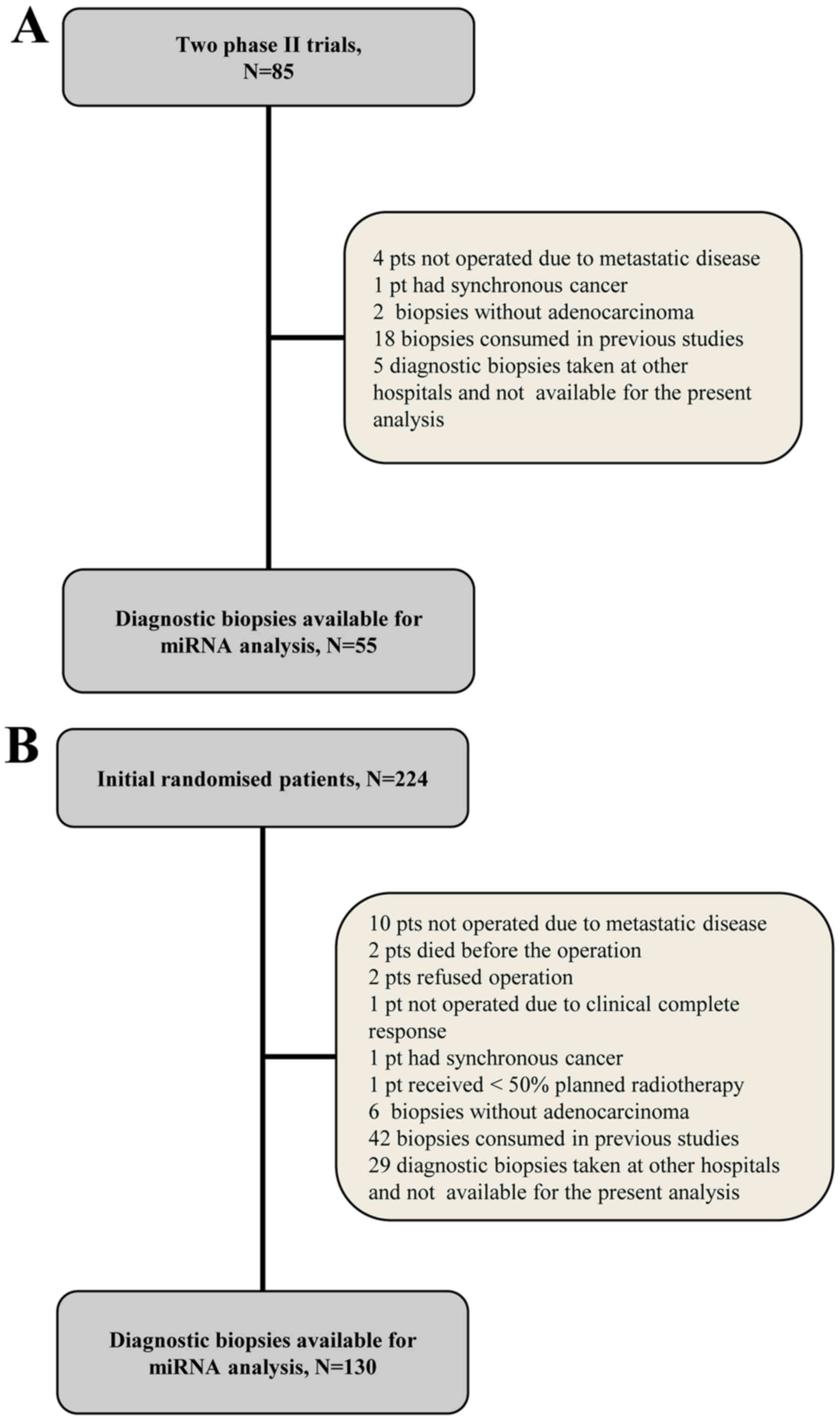
publications (28–30). Briefly, the test cohort began with 85
patients treated in one of two following trials: i) 50 patients
(enrollment, November 2002-June 2004) with resectable T3 rectal
cancer treated with preoperative CRT (60 Gy/30 fractions, and
concurrent tegafur-uracil (300 mg/m2) and L-leucovorin
(22.5 mg/day) combined with an endorectal boost (5 Gy/1 fraction)
(28); ii) 35 patients (enrollment,
June 2004-January 2005) with resectable T3-4 rectal cancer
receiving similar treatment combined with the
cyclooxygenase-2-inhibitor celecoxib (400 mg twice a day) (30). The validation cohort (enrollment,
March 2005-November 2009) was based on a previous randomized phase
III trial (29) of 224 patients with
resectable T3-4 rectal cancer receiving neoadjuvant CRT (50.4 Gy/28
fractions, and concomitant tegafur-uracil (300 mg/m2)
and L-leucovorin (22.5 mg/day) with (arm B) or without (arm A) an
endorectal boost (10 Gy/2 fractions)).
The present study was approved by the Regional
Committee on Health Research Ethics of Southern Denmark and the
requirement for written informed consent was waived (protocol ID
no. S-20140083). The study was registered with The Danish Data
Protection Agency and The Danish Registry of Human Tissue
Utilization was consulted prior to any tissue samples being used.
The inclusion criteria were the availability of formalin-fixed
paraffin-embedded (FFPE) diagnostic biopsies containing
adenocarcinoma tissue and tumor specimens for the pathological
evaluation of tumor regression grade (TRG). The exclusion criteria
were the presence of synchronous malignant diseases (with the
exception of non-melanoma skin cancer and carcinoma in situ
of the cervical uterus). A total of 55 and 130 patients from the
test and validation cohorts, respectively, met the criteria for the
present investigation. Flowcharts of the study populations are
presented in Fig. 1 and a summary of
their clinicopathological characteristics are presented in Table I.
 | Table I.Clinicopathological characteristics
and response to chemoradiotherapy of patients in the test and
validation study cohorts. |
Table I.
Clinicopathological characteristics
and response to chemoradiotherapy of patients in the test and
validation study cohorts.
| Clinicopathological
characteristic | Test cohort (N=55),
no. of patients (%) | Validation cohort
(N=130), no. of patients (%) |
|---|
| Gender |
|
|
|
Female | 19 (34.5) | 50 (38) |
|
Male | 36 (65.5) | 80 (62) |
| T-stage |
|
|
| T3 | 48 (87) | 110 (85) |
| T4 | 7 (13) | 20 (15) |
| N-stage |
|
|
| N0 | 16 (29) | 14 (11) |
|
N+ | 39 (71) | 116 (89) |
| TRG |
|
|
|
TRG1 | 13 (23.5) | 26 (20) |
|
TRG2 | 13 (23.5) | 17 (13) |
|
TRG3 | 28 (51) | 75 (58) |
|
TRG4 | 1 (2) | 12 (9) |
Samples
Diagnostic biopsies from the rectal tumors followed
routine preservation (FFPE), and were transported and stored at
room temperature. The median storage duration from archiving to
analysis was 11.5 years in the test cohort and 9 years in the
validation cohort. Histological sections stained with hematoxylin
and eosin (H&E) were examined by a pathologist in order to
ensure the presence of tumor cells in the analyzed sections. From
the corresponding tissue blocks, 8-µm-thick sections were cut for
use in subsequent reverse transcription-quantitative polymerase
chain reaction (RT-qPCR) analysis. In the test population, areas of
tumor cells were identified by a pathologist and encircled as
regions of interest (ROI) on an image of the H&E-stained
section. The ROI were isolated through membrane-based laser
microdissection (LMD) using a Leica LMD6500 Microsystems (Leica
Microsystems GmbH, Wetzlar, Germany) and collected in the caps of
0.5 ml RNase-free PCR tubes with a drop of ethanol (99%). In the
validation population, LMD was performed as described above.
However, in a subset of cases in the validation cohort LMD was not
required, since the whole biopsy consisted of tumor tissue without
marked inflammation. In these cases (N=26) the tissue was removed
from the slides using a scalpel and collected into 1.5 ml
RNase-free PCR tubes containing a drop of ethanol (99%).
Expression analysis via RT-qpcr
Normalizer miRNAs
Recently, our group performed a study (17) on miRNA expression profiling to
identify and validate reference genes for the relative
quantification of miRNAs in rectal cancer tissue. miR-193a-5p,
miR-27a and let-7g were identified as the most stably expressed
miRNAs in rectal cancer tissue, and the mean expression value of
these three miRNAs were subsequently used for normalization in the
present study.
RNA extraction
Total RNA was isolated from the FFPE tissue using
the miRNeasy FFPE kit for the test study and the AllPrep DNA/RNA
FFPE kit (both Qiagen GmbH, Hilden, Germany) for the validation
study according to the manufacturer's protocol. Total RNA was
eluted into 14 µl RNase-free water.
RT-qPCR
RT and preamplification were performed as previously
described (18). Custom
TaqMan® MicroRNA Single Assays (Thermo Fisher
Scientific, Inc., Waltham, MA, USA) for hsa-let-7g (cat. no.
002282), hsa-miR-193a-5p (cat. no. 002281), hsa-miR-27a (cat. no.
000408), hsa-miR-21 (cat. no. 000397), hsa-miR-31 (cat. no.
002279), hsa-miR-125b (cat. no. 000449), hsa-miR-145 (cat. no.
002278) and hsa-miR-630 (cat. no. 001563) were used, according to
the manufacturer's protocol. The qPCR analyses were carried out
using a QuantStudio™ 12K Flex Real-Time PCR system (Thermo Fisher
Scientific, Inc.) with 1 µl diluted preamplification product,
TaqMan MicroRNA Assays and TaqMan® Universal Master Mix
II NoAmpErase® UNG in a total reaction volume of 20 µl.
All reactions were performed in triplicate. Data analysis was
performed using Quantstudio 12K Flex software (version 1.2.2;
Thermo Fisher Scientific, Inc.) and relative quantification was
performed using the 2−ΔΔCq method as described by
Eriksen et al (18). The mean
expression value of miR-193a-5p, miR-27a and let-7g was used as the
normalization factor. Water was used as the negative control. A
no-template control was included in the entire process and analyzed
together with samples. The analyses were performed by staff blinded
to the patient outcome.
Endpoints
The primary endpoint of tumor regression was
determined by assessing the operative specimens according to
Mandard's Tumor Regression Grade (TRG) system (31,32), as
follows: TRG1, no residual tumor; TRG2, microscopic residual tumor;
TRG3, moderate response; TRG4, minor response; and TRG5, no
response. Overall survival (OS) and disease-free survival (DFS)
were the secondary endpoints. OS was defined as the time from
inclusion in the primary study until mortality from any cause. DFS
was calculated as the time from inclusion in the primary study
until the first documented tumor progression or mortality from any
cause.
Statistical analysis
Wilcoxon rank-sum test was used for comparison of
medians between groups. The prognostic value of variables was
analyzed using the log rank-test and survival curves were produced
using the Kaplan-Meier estimator. Patients with additional
malignancies were excluded from the DFS analysis (test study, N=9;
validation study, N=16). Clinical outcome data were last updated in
April 2016. Possible correlations between continuous data were
analyzed and visualized using the parametric linear regression
analysis. Univariate Cox's regression analysis was used to estimate
the prognostic value of individual variables and those with
P<0.1 were included in a multivariate Cox's regression analysis
for independent prognostic value. All tests were two-tailed. All
statistical analyses were performed using NCSS Statistical Software
2007 (version 07.1.20; NSCC, LLC, Kaysville, UT, USA). P<0.05
was considered to indicate a statistically significant
difference.
Results
Test population
The test cohort analyses consisted of 55 patients
and the treatment compliance was high (28). The median follow-up time was 11.6
years. All patients were responsive to preoperative CRT. Patient
characteristics and treatment responses are presented in Table I. An expression of miR-145 below the
median expression level was significantly associated with major
response (TRG1+2) to treatment (P<0.001; Table II). For the other investigated miRNAs
no significant association between their expression and TRG was
identified (Table II). For survival
analysis, patients were grouped according to their median
expression of the miRNA of interest [above (high) or below (low)
the median]. No significant difference was identified between the
high and low expression groups for the investigated miRNAs
following OS and DFS analyses (data not shown).
| Table II.Association between TRG and miR
expression in the test cohort (N=55). |
Table II.
Association between TRG and miR
expression in the test cohort (N=55).
|
| miR expression,
median (95% CI) |
|
|---|
|
|
|
|
|---|
| miR | TRG1+2 | TRG3+4 | P-value |
|---|
| 125b | 1.030
(0.558–1.447) | 1.708
(0.951–2.026) | 0.174 |
| 145 | 9.854
(5.501–13.846) | 17.370
(13.515–25.070) |
<0.001 |
| 21 | 14.206
(11.529–18.195) | 17.463
(16.057–19.450) | 0.062 |
| 3 | 0.128
(0.051–0.235) | 0.136
(0.066–0.262) | 0.463 |
| 630 | 0.004
(0.003–0.005) | 0.003
(0.002–0.004) | 0.104 |
Validation population
A total of 130 patients were included in the
validation population. Patient characteristics and treatment
responses are presented in Table I.
All patients, with the exception of two, received >80% of the
planned radiotherapy with a curative intent. Eight patients had
developed distant metastases in the liver and/or lungs at the time
of surgery. In the validation population, an expression of miR-21
above the median expression level was significantly associated
(P=0.035) with major response (TRG1+2) to treatment (Table III). The association between miR-145
expression and TRG was similar to the one demonstrated in the test
cohort, although it did not reach statistical significance
(P=0.085). For miR-125b, −31 and −630, no significant association
between expression and TRG was detected (Table III).
| Table III.Association between TRG and miR
expression in the validation cohort (N=130). |
Table III.
Association between TRG and miR
expression in the validation cohort (N=130).
|
| miR expression,
median (95% CI) |
|
|---|
|
|
|
|
|---|
| miR | TRG1+2 | TRG3+4 |
P-valuea |
|---|
| 125b | 1.338
(1.009–1.729) | 1.566
(1.252–1.922) | 0.337 |
| 145 | 18.735
(13.923–22.943) | 22.015
(18.164–27.612) | 0.085 |
| 21 | 26.861
(22.906–29.768) | 21.107
(19.615–22.829) | 0.035 |
| 3 | 0.109
(0.072–0.391) | 0.135
(0.090–0.210) | 0.761 |
| 630 | 0.001
(0.001–0.002) | 0.001
(0.001–0.001) | 0.333 |
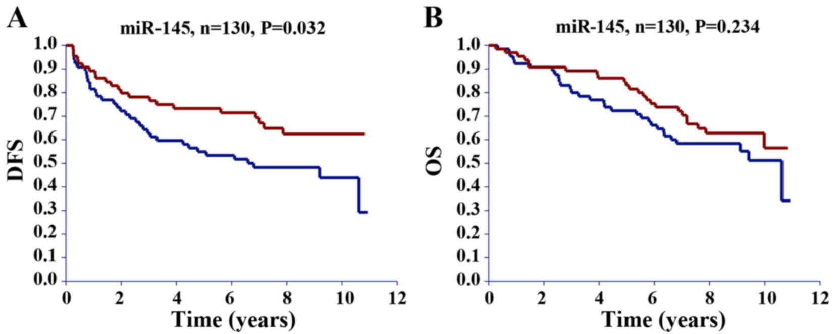
Grouping patients in the validation cohort according
to their median expression of the miRNA of interest revealed that
patients with miR-125b and miR-145 expression below the median had
a significantly better DFS compared with those with expression
above the median (Table IV).
Survival curves for miR-145 are illustrated in Fig. 2. No significant difference in DFS was
detected for miR-21, −31 and −630 (Table
IV). No significant difference was demonstrated between the
high and low expression of any of the investigated miRNAs and OS
(Table IV).
| Table IV.Univariate Cox's regression analysis
of the association between clinicopathological characteristics and
DFS and OS in the validation cohort (N=130). |
Table IV.
Univariate Cox's regression analysis
of the association between clinicopathological characteristics and
DFS and OS in the validation cohort (N=130).
|
| DFS | OS |
|---|
|
|
|
|
|---|
| Clinicopathological
characteristic | HR | 95% CI | P-value | HR | 95% CI | P-value |
|---|
| Gender |
| 0.50–1.42 | 0.517 |
| 0.64–1.93 | 0.711 |
|
Female | 1 |
|
| 1 |
|
|
|
Male | 0.84 |
|
| 1.11 |
|
|
| Age |
| 0.84–2.41 | 0.187 |
| 1.22–3.74 | 0.008 |
|
<64 | 1 |
|
| 1 |
|
|
|
≥64 | 1.42 |
|
| 2.14 |
|
|
| T-stage |
| 0.78–2.92 | 0.220 |
| 0.47–2.09 | 0.973 |
| T3 | 1 |
|
| 1 |
|
|
| T4 | 1.51 |
|
| 0.98 |
|
|
| N-stage |
| 0.44–2.16 | 0.954 |
| 0.42–2.04 | 0.837 |
| N0 | 1 |
|
| 1 |
|
|
| N+ | 0.98 |
|
| 0.92 |
|
|
| Distance from anal
verge (cm) |
| 0.42–2.65 | 0.913 |
| 0.54–3.50 | 0.497 |
| ≤5 | 1 |
|
| 1 |
|
|
|
>5 | 1.05 |
|
| 1.38 |
|
|
| TRG |
| 0.28–0.94 | 0.032 |
| 0.46–1.44 | 0.473 |
|
TRG1-2 | 0.52 |
|
| 0.81 |
|
|
|
TRG3-4 | 1 |
|
| 1 |
|
|
| Resection
status |
| 1.81–7.19 | <0.001 |
| 1.78–7.59 | <0.001 |
| R0 | 1 |
|
| 1 |
|
|
| Not
R0 | 3.61 |
|
| 3.68 |
|
|
| miR-125b
expression |
| 1.14–3.34 | 0.015 |
| 0.74–2.15 | 0.404 |
| Above
median | 1 |
|
| 1 |
|
|
| Below
median | 1.95 |
|
| 1.26 |
|
|
| miR-145
expression |
| 1.04–3.03 | 0.035 |
| 0.81–2.37 | 0.237 |
| Above
median | 1 |
|
| 1 |
|
|
| Below
median | 1.78 |
|
| 1.38 |
|
|
| miR-21
expression |
| 0.48–1.36 | 0.418 |
| 0.43–1.25 | 0.255 |
| Above
median | 1 |
|
| 1 |
|
|
| Below
median | 0.81 |
|
| 0.73 |
|
|
| miR-31
expression |
| 0.75–2.12 | 0.389 |
| 0.74–2.15 | 0.402 |
| Above
median | 1 |
|
| 1 |
|
|
| Below
median | 1.26 |
|
| 1.26 |
|
|
| miR-630
expression |
| 0.68–1.96 | 0.602 |
| 0.79–2.32 | 0.269 |
| Above
median | 1 |
|
| 1 |
|
|
| Below
median | 1.15 |
|
| 1.35 |
|
|
For a parameter to be included in the multivariate
Cox's regression analysis, a cut-off significance level of P<0.1
in the univariate analysis was pre-specified. In the linear
regression analysis, a significant positive correlation was
demonstrated between miR-125b and miR-145 expression (r=0.73;
P<0.0001; Fig. 3), and therefore
they were included in a separate multivariate analysis (Table V). Resection status (P=0.002, Table V) and miR-125b expression (P=0.026,
Table V) remained significant in
predicting DFS, and a borderline significance was demonstrated for
miR-145 expression (P=0.053). Multivariate analysis was not
performed for OS, as none of the investigated miRNAs were
significantly associated with OS in the univariate analysis.
| Table V.Multivariate Cox's regression
analysis of the association between clinicopathological
characteristics and disease-free survival in the validation cohort
(N=130). |
Table V.
Multivariate Cox's regression
analysis of the association between clinicopathological
characteristics and disease-free survival in the validation cohort
(N=130).
|
| Multivariate
analysis including miR-125b | Multivariate
analysis including miR-145 |
|---|
|
|
|
|
|---|
| Clinicopathological
characteristic | HR | 95% CI | P-value | HR | 95% CI | P-value |
|---|
| TRG |
| 0.36–1.28 | 0.232 |
| 0.34–1.20 | 0.168 |
|
TRG1-2 | 0.68 |
|
| 0.64 |
|
|
|
TRG3-4 | 1 |
|
| 1 |
|
|
| Resection
status |
| 1.54–6.47 | 0.002 |
| 1.49–6.22 | 0.002 |
| R0 | 1 |
|
| 1 |
|
|
| Not
R0 | 3.15 |
|
| 3.04 |
|
|
| miR-125b
expression |
| 1.08–3.20 | 0.026 |
|
|
|
| Above
median | 1 |
|
|
|
|
|
| Below
median | 1.86 |
|
|
|
|
|
| miR-145
expression |
|
|
|
| 0.99–2.91 | 0.053 |
| Above
median |
|
|
| 1 |
|
|
| Below
median |
|
|
| 1.70 |
|
|
Discussion
The balance between treatment efficacy and toxicity
is a major issue in the clinical management of patients with LARC.
Identifying molecular biomarkers capable of predicting the response
of patients to preoperative CRT is therefore important. The
‘watchful waiting’ strategy is emerging as an alternative the
typical preoperative CRT followed by surgery; thus far, a number of
studies have revealed encouraging outcomes for this strategy
(33–37). If tumor response to neoadjuvant CRT
could be predicted it would facilitate in providing more
individualized treatment planning, whereby patients with pCR could
avoid the standard resection procedure, which is frequently
followed by a high risk of morbidity.
It has previously been demonstrated by Ryan et
al (38) that patients with TRG1
and TRG2 tumors can be regarded as having pCR to preoperative CRT.
In addition, Lindebjerg et al (39) demonstrated that 28% of tumors
originally classified as TRG1 were reclassified as TRG2 tumors
following step sectioning (39).
Based on these results, patients with TRG1 and TRG2 were pooled as
complete responders in the present study and compared with patients
with a poorer response (TRG3+4).
In the present study, the expression of selected
miRNAs in diagnostic biopsies from patients with locally advanced
T3-4 rectal cancer was analyzed. The results demonstrated a
significant association between miR-145 expression and the overall
response of patients with rectal cancer to preoperative CRT.
Furthermore, the results suggest a possible association between
miR-21 and TRG, and between miR-125b and DFS.
In the test cohort, a significant association
between low miR-145 expression and major treatment response
(TRG1+2) was detected. Similarly, this association was identified
in the validation cohort; however, it did not reach statistical
significance (P=0.085). Drebber et al (19) reported a significant correlation
between a major response to neoadjuvant CRT and a high level of
miR-145 expression. However, the results of this study are
difficult to compare with those of the present study, since they
used a different tumor regression grading system (40) and performed macrodissection, whereas
the majority of the samples in the current study underwent laser
microdissection.
The significant association between a high
expression of miR-21 and TRG1+2 in the validation population is in
accordance with a previous study by Lopes-Ramos et al
(22), which revealed that the
overexpression of miR-21-5p is predictive of complete tumor
regression following neoadjuvant CRT in patients with rectal cancer
with a sensitivity and specificity of 78 and 86%, respectively.
Furthermore, Bandres et al (41) revealed that an upregulation of miR-21
was associated with TRG1 or TRG2 tumors. The results of the test
study population in the present study did not identify the same
association between miR-21 expression and treatment response.
Results from the test cohort suggested an association between a low
expression of miR-21 and a major treatment response, but this did
not reach statistical significance. However, in the validation
population there was a significant association between a high
miR-21 expression and a major response to preoperative CRT, in
agreement with Lopes-Ramos et al (22) and Bandres et al (41).
miR-125b and miR-31 were included in the present
study based on previous literature on miRNAs associated with rectal
cancer (23–25). In the current study, no association
between their expression and response to preoperative CRT was
detected. However, the significant association between a low
expression of miR-125b and increased DFS in the validation cohort
requires further investigation.
The results from a study by Della Vittoria Scarpati
et al (26), which
demonstrated that miR-630 had 100% sensitivity and specificity in
selecting patients having pCR after undergoing CRT (N=38), were not
confirmed in the current study. The expression of miR-630 was
analyzed in diagnostic biopsies from a total of 185 patients in the
present study. In general, miR-630 was sparsely expressed, and it
was undetected in 24 samples. Previously, high intratumoral
heterogeneity has been reported regarding miR-630 expression in
rectal cancer (18).
In conclusion, the partially diverging results
between the test and the validation cohorts in the present study
underline the importance of the validation of biomarker studies.
This issue has not been adequately addressed in the previous
literature. The majority of studies include <50 patients with no
validation. The risk of positive results being considered false
positives in investigations with small cohorts is high. This
approach has hampered or delayed the clinical application of
diagnostic and prognostic biomarkers. The results of the present
study underline the necessity for large prospective trials of miRNA
biomarkers in the future.
Acknowledgements
The authors would like to thank Laboratory
Technician Birgit Roed Sørensen (Department of Clinical Pathology,
Vejle Hospital, Denmark) and Laboratory Technician Pia Nielsen
(Department of Clinical Biochemistry, Vejle Hospital) for their
technical assistance, Research Secretary, PA Karin Larsen
(Department of Oncology, Vejle Hospital) for the linguistic editing
of the manuscript and University Academic Fellow in Radiotherapy
Research, Ane Lindegaard Appelt (Faculty of Medicine and Health,
University of Leeds, & St James Institute of Oncology, St
James's Hospital, Leeds, UK), for the initial data collection.
References
|
1
|
Cellini F and Valentini V: Current
perspectives on preoperative integrated treatments for locally
advanced rectal cancer: A review of agreement and controversies.
Oncology (Williston Park). 26:730–735. 2012.PubMed/NCBI
|
|
2
|
Valentini V, Glimelius B, Haustermans K,
Marijnen CA, Rödel C, Gambacorta MA, Boelens PG, Aristei C and van
de Velde CJ: EURECCA consensus conference highlights about rectal
cancer clinical management: The radiation oncologist's expert
review. Radiother Oncol. 110:195–198. 2014. View Article : Google Scholar : PubMed/NCBI
|
|
3
|
Julien LA and Thorson AG: Current
neoadjuvant strategies in rectal cancer. J Surg Oncol. 101:321–326.
2010. View Article : Google Scholar : PubMed/NCBI
|
|
4
|
Sauer R, Becker H, Hohenberger W, Rödel C,
Wittekind C, Fietkau R, Martus P, Tschmelitsch J, Hager E, Hess CF,
et al: Preoperative versus postoperative chemoradiotherapy for
rectal cancer. N Engl J Med. 351:1731–1740. 2004. View Article : Google Scholar : PubMed/NCBI
|
|
5
|
Kapiteijn E, Marijnen CA, Nagtegaal ID,
Putter H, Steup WH, Wiggers T, Rutten HJ, Pahlman L, Glimelius B,
van Krieken JH, et al: Preoperative radiotherapy combined with
total mesorectal excision for resectable rectal cancer. N Engl J
Med. 345:638–646. 2001. View Article : Google Scholar : PubMed/NCBI
|
|
6
|
Kim JC, Kim TW, Kim JH, Yu CS, Kim HC,
Chang HM, Ryu MH, Park JH, Ahn SD, Lee SW, et al: Preoperative
concurrent radiotherapy with capecitabine before total mesorectal
excision in locally advanced rectal cancer. Int J Radiat Oncol Biol
Phys. 63:346–353. 2005. View Article : Google Scholar : PubMed/NCBI
|
|
7
|
Lim SH, Chua W, Henderson C, Ng W, Shin
JS, Chantrill L, Asghari R, Lee CS, Spring KJ and de Souza P:
Predictive and prognostic biomarkers for neoadjuvant
chemoradiotherapy in locally advanced rectal cancer. Crit Rev Oncol
Hematol. 96:67–80. 2015. View Article : Google Scholar : PubMed/NCBI
|
|
8
|
Kuremsky JG, Tepper JE and McLeod HL:
Biomarkers for response to neoadjuvant chemoradiation for rectal
cancer. Int J Radiat Oncol Biol Phys. 74:673–688. 2009. View Article : Google Scholar : PubMed/NCBI
|
|
9
|
Maas M, Nelemans PJ, Valentini V, Das P,
Rödel C, Kuo LJ, Calvo FA, García-Aguilar J, Glynne-Jones R,
Haustermans K, et al: Long-term outcome in patients with a
pathological complete response after chemoradiation for rectal
cancer: A pooled analysis of individual patient data. Lancet Oncol.
11:835–844. 2010. View Article : Google Scholar : PubMed/NCBI
|
|
10
|
Fokas E, Liersch T, Fietkau R, Hohenberger
W, Beissbarth T, Hess C, Becker H, Ghadimi M, Mrak K, Merkel S, et
al: Tumor regression grading after preoperative chemoradiotherapy
for locally advanced rectal carcinoma revisited: Updated results of
the CAO/ARO/AIO-94 Trial. J Clin Oncol. 32:1554–1562. 2014.
View Article : Google Scholar : PubMed/NCBI
|
|
11
|
Rödel C, Martus P, Papadoupolos T, Füzesi
L, Klimpfinger M, Fietkau R, Liersch T, Hohenberger W, Raab R,
Sauer R and Wittekind C: Prognostic significance of tumor
regression after preoperative chemoradiotherapy for rectal cancer.
J Clin Oncol. 23:8688–8696. 2005. View Article : Google Scholar : PubMed/NCBI
|
|
12
|
Garcia-Aguilar J, de Anda E Hernandez,
Sirivongs P, Lee SH, Madoff RD and Rothenberger DA: A pathologic
complete response to preoperative chemoradiation is associated with
lower local recurrence and improved survival in rectal cancer
patients treated by mesorectal excision. Dis Colon Rectum.
46:298–304. 2003. View Article : Google Scholar : PubMed/NCBI
|
|
13
|
Slaby O, Svoboda M, Michalek J and Vyzula
R: MicroRNAs in colorectal cancer: Translation of molecular biology
into clinical application. Mol Cancer. 8:1022009. View Article : Google Scholar : PubMed/NCBI
|
|
14
|
Riordan AM, Thomas MK, Ronnekleiv-Kelly S,
Warner T, Geiger PG and Kennedy GD: Utility of micro-ribonucleic
acid profile for predicting recurrence of rectal cancer. J Surg
Res. 177:87–92. 2012. View Article : Google Scholar : PubMed/NCBI
|
|
15
|
Mestdagh P, van Vlierberghe P, De Weer A,
Muth D, Westermann F, Speleman F and Vandesompele J: A novel and
universal method for microRNA RT-qPCR data normalization. Genome
Biol. 10:R642009. View Article : Google Scholar : PubMed/NCBI
|
|
16
|
Nugent M, Miller N and Kerin MJ: MicroRNAs
in colorectal cancer: Function, dysregulation and potential as
novel biomarkers. Eur J Surg Oncol. 37:649–654. 2011. View Article : Google Scholar : PubMed/NCBI
|
|
17
|
Eriksen AH, Andersen RF, Pallisgaard N,
Sørensen FB, Jakobsen A and Hansen TF: MicroRNA expression
profiling to identify and validate reference genes for the relative
quantification of microRNA in rectal cancer. PLoS One.
11:e01505932016. View Article : Google Scholar : PubMed/NCBI
|
|
18
|
Eriksen AH, Andersen RF, Nielsen BS,
Sorensen FB, Appelt AL, Jakobsen A and Hansen TF: Intratumoral
heterogeneity of MicroRNA expression in rectal cancer. PloS One.
11:e01569192016. View Article : Google Scholar : PubMed/NCBI
|
|
19
|
Drebber U, Lay M, Wedemeyer I, Vallböhmer
D, Bollschweiler E, Brabender J, Mönig SP, Hölscher AH, Dienes HP
and Odenthal M: Altered levels of the onco-microRNA 21 and the
tumor-supressor microRNAs 143 and 145 in advanced rectal cancer
indicate successful neoadjuvant chemoradiotherapy. Int J Oncol.
39:409–415. 2011.PubMed/NCBI
|
|
20
|
Li T, Leong MH, Harms B, Kennedy G and
Chen L: MicroRNA-21 as a potential colon and rectal cancer
biomarker. World J Gastroenterol. 19:5615–5621. 2013. View Article : Google Scholar : PubMed/NCBI
|
|
21
|
Slaby O, Svoboda M, Fabian P, Smerdova T,
Knoflickova D, Bednarikova M, Nenutil R and Vyzula R: Altered
expression of miR-21, miR-31, miR-143 and miR-145 is related to
clinicopathologic features of colorectal cancer. Oncology.
72:397–402. 2007. View Article : Google Scholar : PubMed/NCBI
|
|
22
|
Lopes-Ramos CM, Habr-Gama A, Bde S
Quevedo, Felicio NM, Bettoni F, Koyama FC, Asprino PF, Galante PA,
Gama-Rodrigues J, Camargo AA, et al: Overexpression of miR-21-5p as
a predictive marker for complete tumor regression to neoadjuvant
chemoradiotherapy in rectal cancer patients. BMC Med Genomics.
7:682014. View Article : Google Scholar : PubMed/NCBI
|
|
23
|
Wang CJ, Zhou ZG, Wang L, Yang L, Zhou B,
Gu J, Chen HY and Sun XF: Clinicopathological significance of
microRNA-31, -143 and -145 expression in colorectal cancer. Dis
Markers. 26:27–34. 2009. View Article : Google Scholar : PubMed/NCBI
|
|
24
|
Laurila EM and Kallioniemi A: The diverse
role of miR-31 in regulating cancer associated phenotypes. Genes
Chromosomes Cancer. 52:1103–1113. 2013. View Article : Google Scholar : PubMed/NCBI
|
|
25
|
Svoboda M, Holla L Izakovicova, Sefr R,
Vrtkova I, Kocakova I, Tichy B and Dvorak J: Micro-RNAs miR125b and
miR137 are frequently upregulated in response to capecitabine
chemoradiotherapy of rectal cancer. Int J Oncol. 33:541–547.
2008.PubMed/NCBI
|
|
26
|
Della Vittoria, Scarpati G, Falcetta F,
Carlomagno C, Ubezio P, Marchini S, De Stefano A, Singh VK,
D'Incalci M, De Placido S and Pepe S: A specific miRNA signature
correlates with complete pathological response to neoadjuvant
chemoradiotherapy in locally advanced rectal cancer. Int J Radiat
Oncol Biol Phys. 83:1113–1119. 2012. View Article : Google Scholar : PubMed/NCBI
|
|
27
|
McShane LM, Altman DG, Sauerbrei W, Taube
SE, Gion M and Clark GM: Statistics Subcommittee of the NCI-EORTC
Working Group on Cancer Diagnostics: Reporting recommendations for
tumor marker prognostic studies (REMARK). J Natl Cancer Inst.
97:1180–1184. 2005. View Article : Google Scholar : PubMed/NCBI
|
|
28
|
Jakobsen A, Mortensen JP, Bisgaard C,
Lindebjerg J, Hansen JW and Rafaelsen SR: Preoperative
chemoradiation of locally advanced T3 rectal cancer combined with
an endorectal boost. Int J Radiat Oncol Biol Phys. 64:461–465.
2006. View Article : Google Scholar : PubMed/NCBI
|
|
29
|
Jakobsen A, Ploen J, Vuong T, Appelt A,
Lindebjerg J and Rafaelsen SR: Dose-effect relationship in
chemoradiotherapy for locally advanced rectal cancer: A randomized
trial comparing two radiation doses. Int J Radiat Oncol Biol Phys.
84:949–954. 2012. View Article : Google Scholar : PubMed/NCBI
|
|
30
|
Jakobsen A, Mortensen JP, Bisgaard C,
Lindebjerg J, Rafaelsen SR and Bendtsen VO: A COX-2 inhibitor
combined with chemoradiation of locally advanced rectal cancer: A
phase II trial. Int J Colorectal Dis. 23:251–255. 2008. View Article : Google Scholar : PubMed/NCBI
|
|
31
|
Mandard AM, Dalibard F, Mandard JC, Marnay
J, Henry-Amar M, Petiot JF, Roussel A, Jacob JH, Segol P and Samama
G: Pathologic assessment of tumor regression after preoperative
chemoradiotherapy of esophageal carcinoma. Clinicopathologic
correlations. Cancer. 73:2680–2686. 1994. View Article : Google Scholar : PubMed/NCBI
|
|
32
|
Bouzourene H, Bosman FT, Seelentag W,
Matter M and Coucke P: Importance of tumor regression assessment in
predicting the outcome in patients with locally advanced rectal
carcinoma who are treated with preoperative radiotherapy. Cancer.
94:1121–1130. 2002. View Article : Google Scholar : PubMed/NCBI
|
|
33
|
Habr-Gama A, Perez RO, Nadalin W, Sabbaga
J, Ribeiro U Jr, e Sousa AH Silva Jr, Campos FG, Kiss DR and
Gama-Rodrigues J: Operative versus nonoperative treatment for stage
0 distal rectal cancer following chemoradiation therapy: Long-term
results. Ann Surg. 240:711–718. 2004.PubMed/NCBI
|
|
34
|
Habr-Gama A, Gama-Rodrigues J, São Julião
GP, Proscurshim I, Sabbagh C, Lynn PB and Perez RO: Local
recurrence after complete clinical response and watch and wait in
rectal cancer after neoadjuvant chemoradiation: Impact of salvage
therapy on local disease control. Int J Radiat Oncol Biol Phys.
88:822–828. 2014. View Article : Google Scholar : PubMed/NCBI
|
|
35
|
Maas M, Beets-Tan RG, Lambregts DM,
Lammering G, Nelemans PJ, Engelen SM, van Dam RM, Jansen RL, Sosef
M, Leijtens JW, et al: Wait-and-see policy for clinical complete
responders after chemoradiation for rectal cancer. J Clin Oncol.
29:4633–4640. 2011. View Article : Google Scholar : PubMed/NCBI
|
|
36
|
Appelt AL, Pløen J, Harling H, Jensen FS,
Jensen LH, Jørgensen JC, Lindebjerg J, Rafaelsen SR and Jakobsen A:
High-dose chemoradiotherapy and watchful waiting for distal rectal
cancer: A prospective observational study. Lancet Oncol.
16:919–927. 2015. View Article : Google Scholar : PubMed/NCBI
|
|
37
|
Habr-Gama A, De Souza PM, Ribeiro U Jr,
Nadalin W, Gansl R, Sousa AH Jr, Campos FG and Gama-Rodrigues J:
Low rectal cancer: Impact of radiation and chemotherapy on surgical
treatment. Dis Colon Rectum. 41:1087–1096. 1998. View Article : Google Scholar : PubMed/NCBI
|
|
38
|
Ryan R, Gibbons D, Hyland JM, Treanor D,
White A, Mulcahy HE, O'Donoghue DP, Moriarty M, Fennelly D and
Sheahan K: Pathological response following long-course neoadjuvant
chemoradiotherapy for locally advanced rectal cancer.
Histopathology. 47:141–146. 2005. View Article : Google Scholar : PubMed/NCBI
|
|
39
|
Lindebjerg J, Rafaelsen S, Pløen J and
Jakobsen A: Step-sectioning of paraffin block from ‘completely
regressed’ rectal carcinoma after preoperative chemorediation. Ann
Oncol. 2009.PubMed/NCBI
|
|
40
|
Schneider PM, Baldus SE, Metzger R, Kocher
M, Bongartz R, Bollschweiler E, Schaefer H, Thiele J, Dienes HP,
Mueller RP and Hoelscher AH: Histomorphologic tumor regression and
lymph node metastases determine prognosis following neoadjuvant
radiochemotherapy for esophageal cancer: Implications for response
classification. Ann Surg. 242:684–692. 2005. View Article : Google Scholar : PubMed/NCBI
|
|
41
|
Bandres E, Arias F, Guerrero D, Lopez I,
Gonzalez-Huarriz M, Dorronsoro ML Gomez, Montes M, Monzon F, Torrea
N, Armendariz P and Balen E: Association between a specific miRNA
signature and pathological response to neoadjuvant
chemoradiotherapy (CRT) in locally advanced rectal cancer (LARC)
patients. ASCO. e14057. 2012.
|