Introduction
Breast cancer (BC) is the most common type of female
malignancy in Saudi Arabia with evidence of an increased annual
incidence from 23.5 cases/100,000 people in 2000 to 34.5
cases/100,000 people in 2010 (1,2). BC
remains a heterogeneous disease and includes a range of clinical
patterns, pathological characteristics, prognostic factors and
responses to different types of treatment. An intrinsic molecular
classification has defined four main BC subtypes: Human epidermal
growth factor receptor 2 (HER 2), triple-negative, and luminal A
and B (3).
Although the luminal types of cancer share
similarities, previously conducted studies using next-generation
sequencing technology have revealed that luminal A and B BC should
be perceived as distinct subtypes, with specific oncogenic drivers,
rather than more proliferative varieties of the luminal tumor
subtype (4). It has been reported
that luminal B BC exhibits a lower expression of hormone receptors,
higher expression of proliferation markers and higher histologic
grade compared with luminal A cancer (3,5).
Furthermore, patients with luminal B cancer exhibit worse prognosis
and have a distinct profile of response to hormonal and
chemotherapy (4), and numerous
efforts have been made to improve survival rates through early
diagnosis and multiple therapies (6).
However, the limitations of the current therapeutic modalities and
advances in molecular diagnostics have resulted in increasing
requirements for defining novel prognostic and predictive tools
(7).
Neoadjuvant chemotherapy (NC) or primary systemic
therapy is considered the standard treatment for locally advanced
BC. However, its utilization has increased to treat patients with
operable BC (8). One of the main
advantages of NC is the reduction in tumor size, allowing for an
increased incidence of conservative surgery with improved cosmetic
outcomes (9). In addition, beyond
initiating an early systemic treatment for clinically undetectable
micrometastases, NC provides an opportunity to evaluate the tumor
sensitivity to different chemotherapeutic regimens (10). Furthermore, the pathological response
to NC may possess prognostic value, as pathological complete
response (pCR) may be correlated with improved disease-free
survival (DFS) and overall survival (OS), particularly in patients
with triple-negative and enriched-HER 2 (11). However, for patients with a luminal B
tumor, factors predicting pCR post-NC appear unclear, and no
significant correlation between pCR and outcomes, including DFS and
OS, has been reported (12).
There is growing interest among researchers towards
‘tumor-specific adaptive immune response’. Infiltrating
inflammatory cells, particularly lymphocytes and macrophages, often
surround tumor cells. Previous studies have demonstrated that cells
of the adaptive immune system perform surveillance and eliminate
nascent tumors, in a process termed immunosurveillance (13). Tumor antigens drive the development of
tumor-specific adaptive immune responses (14).
Cluster of differentiation 8+
(CD8+) T lymphocytes are essential components of
tumor-specific cellular adaptive immunity that attack tumor cells
presenting tumor-associated antigen peptide with major
histocompatibility complex class I on their surface.
CD8+ T cells produce interferon-γ following interactions
with their tumor targets, which subsequently lead to cell cycle
inhibition, apoptosis, angiostasis and induction of macrophage
tumoricidal activity (13–15). When the immune system fails to
eliminate all tumor cells, tumors with reduced immunogenicity may
emerge with the capability to escape immune attacks. This
combination of host-protective and tumor-promoting functions of
inflammatory/immune cells has led to the concept of ‘cancer
immuno-editing’ (13,16).
Despite the fact that high levels of tumor
infiltrating lymphocytes (TILs) have been suggested to be
predictive of pCR post-NC in certain BC, the predictive value of
TIL subtypes for pCR in different molecular subtypes, particularly
in luminal B BC remains unclear (17–21).
Additionally, studies on the association between CD8+
T-cell infiltration and OS in BC have presented conflicting results
(22–28).
In contrast, CD8+ TILs were extensively
studied for their prognostic significance in different types of
cancer. Immunohistochemistry (IHC) studies have demonstrated that
CD8+ TILs have a favorable effect on OS in different
types of solid tumors, including ovarian (29), renal (30), lung (31) and pancreatic (32) cancer. Taking into consideration the
heterogeneity of patients with BC, it is necessary to address the
subtype-specific immunobiology of distinct molecular and
histological subtypes. The interaction between the immune response,
intrinsic tumor subtype, and treatment strategy are all potential
contributors to the outcome of the disease (33,34).
Notably, the immunobiology of patients with certain
BC subtypes, including those with luminal cancers, remains to be
elucidated. Few studies have addressed the predictive and/or
prognostic significance of CD8+ TIL in patients with the
luminal subtypes of BC (3,4,19,20).
In the present study, the predictive and prognostic
significance of CD8+ TIL expression was retrospectively
evaluated in a cohort of patients with luminal B/HER 2-negative BC
treated with anthracycline-based NC.
Patients and methods
Study population
The present study included 31 female patients with
stage II (n=12) and stage III (n=19) luminal B tumors treated with
NC followed by breast-conserving surgery or mastectomy. The median
age of the patients was 53 years (range, 31–68) Follow-ups were
performed at the King Khalid University Hospital (Riyadh, Saudi
Arabia) between December 2009 and December 2014.
All patients underwent a true-cut core needle biopsy
prior to NC for diagnosis of invasive breast carcinoma according to
the World Health Organization histopathological diagnostic criteria
and confirmation of the luminal B subtype (35). NC consisted, for 14 patients (45.2 %),
of a sequential dose dense AC-T chemotherapy regimen [doxorubicin
intravenous (IV) 60 mg/m2/cyclophosphamide IV 600
mg/m2 every 2 weeks for 4 cycles followed by docetaxel
75 mg/m2 q. 2 weeks for 4 cycles, with granulocyte
colony stimulating factor support]. The remaining 17 patients
(54.8%) received a fluorouracil epirubicin cyclophosphamide 100-T
chemotherapy regimen [5-fluorouracil 500 mg/m2,
epirubicin 100 mg/m2, cyclophosphamide 500
mg/m2 q. 3 weeks for 3 cycles followed by docetaxel 100
mg/m2 q. 3 weeks for 3 cycles]. All patients underwent
breast surgery within 4 weeks following the last course of NC.
Medical records were collected for all patients in
order to acquire, and review the clinical information including
age, sex, and initial clinical T and N stage as recommended by the
7th American Joint Committee on Cancer (36). A pair of formalin-fixed
paraffin-embedded tumor samples, consisting of a pre-chemotherapy
biopsy and post-chemotherapy resection specimens were collected
from each patient. Pathology reports, hematoxylin and eosin
(H&E)-stained sections, and IHC slides for basic biomarkers
were reviewed to acquire pathological information including
pathological T and N stage following NC, histologic tumor type
(37), histologic grade according to
Scarff-Bloom-Richardson (38), Ki-67
score, presence of carcinoma in situ, lymphatic invasion,
tumor margins, estrogen receptor (ER) and progesterone receptor
(PR), and HER 2 status. BC was identified as luminal B subtype
based on positive hormonal expression ER and/or PR >1%, HER
2-negative (score 0 or 1 through IHC or fluorescence in situ
hybridization-negative if IHC was score 2), and a Ki-67 score
>14%. (5,35).
pCR was defined as the complete disappearance of all
invasive tumor cells from breast tissue and regional lymph nodes
regardless of the presence of residual ductal carcinoma in
situ (17). The present study was
approved by the Institutional Review Board of King Khalid
University Hospital (Riyadh, Saudi Arabia) and written informed
consent was obtained from all participants.
IHC and CD8+ TIL
quantification
The paraffin blocks from all 31 patients were
retrieved from the archives of the histopathology unit at the King
Khalid University Hospital (Riyadh, Saudi Arabia). Then, 4-µm thick
sections were cut from the pre-chemotherapy biopsies. The sections
obtained were processed and stained with H&E in order to
identify the areas with dense lymphocytic infiltration.
Furthermore, unstained sections were subsequently obtained and
stained using an IHC method for CD8+ T cells. First the
cells were incubated in hot air oven for 25 to 30 min at 60 to
65°C. The slides were loaded on automated IHC stainer (Ventana
Medical system BenchMark XT; Roche Applied Science, Penzberg,
Germany), Ultra View Universal DAB Detection system according to
manufacturers protocol, (#760-500; Ventana Benchmark XT; Roche
Applied Science). The slides were deparaffinized and the endogenous
peroxidase activity was blocked within the closed detection system
according to the manufacturers protocol. Antigen epitope retrieval
was performed by unmasking with standard CC1 (cell conditioning)
ready to use solution for 60 min at 95°C. Incubation was performed
with primary antibody, CD8 rabbit monoclonal antibody, RTU
(#REF.790-4460; Ventana Benchmark XT; Roche Applied Science) for 24
min at 37°C. The secondary antibody from the kit was the used for
the second incubation for ~20 min at 36°C. The staining was
visualized by using the light microscopy (Nikon-Eclipse-80 i) and
only cells expressing strong membranous and cytoplasmic staining
for CD8+ were counted as positive.
CD8+ lymphocytic infiltration was
interpreted as intratumoral if they were encountered within the
tumor cell nests, whereas it was interpreted as peritumoral if they
were encountered within the adjacent stroma; defined as
CD8+ cells within one tumor cell diameter of the tumor
(26).
A semi-quantitative analysis of the intratumoral and
peritumoral CD8+ positive lymphocytes was performed
using a Nikon Eclipse 80i microscope (Nikon Corporation, Tokyo,
Japan) by two experienced histopathologists (A.R and S.H) who were
blind to the patients' clinical background and survival data. The
numbers of both intratumoral and peritumoral lymphocytes were
manually counted with the help of a numbered grid and eyepiece
graticules.
The number of intratumoral and peritumoral
lymphocytes was counted in three high power fields (magnification,
×400). The counts were performed in areas of maximum lymphocytic
infiltration. Foci demonstrating hemorrhage and/or necrosis were
excluded. The mean of the three counts was calculated and recorded
for intratumoral and peritumoral zones in each patient.
Intratumoral and peritumoral CD8+ lymphocytes expression
levels were classified as either high and low based on values ≤ or
> the median value of each, respectively.
Statistical analysis
To achieve the aims of the present study, the effect
of CD8+ TIL expression was evaluated using two main end
points: pCR post-NC and the patients' outcome as indicated by the
DFS and OS. The associations between clinicopathological
characteristics and pCR were analyzed using the chi-squared test
for categorical variables. Mean differences were analyzed using a
paired t-test. DFS was measured as the time between the date of
diagnosis and the date of the last follow-up or disease relapse. OS
was measured as the time between the date of diagnosis and the date
of the last follow-up or mortality. OS and DFS rates were analyzed
using the Kaplan-Meier estimator method and differences in DFS or
OS among subgroups were evaluated for significance using the
log-rank test. Cox's proportional hazards regression models were
used for multivariate survival analyses to estimate the hazard
ratio (HR) of CD8+ TILs adjusted by potential
confounding factors, including age at diagnosis, tumor histological
type, tumor grade, disease stage, pCR and lymphovascular invasion.
The Wald test was used to evaluate the significance of individual
coefficients in the model. All statistical analyses were performed
using SPSS software (version 16; SPSS, Inc., Chicago, IL, USA).
P<0.05 (two-tailed) was considered to indicate a statistically
significant difference.
Results
A total of 31 patients with luminal B/HER 2-negative
invasive BC were enrolled into the present study. The demographic
and clinicopathological data of patients are presented in Table I. Characteristically, 11/31 (35.5%)
patients achieved pCR post-NC, while 20/31 (64.5%) did not. The
median value was 9 (range, 3–53) for intratumoral CD8+
TIL expression and 50 (range, 28–116) for peritumoral expression.
Intratumoral CD8+ TIL expression was high in 16 patients
(58.1%) and low in 15 patients (41.9%). High and low peritumoral
CD8+ TIL expression levels were recorded in 20 (64.5%)
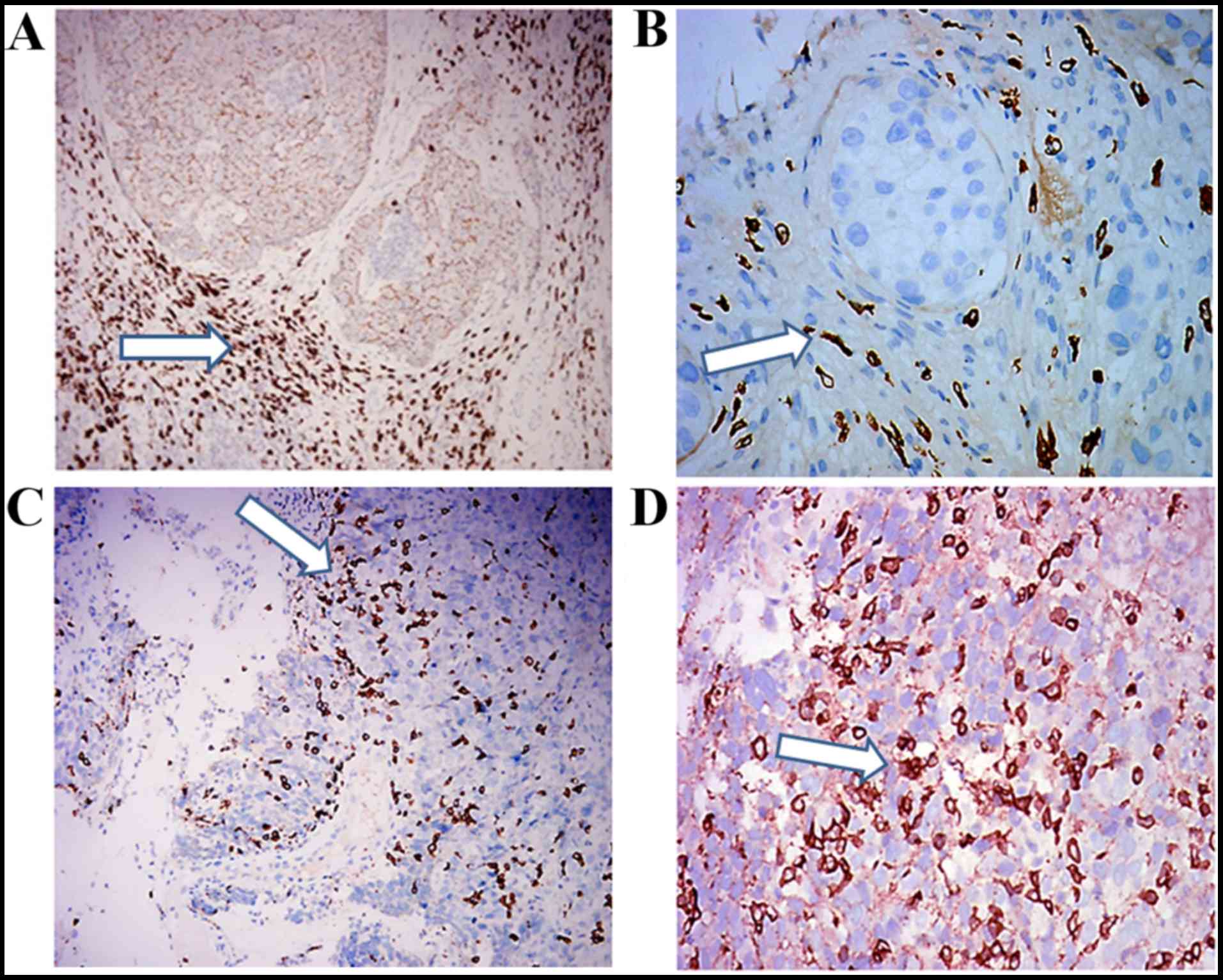
and 11 (35.5%) patients, respectively (Table I). Intra- and peri-tumoral
CD8+ TIL infiltration of luminal B BC samples were
illustrated in Fig. 1.
 | Table I.Clinicopathological characteristics
of patients with luminal B breast cancer, including expression of
CD8+ TIL. |
Table I.
Clinicopathological characteristics
of patients with luminal B breast cancer, including expression of
CD8+ TIL.
| Clinicopathological
characteristic | Value (%) |
|---|
| Age, years |
|
| Mean ± standard
deviation | 50.58±1.58 |
|
>50 | 13 (42) |
|
≤50 | 18 (58) |
| Histological
type |
|
|
Infiltrating ductal | 26 (83.9) |
|
Lobular | 5 (16.1) |
| Tumor grade at
initial biopsy |
|
| Grade
II | 16 (51.6) |
| Grade
III | 15 (48.4) |
| Lymphovascular
invasion |
|
|
Present | 9 (29) |
|
Absent | 22 (71) |
| Disease stage |
|
| Stage
II | 12 (27.5) |
| Stage
III | 19 (72.5) |
| pCR |
|
|
Present | 11 (35.5) |
|
Absent | 20 (64.5) |
| Peritumoral
CD8+TIL |
|
|
High | 20 (64.5) |
|
Low | 11 (35.5) |
| Intratumoral
CD8+TIL |
|
|
High | 16 (58.1) |
|
Low | 15 (41.9) |
Correlations between clinicopathological features
and pCR were only identified to be significant for intratumoral
expression of CD8+ TIL (Table
II). A total of 9/16 patients (56%) with high intratumoral
CD8+ TIL expression achieved a pCR, in contrast with
only 2/15 patients (13.3%) with low intratumoral CD8+
TIL expression (P=0.016). No significant correlation was identified
between peritumoral CD8+ TIL expression and pCR
(P=0.135).
| Table II.Prediction of pCR according to the
clinicopathological characteristics of patients with luminal B
breast cancer. |
Table II.
Prediction of pCR according to the
clinicopathological characteristics of patients with luminal B
breast cancer.
|
|
| Pathological
response |
|
|---|
|
|
|
|
|
|---|
| Clinicopathological
characteristic |
| Non pCR No.
(%) | pCR No. (%) | P-value |
|---|
| Age, years |
|
|
|
|
|
>50 | 13 | 9
(29) | 4
(13) | 0.654 |
|
<50 | 18 | 11 (35) | 7
(23) |
|
| Histological
type |
|
|
|
|
|
Infiltrating ductal | 26 | 17 (55) | 9
(29) | 0.595 |
|
Lobular | 5 | 3
(10) | 2 (6) |
|
| Tumor grade |
|
|
|
|
| Grade
II | 16 | 12 (39) | 4
(13) | 0.189 |
| Grade
III | 15 | 8
(26) | 7
(22) |
|
| Lymphovascular
invasion |
|
|
|
|
|
Present | 9 | 5
(16) | 4
(13) | 0.581 |
|
Absent | 22 | 15 (48) | 7
(23) |
|
| Disease stage |
|
|
|
|
| Stage
II | 12 | 10 (32) | 2 (7) | 0.086 |
| Stage
III | 19 | 10 (32) | 9
(29) |
|
| Peritumoral
CD8+TIL |
|
|
|
|
|
High | 20 | 11 (35) | 9
(29) | 0.135 |
|
Low | 11 | 9
(29) | 2 (7) |
|
| Intratumoral
CD8+TIL |
|
|
|
|
|
High | 16 | 7 (23) | 9
(29) | 0.016 |
|
Low | 15 | 13 (42) | 2 (6) |
Correlations were studied between peri- and
intra-tumoral expression of CD8+ TIL, and
clinicopathological characteristics, in addition to pCR. The
results confirmed that only intratumoral CD8+ TIL
expression was significantly correlated with pCR (Table III).
| Table III.Correlations between
clinicopathological characteristics of patients with luminal B
breast cancer and the expression of CD8+ TIL. |
Table III.
Correlations between
clinicopathological characteristics of patients with luminal B
breast cancer and the expression of CD8+ TIL.
|
|
| Intratumoral
CD8+ No. (%) |
| Peritumoral
CD8+ No. (%) |
|
|---|
|
|
|
|
|
|
|
|---|
| Clinicopathological
characteristic |
| High | Low | P-value | High | Low | P-value |
|---|
| Age, years |
|
|
| 0.677 |
|
| 0.2 |
|
>50 | 13 | 5 (16) | 8 (26) |
| 10 (32) | 3 (10) |
|
|
<50 | 18 | 11 (35) | 7 (23) |
| 10 (32) | 8 (26) |
|
| Histological
type |
|
|
| 0.186 |
|
| 0.405 |
|
Infiltrating ductal | 26 | 12 (39) | 14 (45) |
| 16 (52) | 10 (32) |
|
|
Lobular | 5 | 4 (13) | 1 (3) |
| 4 (13) | 1 (3) |
|
| Tumor grade |
|
|
| 0.838 |
|
| 0. 553 |
| Grade
II | 16 | 9 (29) | 7 (23) |
| 10 (32) | 6 (19) |
|
| Grade
III | 15 | 7 (23) | 8 (25) |
| 10 (32) | 5 (16) |
|
| Disease stage |
|
|
| 0.862 |
|
| 0.423 |
| Stage
II | 12 | 5 (16) | 7 (23) |
| 7 (23) | 5 (16) |
|
| Stage
III | 19 | 11 (35) | 8 (26) |
| 13 (42) | 6 (19) |
|
| Lymphovascular
invasion |
|
|
| 0.204 |
|
| 0.287 |
|
Present | 9 | 6 (19) | 3 (10) |
| 7 (23) | 2 (6) |
|
|
Absent | 22 | 10 (32) | 12 (39) |
| 13 (42) | 9 (29) |
|
| pCR |
|
|
| 0.016 |
|
| 0.135 |
|
Present | 11 | 9 (29) | 2 (6) |
| 9 (29) | 2 (7) |
|
|
Absent | 20 | 7 (23) | 13 (42) |
| 11 (35) | 9 (29) |
|
Univariate analysis revealed that high expression of
intratumoral CD8+ TILs was significantly associated with
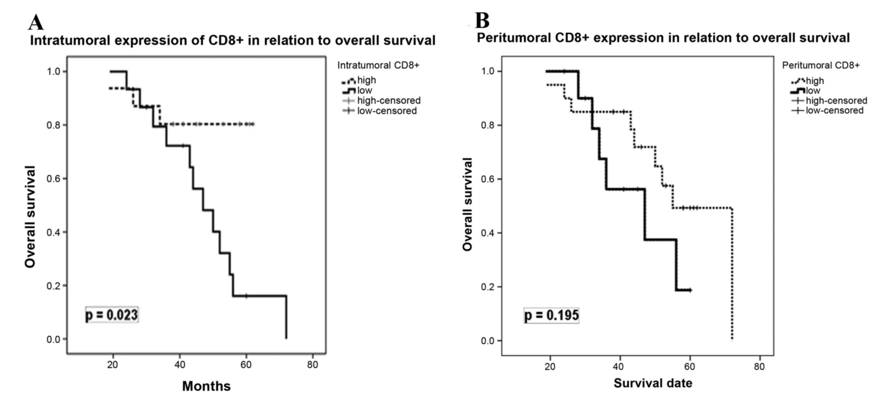
OS (P=0.023), but not with DFS (P=0.869). In contrast, peritumoral
CD8+ TIL expression exhibited no significant effect on
OS (P=0.195) and DFS (P=0.651). Fig.
2 depicts the correlations between intra-and peri-tumoral
CD8+ TIL expression, and OS. A multivariate Cox's
regression model was performed to assess the correlation between
the expression of CD8+ TIL, and DFS and OS, including
the following covariates: Age at diagnosis, tumor histological
type, tumor grade, disease stage, pCR and lymphovascular invasion.
As Table IV demonstrates,
intratumoral expression of CD8+ TIL was identified as an
independent prognostic factor for OS [HR=2.82; 95% confidence
interval (CI)=0.911–4.833; P=0.007], but not for DFS (HR=1.11; 95%
CI=0.282–2.078; P=0.508). No statistically significant effect of
any of other covariates was observed for DFS and OS.
| Table IV.Disease-free and overall survival
according to clinicopathological characteristics of patients with
luminal B breast cancer and pCR. |
Table IV.
Disease-free and overall survival
according to clinicopathological characteristics of patients with
luminal B breast cancer and pCR.
|
| Disease-free
survival | Overall
survival |
|---|
|
|
|
|
|---|
| Clinicopathological
characteristic | HR | 95% CI | P-value | HR | 95% CI | P-value |
|---|
| Age: <50 vs.
>50 years | 1.67 | 0.187–1.591 | 0.268 | 1.55 | 0.078–2.248 | 0.309 |
| Histological
type: | 1.1 | 0.238–1.984 | 0.688 | 1.62 | 0.376–3.867 | 0.211 |
| Ductal vs.
lobular |
|
|
|
|
|
|
| Tumor grade: II vs.
III | 0.88 | 0.369–2.376 | 0.891 | 1.76 | 0.051–1.466 | 0.131 |
| Disease stage: II
vs. III | 0.96 | 0.338–2.251 | 0.778 | 1.98 | 0.920–4.689 | 0.064 |
| Lymphovascular
invasion: | 1.02 | 0.330–2.196 | 0.74 | 1.32 | 0.455–2.974 | 0.245 |
| Absent vs.
present |
|
|
|
|
|
|
| pCR: Absent vs.
present | 1.12 | 0.211–1.434 | 0.69 | 1.66 | 0.011–1.408 | 0.072 |
| Peritumoral
CD8+ TIL: | 0.89 | 0.455–3.974 | 0.607 | 0.78 | 0.193–1.738 | 0.956 |
| Low vs. high |
|
|
|
|
|
|
| Intratumoral
CD8+ TIL: | 1.11 | 0.228–2.078 | 0.508 | 2.82 | 0.911–4.883 | 0.007 |
| Low vs. high |
|
|
|
|
|
|
Discussion
The traditional prognostic markers for BC include
clinical stage, lymph node involvement, tumor grade, ER and PR
status, and the presence of HER 2 abnormalities (39). However, the heterogeneity of the
disease, together with limitations of current therapeutic
modalities and advances in molecular diagnostics have led to
increasing interest in identifying novel prognostic and predictive
tools (7). Therefore, in the present
study, the expression of CD8+ TILs was evaluated in
patients with luminal B/HER 2-negative BC, in order to identify its
potential predictive and/or prognostic value. The present study
involved a cohort of 31 patients treated with
anthracycline-taxane-based NC, which is considered the standard
treatment for early BC, particularly for this molecular
subtype.
The contribution of the different TIL subpopulations
to the clinical and biological characteristics of BC remains
unclear, particularly in terms of prediction of chemotherapy
efficacy. The results of the present study revealed that the
intratumoral expression of CD8+ TIL may be predictive of
pCR in this patient population. Similar to previous studies
(17,21,22,40–42),
56% of patients with high intratumoral expression of
CD8+ TIL achieved a pCR, in contrast with 13.3% for
patients with low intratumoral CD8+ TIL.
Denkert et al (17) investigated intratumoral and stromal
lymphocytes in a total of 1,058 pre-therapeutic BC core biopsies
from two neoadjuvant anthracycline/taxane-based studies. It was
concluded that the presence of tumor-associated lymphocytes in BC
is an independent predictor of response to
anthracycline/taxane-based chemotherapy, and provides useful
information for oncologists to identify the subgroup of patients
that are most likely to benefit from this type of chemotherapy. A
multicentric neoadjuvant pilot study by Nabholtz et al
(21) investigated the value of
CD8+ expression as a predictor of pCR in a series of 60
patients with operable stage II–III triple-negative BC (TNBC),
treated with anthracycline-taxane-based chemotherapy plus the
anti-HER 1 monoclonal antibody panitumumab. It was reported that
high CD8+ TILs counts (≥118) predicted an 84%
probability of pCR, as opposed to low counts (<118) yielding a
10% probability. In addition, West et al (40) demonstrated that higher TIL counts
detected by eight-gene expression profiling were correlated with
the pCR rate in TNBC- and HER 2-positive tumors. In a previous
systematic review and meta-analysis, Mao et al (41) observed that a high number of TILs,
either stromal or intra-tumoral or both, is a significant predictor
of pCR for patients treated with NC. TILs detected in the
pre-treatment biopsy indicated a 2.5 and 5 times increased
probability of pCR in patients that were TNBC- and HER 2-positive,
respectively. Notably, no reported effects were observed in
ER-positive patients (41).
It was also observed that chemotherapy induced a
high rate of pCR in TIL-positive patients and was able to convert a
TIL-negative tumor into a TIL-positive one. It has previously been
reported that taxane-based chemotherapy converted 7/21 breast
tumors from TIL-negative to TIL-positive (42). Post-chemotherapy TIL status has been
identified to be associated with an improvement in clinical
response (42). Furthermore, a
comprehensive analysis of the immune characteristics in a small
group of patients with breast carcinoma demonstrated an increase in
CD8 and a decrease in CD4 and CD20 lymphocytes following
chemotherapy (43). Notably, the
results of the present study revealed that 86.7% of patients with
low intratumoral CD8+ TIL counts did not achieve a pCR.
This finding may have an important clinical impact regarding
neoadjuvant management of patients with luminal B BC. The potential
capability to predict a low probability of pCR may allow for the
consideration of alternative therapeutic strategies, including
endocrine therapy or the potential addition of immunotherapy.
Cytotoxic T cells, identifiable by CD8+
expression, form a major component of the adaptive immune system.
Cells that present foreign antigens in association with the major
histocompatibility complex class I molecule are recognized by
cytotoxic T lymphocytes through a specific interaction between the
presented antigen and the T-cell receptor (44). This interaction causes the activated T
cell to release proteins, including perforin and granzyme, enabling
cytotoxic activity through membranolysis (44). These mechanisms act on tumor cells
which, unlike normal cells, present atypical antigens (45). However, regulatory T cells, which
express forkhead box P3, act by decreasing the immune response to
self-antigens. The hypothesis that regulatory T cells may be
recruited by tumors to evade immune destruction is supported by the
observation that T cell ablation in mice enables an effective
anti-tumor response (46). Thus, it
is not surprising that the prognostic importance of lymphocytic
infiltration has been demonstrated in different types of solid
tumor.
Previous IHC studies have suggested that tumor
infiltrating CD8+ TILs may exhibit antitumor activity,
as indicated by their favorable effect on patient survival in
colorectal (47), ovarian (29), renal (30), lung (31) and pancreatic (32) cancer. Furthermore, in colorectal
cancer, the density and location of CD8+ TILs possess
prognostic value superior to and independent of the International
Union against Cancer tumor node metastasis classification (47). However, such findings have not been
confirmed in patients with BC, and in contrast to the unique
predictive function of CD8+ TILs for pCR, reports on the
association between CD8+ T-cell infiltration and BC
survival have presented conflicting results (22–28).
Nevertheless, two previous studies have investigated a larger
series of cases. Mahmoud et al (23) used a retrospective cohort of 1,334
patients with primary BC to demonstrate that total CD8+
TILs were independently associated with an improved survival rate
in BC. Baker et al (48),
investigated 1,953 BC cases and demonstrated that the independent
favorable prognostic effect of total CD8+ TILs was
observed only in tumors that were ER-negative.
These controversial results with regards to the
contribution of TIL to tumor progression and clinical outcome in BC
may be partially due to the fact that this effect may be restricted
to certain tumor subtypes. Another potential explanation may be
associated with the method of TIL analysis. Numerous studies have
used H&E-stained sections in their evaluation (22,27,28).
Evidently, the use of IHC to detect TILs is advantageous for
several reasons, including the ability to directly quantify cells
that express a given marker, in addition to accurately determining
their specific localization within a tissue (23). It is not possible to acquire this
information using conventional methods of gene-expression analysis
(23,25). Thus, this is why IHC was used in the
present study to address the potential value of CD8+
TILs in a well-characterized group of patients with luminal B BC.
The results revealed that the intratumoral expression of
CD8+ TIL was an independent prognostic factor for
improved OS, as patients with high intratumoral CD8+ TIL
counts had a 2.82 times higher probability of OS, compared with
those with low intratumoral CD8+ TIL counts.
Overall, the results of the present study may have
important clinical implications. Considering the prognosis of
luminal B/HER 2-negative cancer and the various profiles of
hormonal and chemotherapy sensitivity, immunobiological
stratification according to the expression of CD8+ TILs
may improve the management of these patients (4,6). In
addition, this type of immunological profiling may represent the
first step towards a more improved understanding of the potential
function of antitumor immune responses in mediating the clinical
outcome of NC. Furthermore, it may direct novel immunotherapeutic
approaches, including the use of programmed death-1 pathway
inhibitors for the treatment of patients with BC (49).
In conclusion, the results of the present study
suggested that high intratumoral CD8+ TILs expression is
significantly predictive of pCR post-NC (56%) and represents an
independent prognostic factor for improved OS in patients with
luminal B/HER 2-negative BC. In contrast, low intratumoral
CD8+ TIL expression was identified as a strong predictor
of lack of pCR to NC (13.3%), in addition to an independent
prognostic factor for poor OS. Assessment of the immune response,
in conjunction with the usual parameters, may aid in the
stratification of patients with luminal B/HER 2-negative BC
regarding the prediction of pCR post-NC and overall prognosis.
Further studies to develop TIL-based predictive and therapeutic
strategies in patients with luminal B BC are warranted.
Acknowledgements
The authors would like to thank and acknowledge the
support they received from the college of Medicine Research Center,
Deanship of Scientific Research, King Saud University and the
laboratory technicians for their help with the IHC analysis.
Glossary
Abbreviations
Abbreviations:
|
AC-T
|
doxorubicin cyclophosphamide
docetaxel
|
|
BC
|
breast cancer
|
|
CD8+
|
cluster of differentiation
8+
|
|
DFS
|
disease-free survival
|
|
ER
|
estrogen receptor
|
|
H&E
|
hematoxylin and eosin
|
|
HER 2
|
human epidermal growth factor receptor
2
|
|
NC
|
neoadjuvant chemotherapy
|
|
OS
|
overall survival
|
|
pCR
|
pathologic complete response
|
|
PR
|
progesterone receptor
|
|
TIL
|
tumor-infiltrating lymphocytes
|
|
TNBC
|
triple-negative breast cancer
|
References
|
1
|
Alghamdi IG, Hussain II, Alghamdi MS and
El-Sheemy MA: The incidence rate of female breast cancer in Saudi
Arabia: An observational descriptive epidemiological analysis of
data from Saudi Cancer Registry 2001–2008. Breast Cancer (Dove Med
Press). 5:103–109. 2013.PubMed/NCBI
|
|
2
|
Al-Rikabi A and Husain S: Increasing
prevalence of breast cancer among Saudi patients attending a
tertiary referral hospital: A retrospective epidemiologic study.
Croat Med J. 53:239–243. 2012. View Article : Google Scholar : PubMed/NCBI
|
|
3
|
Sørlie T, Perou CM, Tibshirani R, Aas T,
Geisler S, Johnsen H, Hastie T, Eisen MB, van de Rijin M, Jeffrey
SS, et al: Gene expression patterns of breast carcinomas
distinguish tumor subclasses with clinical implications. Proc Natl
Acad Sci USA. 98:pp. 10869–10874. 2001; View Article : Google Scholar : PubMed/NCBI
|
|
4
|
Ades F, Zardavas D, Bozovic-Spasojevic I,
Pugliano L, Fumagalli D, De Azambuja E, Viale G, Sotiriou C and
Piccart M: Luminal B breast cancer: Molecular characterization,
clinical management, and future perspectives. J Clin Oncol.
32:2794–2803. 2014. View Article : Google Scholar : PubMed/NCBI
|
|
5
|
Prat A, Cheang MC, Martin M, Parker JS,
Carrasco E, Caballero R, Tyldesley S, Gelmon K, Bernard PS, Nielsen
TO and Perou CM: Prognostic significance of progesterone
receptor-positive tumor cells within immunohistochemically defined
luminal a breast cancer. J Clin Oncol. 31:203–209. 2013. View Article : Google Scholar : PubMed/NCBI
|
|
6
|
Ladoire S, Mignot G, Dabakuyo S, Amould L,
Apetoh L, Rébé C, Coudert B, Martin F, Bizollon MH, Vanoli A, et
al: In situ immune response after neoadjuvant chemotherapy for
breast cancer predicts survival. J Pathol. 224:389–400. 2011.
View Article : Google Scholar : PubMed/NCBI
|
|
7
|
Cleator S and Ashworth A: Molecular
profiling of breast cancer: Clinical implications. Br J Cancer.
90:1120–1124. 2004. View Article : Google Scholar : PubMed/NCBI
|
|
8
|
Bear HD, Anderson S, Brown A, Smith R,
Mamounas EP, Fisher B, Margolese R, Theoret H, Soran A, Wickerham
DL and Wolmark N: National Surgical Adjuvant Breast and Bowel
Project Protocol B-27. The effect on tumor response of adding
sequential preoperative docetaxel to preoperative doxorubicin and
cyclophosphamide: Preliminary results from national surgical
adjuvant breast and bowel project protocol B-27. J Clin Oncol.
21:4165–4174. 2003. View Article : Google Scholar : PubMed/NCBI
|
|
9
|
Mauri D, Pavlidis N and Ioannidis JP:
Neoadjuvant versus adjuvant systemic treatment in breast cancer: A
meta-analysis. J Natl Cancer Inst. 97:188–194. 2005. View Article : Google Scholar : PubMed/NCBI
|
|
10
|
Chollet P, Amat S, Cure H, De Latour M, Le
Bouedec G, Mouret-Reynier MA, Ferriere JP, Achard JL, Dauplat J and
Penault-Llorca F: Prognostic significance of a complete
pathological response after induction chemotherapy in operable
breast cancer. Br J Cancer. 86:1041–1046. 2002. View Article : Google Scholar : PubMed/NCBI
|
|
11
|
von Minckwitz G, Untch M, Blohmer JU,
Costa SD, Eidtmann H, Fasching PA, Gerber B, Eiermann W, Hilfrich
J, Huober J, et al: Definition and impact of pathologic complete
response on prognosis after neoadjuvant chemotherapy in various
intrinsic breast cancer subtypes. J Clin Oncol. 30:1796–1804. 2012.
View Article : Google Scholar : PubMed/NCBI
|
|
12
|
Seo AN, Lee HJ, Kim EJ, Kim HJ, Jang MH,
Lee HE, Kim YJ, Kim JH and Park SY: Tumour-infiltrating CD8
lymphocytes as an independent predictive factor for pathological
complete response to primary systemic therapy in breast cancer. Br
J Cancer. 109:2705–2713. 2013. View Article : Google Scholar : PubMed/NCBI
|
|
13
|
Smyth MJ, Dunn GP and Schreiber RD: Cancer
immunosurveillance and immunoediting: The roles of immunity in
suppressing tumor development and shaping tumor immunogenicity. Adv
Immunol. 90:1–50. 2006. View Article : Google Scholar : PubMed/NCBI
|
|
14
|
Boon T, Cerottini JC, Van den Eynde B, van
der Bruggen P and Van Pel A: Tumor antigens recognized by T
lymphocytes. Annu Rev Immunol. 12:337–365. 1994. View Article : Google Scholar : PubMed/NCBI
|
|
15
|
Melichar B, Študentova H, Kalábová H,
Vitásková D, Čermáková P, Hornychová H and Ryška A: Predictive and
Prognostic significance of tumor-infiltrating lymphocytes in
patients with breast cancer treated with neoadjuvant systemic
therapy. Anticancer Res. 34:1115–1125. 2014.PubMed/NCBI
|
|
16
|
Dunn GP, Bruce AT, Ikeda H, Old LJ and
Schreiber RD: Cancer immunoediting: From immunosurveillance to
tumor escape. Nat Immunol. 3:991–998. 2002. View Article : Google Scholar : PubMed/NCBI
|
|
17
|
Denkert C, Loibl S, Noske A, Roller M,
Müller BM, Komor M, Budczies J, Darb-Esfahani S, Kronenwett R,
Hanusch C, et al: Tumor-associated lymphocytes as an independent
predictor of response to neoadjuvant chemotherapy in breast cancer.
J Clin Oncol. 28:105–113. 2010. View Article : Google Scholar : PubMed/NCBI
|
|
18
|
Lee HJ, Seo JY, Ahn JH, Ahn SH and Gong G:
Tumor-associated lymphocytes predict response to neoadjuvant
chemotherapy in breast cancer patients. J Breast Cancer. 16:32–39.
2013. View Article : Google Scholar : PubMed/NCBI
|
|
19
|
Ladoire S, Arnould L, Apetoh L, Coudert B,
Martin F, Chauffert B, Fumoleau P and Ghiringhelli F: Pathologic
complete response to neoadjuvant chemotherapy of breast carcinoma
is associated with the disappearance of tumor-infiltrating foxp3+
regulatory T cells. Clin Cancer Res. 14:2413–2420. 2008. View Article : Google Scholar : PubMed/NCBI
|
|
20
|
Houssami N, Macaskill P, von Minckwitz G,
Marinovich ML and Mamounas E: Meta-analysis of the association of
breast cancer subtype and pathologic complete response to
neoadjuvant chemotherapy. Eur J Cancer. 48:3342–3354. 2012.
View Article : Google Scholar : PubMed/NCBI
|
|
21
|
Nabholtz JM, Abrial C, Mouret-Reynier MA,
Dauplat MM, Weber B, Gligorov J, Forest AM, Tredan O, Vanlemmens L,
Petit T, et al: Multicentric neoadjuvant phase II study of
panitumumab combined with an anthracycline/taxane based
chemotherapy in operable triple negative breast cancer:
Identification of biologically-defined signatures predicting
treatment impact. Ann Oncol. 25:1570–1577. 2014. View Article : Google Scholar : PubMed/NCBI
|
|
22
|
Lee AH, Gillett CE, Ryder K, Fentiman IS,
Miles DW and Millis RR: Different patterns of inflammation and
prognosis in invasive carcinoma of the breast. Histopathology.
48:692–701. 2006. View Article : Google Scholar : PubMed/NCBI
|
|
23
|
Mahmoud SM, Paish EC, Powe DG, Macmillan
RD, Grainge MJ, Lee AH, Ellis IO and Green AR: Tumor-Infiltrating
CD8+ lymphocytes predict clinical outcome in breast cancer. J Clin
Oncol. 29:1949–1955. 2011. View Article : Google Scholar : PubMed/NCBI
|
|
24
|
Liu S, Lachapelle J, Leung S, Gao D,
Foulkes WD and Nielsen TO: CD8+ lymphocyte infiltration is an
independent favorable prognostic indicator in basal-like breast
cancer. Breast Cancer Res. 14:R482012. View Article : Google Scholar : PubMed/NCBI
|
|
25
|
Ali HR, Provenzano E, Dawson SJ, Blows FM,
Liu B, Shah M, Earl HM, Poole CJ, Hiller L, Dunn JA, et al:
Association between CD8+ T-cell infiltration and breast cancer
survival in 12,439 patients. Ann Oncol. 25:1536–1543. 2014.
View Article : Google Scholar : PubMed/NCBI
|
|
26
|
Matkowski R, Gisterek I, Halon A, Lacko A,
Szewczyk K, Staszek U, Pudelko M, Szynglarewicz B, Szelachowska J,
Zolnierek A and Kornafel J: The prognostic role of
tumor-infiltrating CD4 and CD8 T lymphocytes in breast cancer.
Anticancer Res. 29:2445–2451. 2009.PubMed/NCBI
|
|
27
|
Carlomagno C, Perrone F, Lauria R, De
Laurentiis M, Gallo C, Morabito A, Pettinato G, Panico L, Bellelli
T, Apicella A, et al: Prognostic significance of necrosis,
elastosis, fibrosis and inflammatory cell reaction in operable
breast cancer. Oncology. 52:272–277. 1995. View Article : Google Scholar : PubMed/NCBI
|
|
28
|
Aaltomaa S, Lipponen P, Eskelinen M, Kosma
VM, Marin S, Alhava E and Syrjänen K: Lymphocyte infiltrates as a
prognostic variable in female breast cancer. Eur J Cancer.
28A:859–864. 1992. View Article : Google Scholar : PubMed/NCBI
|
|
29
|
Sato E, Olson SH, Ahn J, Bundy B,
Nishikawa H, Qian F, Jungbluth AA, Frosina D, Gnjatic S, Ambrosone
C, et al: Intraepithelial CD8+ tumor-infiltrating lymphocytes and a
high CD8+/regulatory T cell ratio are associated with favorable
prognosis in ovarian cancer. Proc Natl Acad Sci USA. 102:pp.
18538–18543. 2005; View Article : Google Scholar : PubMed/NCBI
|
|
30
|
Nakano O, Sato M, Naito Y, Suzuki K,
Orikasa S, Aizawa M, Suzuki Y, Shintaku I, Nagura H and Ohtani H:
Proliferative activity of intratumoral CD8(+) T-lymphocytes as a
prognostic factor in human renal cell carcinoma: Clinicopathologic
demonstration of antitumor immunity. Cancer Res. 61:5132–5136.
2001.PubMed/NCBI
|
|
31
|
Kawai O, Ishii G, Kubota K, Murata Y,
Naito Y, Mizuno T, Aokage K, Saijo N, Nishiwaki Y, Gemma A, et al:
Predominant infiltration of macrophages and CD8(+) T cells in
cancer nests is a significant predictor of survival in stage IV
nonsmall cell lung cancer. Cancer. 113:1387–1395. 2008. View Article : Google Scholar : PubMed/NCBI
|
|
32
|
Fukunaga A, Miyamoto M, Cho Y, Murakami S,
Kawarada Y, Oshikiri T, Kato K, Kurokawa T, Suzuoki M, Nakakubo Y,
et al: CD8+ tumor-infiltrating lymphocytes together with CD4+
tumor-infiltrating lymphocytes and dendritic cells improve the
prognosis of patients with pancreatic adenocarcinoma. Pancreas.
28:e26–e31. 2004. View Article : Google Scholar : PubMed/NCBI
|
|
33
|
Camp BJ, Dyhrman ST, Memoli VA, Mott LA
and Barth RJ Jr: In situ cytokine production by breast cancer
tumor-infiltrating lymphocytes. Ann Surg Oncol. 3:176–184. 1996.
View Article : Google Scholar : PubMed/NCBI
|
|
34
|
Muraro E, Martorelli D, Turchet E, Miolo
G, Scalone S, Comaro E, Talamini R, Mastorci K, Lombardi D, Perin
T, et al: A different immunologic profile characterizes patients
with HER 2-overexpressing and HER 2-negative locally advanced
breast cancer: Implications for immune-based therapies. Breast
Cancer Res. 13:R1172011. View Article : Google Scholar : PubMed/NCBI
|
|
35
|
Lakhani SR, Ellis IO, Schnitt SJ, Tan PH
and van de Vijver MJ: WHO classification of tumors of the breast.
4. 4th. Lyon: International Agency for Research on Cancer; 2012
|
|
36
|
Edge S, Byrd DR, Compton CC, Fritz AG,
Greene FL and Trotti A: AJCC cancer staging Manual. 7th. New York:
Springer; 2010
|
|
37
|
Ellis IO, Galea M, Broughton N, Locker A,
Blamey RW and Elston CW: Pathological prognostic factors in breast
cancer: II. Histological type. relationship with survival in a
large study with long-term follow-up. Histopathology. 20:479–489.
1992. View Article : Google Scholar : PubMed/NCBI
|
|
38
|
Elston CW and Elli IO: Pathological
prognostic factors in breast cancer. I. The value of histological
grade in breast cancer: experience from a large study with
long-term follow-up. Histopathology. 41:151–153. 2002. View Article : Google Scholar : PubMed/NCBI
|
|
39
|
Schnitt SJ: Classification and prognosis
of invasive breast cancer: From morphology to molecular taxonomy.
Mod Pathol. 23 Suppl 2:S60–S64. 2010. View Article : Google Scholar : PubMed/NCBI
|
|
40
|
West NR, Milne K, Truong PT, Macpherson N,
Nelson BH and Watson PH: Tumor-infiltrating lymphocytes predict
response to anthracycline-based chemotherapy in estrogen
receptor-negative breast cancer. Breast Cancer Res. 13:R1262011.
View Article : Google Scholar : PubMed/NCBI
|
|
41
|
Mao Y, Qu Q, Zhang Y, Liu J, Chen X and
Shen K: The value of tumor infiltrating lymphocytes (TILS) for
predicting response to neoadjuvant chemotherapy in breast cancer: A
systematic review and meta-analysis. PLoS One. 9:e1151032014.
View Article : Google Scholar : PubMed/NCBI
|
|
42
|
Demaria S, Volm MD, Shapiro RL, Yee HT,
Oratz R, Formenti SC, Muggia F and Symmans WF: Development of
tumor-infiltrating lymphocytes in breast cancer after neoadjuvant
paclitaxel chemotherapy. Clin Cancer Res. 7:3025–3030.
2001.PubMed/NCBI
|
|
43
|
Ruffell B, Au A, Rugo HS, Esserman LJ,
Hwang ES and Coussens LM: Leukocyte composition of human breast
cancer. Proc Natl Acad Sci USA. 109:pp. 2796–2801. 2012; View Article : Google Scholar : PubMed/NCBI
|
|
44
|
Berke G: The binding and lysis of target
cells by cytotoxic lymphocytes: Molecular and cellular aspects.
Annu Rev Immunol. 12:735–773. 1994. View Article : Google Scholar : PubMed/NCBI
|
|
45
|
Zitvogel L, Kepp O and Kroemer G: Immune
parameters affecting the efficacy of chemotherapeutic regimens. Nat
Rev Clin Oncol. 8:151–160. 2011. View Article : Google Scholar : PubMed/NCBI
|
|
46
|
Shimizu J, Yamazaki S and Sakaguchi S:
Induction of tumor immunity by removing CD25+CD4+ T cells: A common
basis between tumor immunity and autoimmunity. J Immunol.
163:5211–5218. 1999.PubMed/NCBI
|
|
47
|
Galon J, Costes A, Sanchez-Cabo F,
Kirilovsky A, Mlecnik B, Lagorce-Pagès C, Tosolini M, Camus M,
Berger A, Wind P, et al: Type, density, and location of immune
cells within human colorectal tumors predict clinical outcome.
Science. 313:1960–1964. 2006. View Article : Google Scholar : PubMed/NCBI
|
|
48
|
Baker K, Lachapelle J, Zlobec I, Bismar
TA, Terracciano L and Foulkes WD: Prognostic significance of CD8+ T
lymphocytes in breast cancer depends upon both oestrogen receptor
status and histological grade. Histopathology. 58:1107–1116.
2011.PubMed/NCBI
|
|
49
|
Lee SM and Chow LQ: A new addition to the
PD-1 checkpoint inhibitors for non-small cell lung cancer-the
anti-PDL1 antibody-MED14736. Transl Lung Cancer Res. 3:408–410.
2014.PubMed/NCBI
|