Spandidos Publications Logo
  • About
    • About Spandidos
    • Aims and Scopes
    • Abstracting and Indexing
    • Editorial Policies
    • Reprints and Permissions
    • Job Opportunities
    • Terms and Conditions
    • Contact
  • Journals
    • All Journals
    • Oncology Letters
      • Oncology Letters
      • Information for Authors
      • Editorial Policies
      • Editorial Board
      • Aims and Scope
      • Abstracting and Indexing
      • Bibliographic Information
      • Archive
    • International Journal of Oncology
      • International Journal of Oncology
      • Information for Authors
      • Editorial Policies
      • Editorial Board
      • Aims and Scope
      • Abstracting and Indexing
      • Bibliographic Information
      • Archive
    • Molecular and Clinical Oncology
      • Molecular and Clinical Oncology
      • Information for Authors
      • Editorial Policies
      • Editorial Board
      • Aims and Scope
      • Abstracting and Indexing
      • Bibliographic Information
      • Archive
    • Experimental and Therapeutic Medicine
      • Experimental and Therapeutic Medicine
      • Information for Authors
      • Editorial Policies
      • Editorial Board
      • Aims and Scope
      • Abstracting and Indexing
      • Bibliographic Information
      • Archive
    • International Journal of Molecular Medicine
      • International Journal of Molecular Medicine
      • Information for Authors
      • Editorial Policies
      • Editorial Board
      • Aims and Scope
      • Abstracting and Indexing
      • Bibliographic Information
      • Archive
    • Biomedical Reports
      • Biomedical Reports
      • Information for Authors
      • Editorial Policies
      • Editorial Board
      • Aims and Scope
      • Abstracting and Indexing
      • Bibliographic Information
      • Archive
    • Oncology Reports
      • Oncology Reports
      • Information for Authors
      • Editorial Policies
      • Editorial Board
      • Aims and Scope
      • Abstracting and Indexing
      • Bibliographic Information
      • Archive
    • Molecular Medicine Reports
      • Molecular Medicine Reports
      • Information for Authors
      • Editorial Policies
      • Editorial Board
      • Aims and Scope
      • Abstracting and Indexing
      • Bibliographic Information
      • Archive
    • World Academy of Sciences Journal
      • World Academy of Sciences Journal
      • Information for Authors
      • Editorial Policies
      • Editorial Board
      • Aims and Scope
      • Abstracting and Indexing
      • Bibliographic Information
      • Archive
    • International Journal of Functional Nutrition
      • International Journal of Functional Nutrition
      • Information for Authors
      • Editorial Policies
      • Editorial Board
      • Aims and Scope
      • Abstracting and Indexing
      • Bibliographic Information
      • Archive
    • International Journal of Epigenetics
      • International Journal of Epigenetics
      • Information for Authors
      • Editorial Policies
      • Editorial Board
      • Aims and Scope
      • Abstracting and Indexing
      • Bibliographic Information
      • Archive
    • Medicine International
      • Medicine International
      • Information for Authors
      • Editorial Policies
      • Editorial Board
      • Aims and Scope
      • Abstracting and Indexing
      • Bibliographic Information
      • Archive
  • Articles
  • Information
    • Information for Authors
    • Information for Reviewers
    • Information for Librarians
    • Information for Advertisers
    • Conferences
  • Language Editing
Spandidos Publications Logo
  • About
    • About Spandidos
    • Aims and Scopes
    • Abstracting and Indexing
    • Editorial Policies
    • Reprints and Permissions
    • Job Opportunities
    • Terms and Conditions
    • Contact
  • Journals
    • All Journals
    • Biomedical Reports
      • Information for Authors
      • Editorial Policies
      • Editorial Board
      • Aims and Scope
      • Abstracting and Indexing
      • Bibliographic Information
      • Archive
    • Experimental and Therapeutic Medicine
      • Information for Authors
      • Editorial Policies
      • Editorial Board
      • Aims and Scope
      • Abstracting and Indexing
      • Bibliographic Information
      • Archive
    • International Journal of Epigenetics
      • Information for Authors
      • Editorial Policies
      • Editorial Board
      • Aims and Scope
      • Abstracting and Indexing
      • Bibliographic Information
      • Archive
    • International Journal of Functional Nutrition
      • Information for Authors
      • Editorial Policies
      • Editorial Board
      • Aims and Scope
      • Abstracting and Indexing
      • Bibliographic Information
      • Archive
    • International Journal of Molecular Medicine
      • Information for Authors
      • Editorial Policies
      • Editorial Board
      • Aims and Scope
      • Abstracting and Indexing
      • Bibliographic Information
      • Archive
    • International Journal of Oncology
      • Information for Authors
      • Editorial Policies
      • Editorial Board
      • Aims and Scope
      • Abstracting and Indexing
      • Bibliographic Information
      • Archive
    • Medicine International
      • Information for Authors
      • Editorial Policies
      • Editorial Board
      • Aims and Scope
      • Abstracting and Indexing
      • Bibliographic Information
      • Archive
    • Molecular and Clinical Oncology
      • Information for Authors
      • Editorial Policies
      • Editorial Board
      • Aims and Scope
      • Abstracting and Indexing
      • Bibliographic Information
      • Archive
    • Molecular Medicine Reports
      • Information for Authors
      • Editorial Policies
      • Editorial Board
      • Aims and Scope
      • Abstracting and Indexing
      • Bibliographic Information
      • Archive
    • Oncology Letters
      • Information for Authors
      • Editorial Policies
      • Editorial Board
      • Aims and Scope
      • Abstracting and Indexing
      • Bibliographic Information
      • Archive
    • Oncology Reports
      • Information for Authors
      • Editorial Policies
      • Editorial Board
      • Aims and Scope
      • Abstracting and Indexing
      • Bibliographic Information
      • Archive
    • World Academy of Sciences Journal
      • Information for Authors
      • Editorial Policies
      • Editorial Board
      • Aims and Scope
      • Abstracting and Indexing
      • Bibliographic Information
      • Archive
  • Articles
  • Information
    • For Authors
    • For Reviewers
    • For Librarians
    • For Advertisers
    • Conferences
  • Language Editing
Login Register Submit
  • This site uses cookies
  • You can change your cookie settings at any time by following the instructions in our Cookie Policy. To find out more, you may read our Privacy Policy.

    I agree
Search articles by DOI, keyword, author or affiliation
Search
Advanced Search
presentation
Oncology Letters
Join Editorial Board Propose a Special Issue
Print ISSN: 1792-1074 Online ISSN: 1792-1082
Journal Cover
July-2017 Volume 14 Issue 1

Full Size Image

Sign up for eToc alerts
Recommend to Library

Journals

International Journal of Molecular Medicine

International Journal of Molecular Medicine

International Journal of Molecular Medicine is an international journal devoted to molecular mechanisms of human disease.

International Journal of Oncology

International Journal of Oncology

International Journal of Oncology is an international journal devoted to oncology research and cancer treatment.

Molecular Medicine Reports

Molecular Medicine Reports

Covers molecular medicine topics such as pharmacology, pathology, genetics, neuroscience, infectious diseases, molecular cardiology, and molecular surgery.

Oncology Reports

Oncology Reports

Oncology Reports is an international journal devoted to fundamental and applied research in Oncology.

Experimental and Therapeutic Medicine

Experimental and Therapeutic Medicine

Experimental and Therapeutic Medicine is an international journal devoted to laboratory and clinical medicine.

Oncology Letters

Oncology Letters

Oncology Letters is an international journal devoted to Experimental and Clinical Oncology.

Biomedical Reports

Biomedical Reports

Explores a wide range of biological and medical fields, including pharmacology, genetics, microbiology, neuroscience, and molecular cardiology.

Molecular and Clinical Oncology

Molecular and Clinical Oncology

International journal addressing all aspects of oncology research, from tumorigenesis and oncogenes to chemotherapy and metastasis.

World Academy of Sciences Journal

World Academy of Sciences Journal

Multidisciplinary open-access journal spanning biochemistry, genetics, neuroscience, environmental health, and synthetic biology.

International Journal of Functional Nutrition

International Journal of Functional Nutrition

Open-access journal combining biochemistry, pharmacology, immunology, and genetics to advance health through functional nutrition.

International Journal of Epigenetics

International Journal of Epigenetics

Publishes open-access research on using epigenetics to advance understanding and treatment of human disease.

Medicine International

Medicine International

An International Open Access Journal Devoted to General Medicine.

Journal Cover
July-2017 Volume 14 Issue 1

Full Size Image

Sign up for eToc alerts
Recommend to Library

  • Article
  • Citations
    • Cite This Article
    • Download Citation
    • Create Citation Alert
    • Remove Citation Alert
    • Cited By
  • Similar Articles
    • Related Articles (in Spandidos Publications)
    • Similar Articles (Google Scholar)
    • Similar Articles (PubMed)
  • Download PDF
  • Download XML
  • View XML
Case Report

Leiomyosarcoma of the splenic vein: A case report

  • Authors:
    • Wenfang Wu
    • Xinxiang Zhao
    • Yan Wang
    • Cong Di
    • Renhui Cai
    • Yingxue Zhang
    • Siying Chen
    • Wei Zhang
    • Xin Yue
  • View Affiliations / Copyright

    Affiliations: Department of Radiology, The Second Affiliated Hospital of Kunming Medical University, Kunming, Yunnan 650101, P.R. China, Department of Pathology, The Second Affiliated Hospital of Kunming Medical University, Kunming, Yunnan 650101, P.R. China
  • Pages: 977-980
    |
    Published online on: May 22, 2017
       https://doi.org/10.3892/ol.2017.6225
  • Expand metrics +
Metrics: Total Views: 0 (Spandidos Publications: | PMC Statistics: )
Metrics: Total PDF Downloads: 0 (Spandidos Publications: | PMC Statistics: )
Cited By (CrossRef): 0 citations Loading Articles...

This article is mentioned in:


Abstract

Leiomyosarcomas (LMSs) originating from the wall of blood vessels are aggressive and rare neoplasms. The current study describes a case of a 52-year-old man who presented with intermittent abdominal pain without weight loss or diarrhea. Computed tomography of the abdomen identified a 4‑cm, solid, heterogeneous tumor in the tail of the pancreas, while a hypodense lesion was also noted in the right hepatic lobe. The patient subsequently underwent splenic pedicle tumor resection, splenectomy and liver tumor resection. Notably, immunohistochemical and histological analyses identified LMS, which had originated from the smooth muscle of the splenic vein. Currently available information regarding LMSs of the splenic vein and their management is also discussed, with the aim of improving diagnostic accuracy.

Introduction

Primary leiomyosarcoma (LMS) of the splenic vein is a rare malignant tumor and there have only been three previously reported cases (1,2). Due to the rarity of the tumor, its imaging features have not yet been described in detail. Venous LMSs predominately occur in middle-aged women in the fifth and sixth decades of life (3). Clinical manifestations depend on the tumor position in the vessel wall (4). The symptoms of LMS include abdominal masses, abdominal pain, nausea and fever (5). The current study presents a case report on the magnetic resonance imaging (MRI) features of primary LMS of the splenic vein. A solid, heterogeneous mass was located in the tail of the pancreas. In portal phase, the splenic vein was embedded in the mass on T1-weighted imaging (T1WI). The patient underwent splenic pedicle tumor resection, splenectomy and liver tumor resection with no postoperative complications. There is a limited amount of evidence demonstrating increased survival following adjuvant radiation, combination radiation and chemotherapy, in addition to surgical resection (6). Further information regarding the prognosis of the present patient is unavailable, as the patient did not undergo further adjuvant therapy or follow-up, and unique treatment recommendations or outcome data for such lesions remains to be established.

Case report

In June 2013, a 52-year-old male was admitted to the Gastroenterology Department of The Second Affiliated Hospital of Kunming Medical University (Kunming, China) presenting with a long history (>1 year) of intermittent epigastric pain. The pain presented as either severe colic lasting for 20–30 min or as dull pain, which would be relieved following squatting. The medical history of the patient was unremarkable. A whole body positron emission tomography scan was performed at Yuxi People's Hospital (Yuxi, China) and revealed an irregular mass in the splenic hilum without increased metabolism.

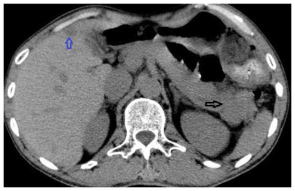
An abdominal computed tomography (CT) scan performed at the Department of Radiology, The Second Affiliated Hospital of Kunming Medical University identified an irregular, hypodense mass measuring ~4.1×3.0 cm in the tail of the pancreas, while a hypodense lesion was also observed in the right hepatic lobe 
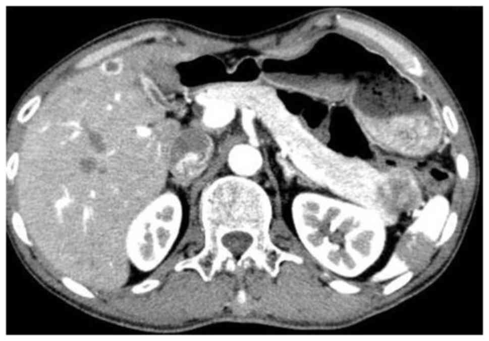
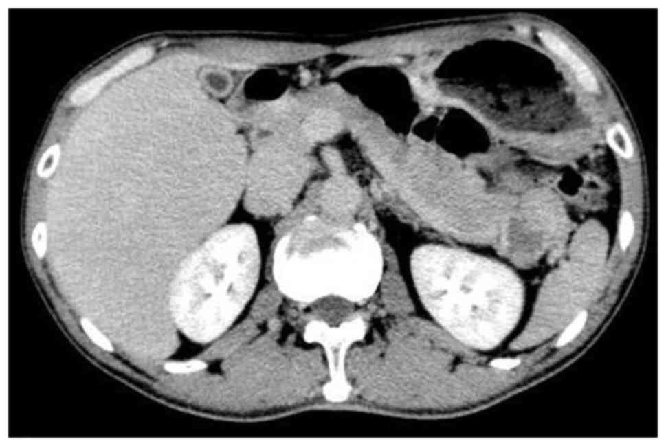
(Fig. 1). Following administration of contrast agent, lesions exhibited peripheral enhancement and there was a low-density zone in the center of the lesion without 
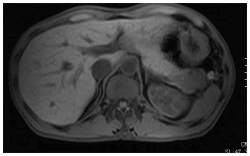
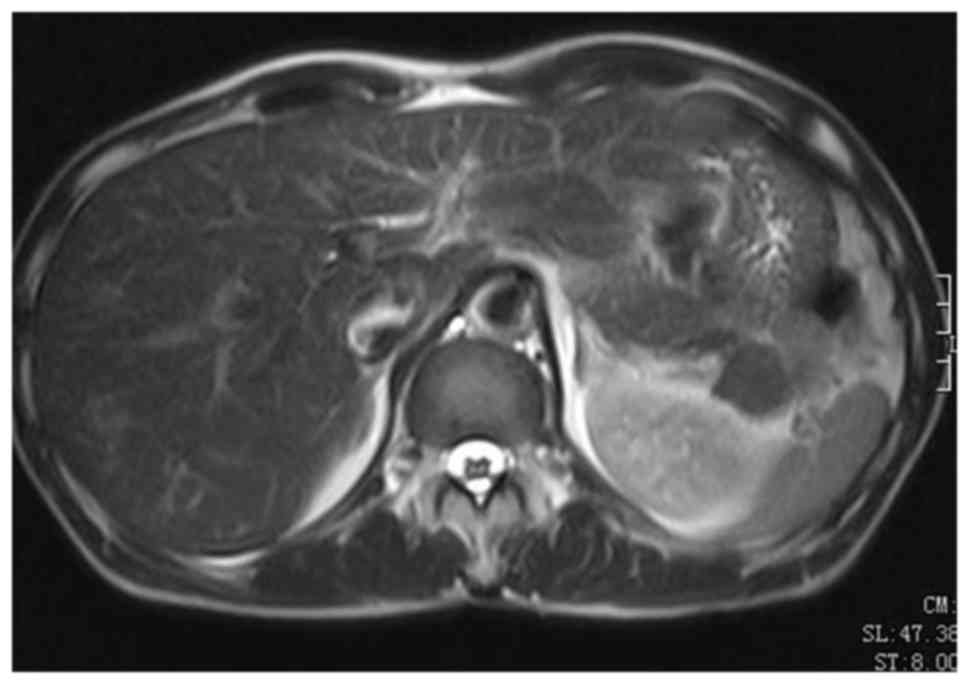
enhancement (Figs. 2 and 3). An MRI scan identified a solid, heterogeneous mass, which had fused with two nodules measuring 1.87×2.44 cm and 2.46×2.34 cm, located in the tail of the pancreas. T1WI (Fig. 4) demonstrated slight hypointensity, while T2-WI exhibited slight hyperintensity 
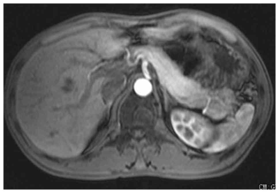
(Fig. 5). Mild inhomogeneous 
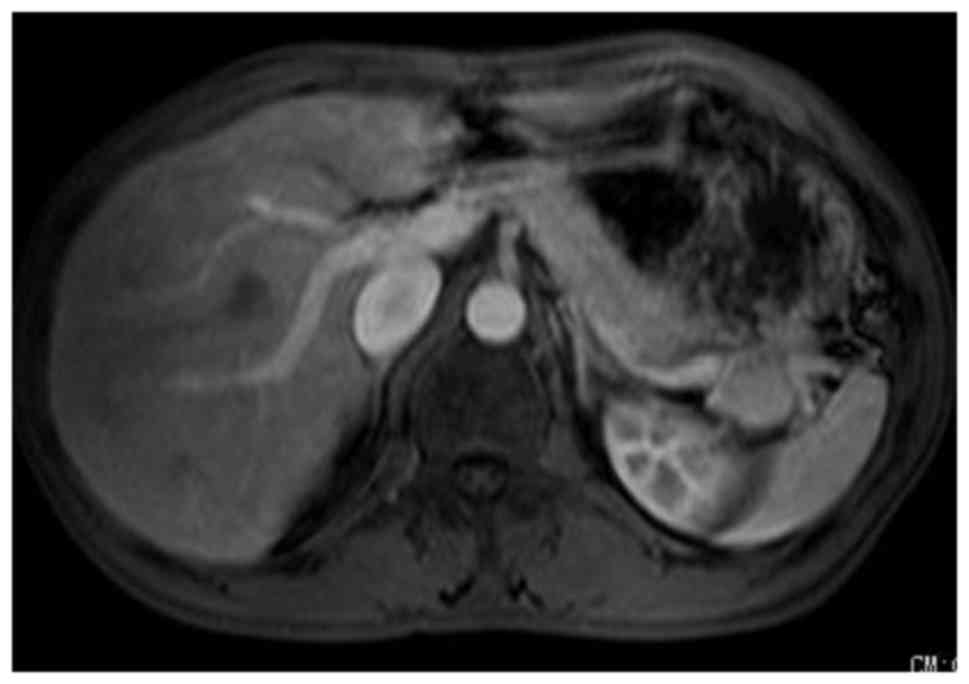
enhancement was observed in the arterial phase (Fig. 6), strengthening in the portal phase (Fig. 7) was higher than the arterial phase and the splenic vein was embedded in the mass on T1WI 
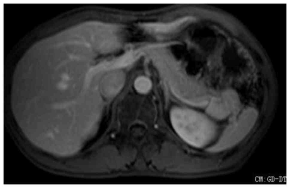
enhanced images. The delay period exhibited inhomogeneous moderate enhancement (Fig. 8) and a small nodule was located between the liver VI (right hepatic lobe lower section) and the liver V (lower right anterior lobe of the liver). The nodule in the liver exhibited slight hypointensity on T1WI, which was similar to the liver signal on T2WI, and the lesion demonstrated peripheral enhancement post-contrast.

Figure 1.

Abdominal computed tomography showed an irregular, hypodense mass measuring 4.1×3.0 cm in the tail of the pancreas (black arrow), while a hypodense lesion was also observed in the right hepatic lobe (blue arrow).

Figure 2.

Post-contrast lesions in the tail of the pancreas exhibited peripheral enhancement. The nodule in the liver demonstrated peripheral enhancement.

Figure 3.

Post-contrast image demonstrating a low-density zone in the center of the lesion (located in the tail of the pancreas) without enhancement.

Figure 4.

A magnetic resonance image showing a solid, heterogeneous mass located in the tail of the pancreas. The lesion demonstrated slight hypointensity on T1-weighted images.

Figure 5.

T2-weighted image of the lesion in the tail of the pancreas, demonstrating slight hyperintensity.

Figure 6.

Lesion located in the tail of the pancreas exhibited inhomogeneous, mild enhancement in the arterial phase.

Figure 7.

In the portal phase, the degree of enhancement of the lesion located in the tail of the pancreas was higher compared with the arterial phase.

Figure 8.

In the delayed phase, the lesion located in the tail of the pancreas exhibited inhomogeneous moderate enhancement.

An initial diagnosis of pancreatic cancer was considered and surgery was scheduled. The patient subsequently underwent splenic pedicle tumor resection, splenectomy and liver tumor resection. Following surgical resection, specimens were fixed in 10% neutral formalin for 12 h, embedded in paraffin and cut into 4 µm-thick sections. These sections were subsequently stained with 
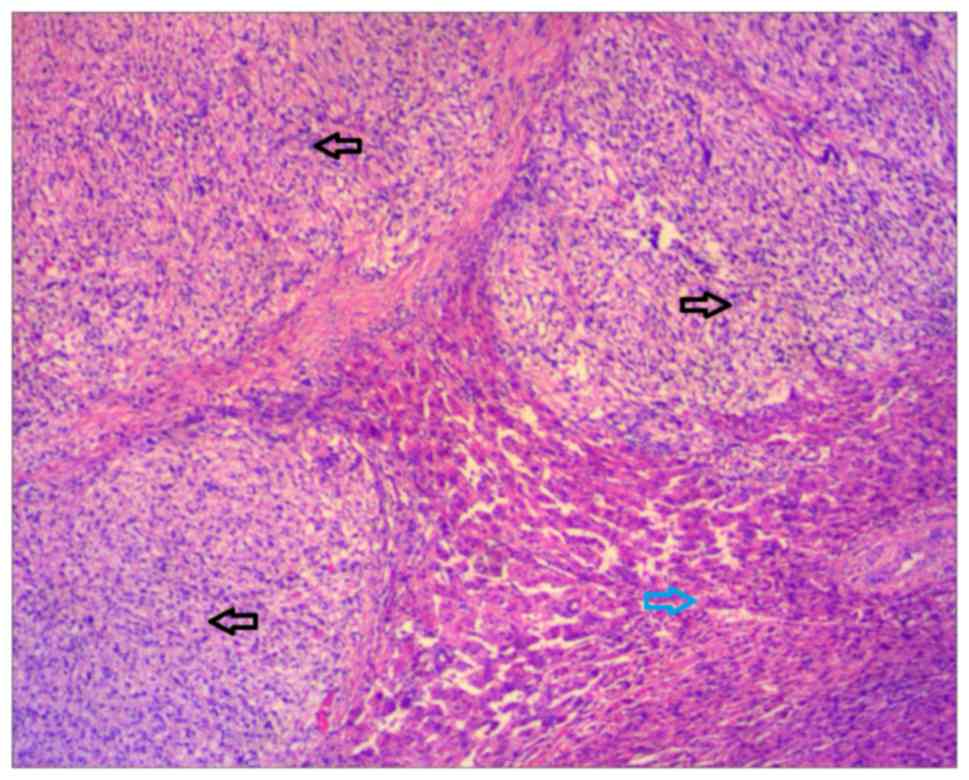
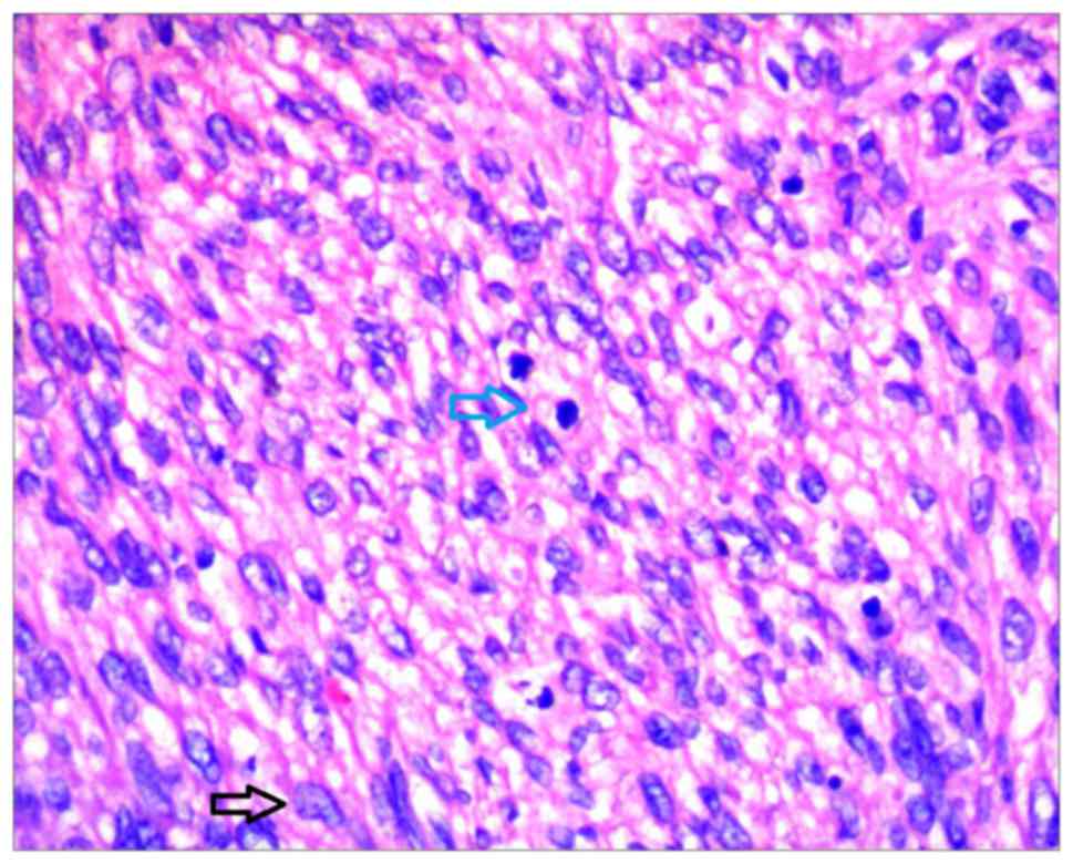
hematoxylin and eosin. The tumor was composed of bundles of staggered and spindle cells with necrosis. Atypia and mitosis were easily observable. For immunohistochemistry, paraffin-embedded sections were dewaxed, rehydrated and incubated with the following primary antibodies for 1 h at 37°C: Anti-vimentin (catalog no. RMA-0547; no dilution), anti-smooth muscle actin (catalog no. Kit-0006; no dilution), anti-desmin (catalog no. Kit-0023; no dilution), anti-H-caldesmon (catalog no. MAB-0634; no dilution) and anti-Ki-67 (catalog no. Kit-0005; no dilution; Fuzhou Maixin Biotech. Co., Ltd., Fuzhou, China). The sections were subsequently incubated with the biotin-labeled secondary antibody (catalog no., Kit-9730; no dilution; Fuzhou Maixin Biotech. Co., Ltd.) for 0.5 h 
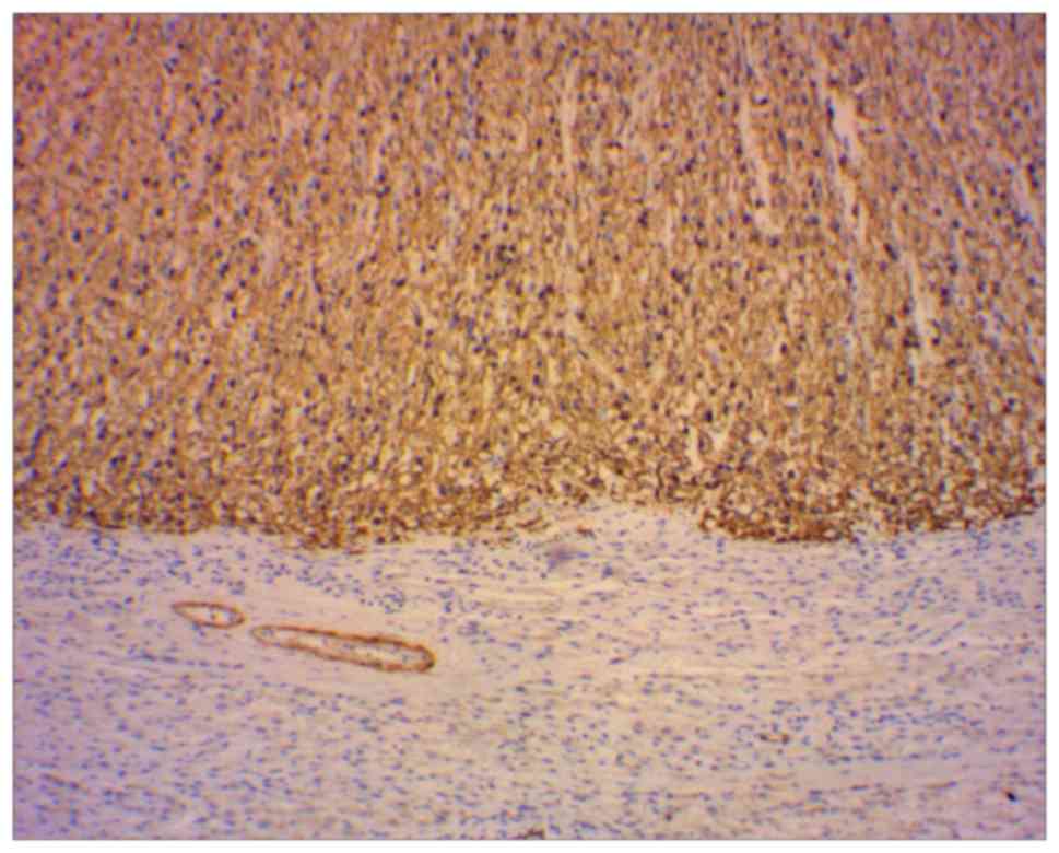
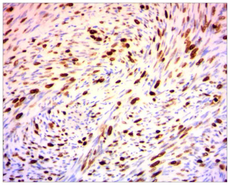
at 37°C. Immunohistochemistry demonstrated positive expression of vimentin, smooth muscle actin, desmin, H-caldesmon, Ki-67 and cluster of differentiation 34 (Figs. 9 and 10). Finally, histopathological examinations confirmed both tumors were intermediate-grade LMSs (Figs. 11 and 12).

Figure 9.

Cells in the mass in the liver were strongly positive for H-caldesmon (magnification, ×2.5).

Figure 10.

Cells within the mass in the splenic hilum were positive for Ki-67 (magnification, ×10).

Figure 11.

Mass in the liver showing tumor tissues (black arrow) and normal liver tissues (blue arrow) (magnification, ×2.5).

Figure 12.

Mass in the splenic hilum showing a large number of spindle tumor cells (black arrow) and a mitotic figure (blue arrow; magnification, ×20).

Discussion

LMSs are rare, malignant, mesenchymal tumors derived from smooth muscle that usually appear in the retroperitoneum space behind the abdominal cavity or the uterus (3). LMSs may occur intra-abdominally in the retroperitoneum, mesentery or omentum (40–45%), in subcutaneous or deep soft tissue of the limbs (20–30%), in the arrector pili muscle of the skin (15–20%), or in blood vessel walls (5%) (1). LMSs are rare and aggressive neoplasms that arise from the blood vessel walls, and vascular LMSs occur more commonly in veins (3). Previous studies have demonstrated that vascular LMSs occur in veins five times more often than in arteries (3). The inferior vena cava locates 50% of large-vessel LMSs and the retroperitoneal veins close to the inferior vena cava, including the iliac, renal, and spermatic and ovarian veins, may be additional sites of occurrence (7). Cases of visceral vein LMSs also exist, however, those derived from the splenic vein are exceedingly rare with only a few previously documented cases (1,2,8).

Venous LMSs predominately occur in middle-aged women in the fifth and sixth decades of life, and usually arise from the inferior vena cava or large veins in the lower extremities (4). Clinical manifestations depend on the tumor position in the vessel wall, venous obstruction and compression of the surrounding organs by the mass, including palpable abdominal masses and symptoms associated with thrombosis or embolism of the splenic vein, such as abdominal pain, nausea and fever (9,10). Sarcomas of the great vessels are classified into luminal and mural tumors according to their site of origin (5). Luminal sarcomas of the blood vessels are more common in arteries than veins and they are characterized by rapid growth and the earlier onset of distant metastases due to intraluminal growth (5,11). By contrast, mural sarcomas of the blood vessels are more common in veins, and unlike luminal sarcomas, they have more positive prognosis due to their slow and extraluminal growth (11). With regard to treatment, the majority of soft-tissue sarcomas undergo surgical resection for localized primary tumors; therefore resection offers the only opportunity for complete cure in the absence of disseminated disease. Additionally, tumor extent and histological examination to determine classification and grade is also important in guiding adjunct treatment.

Radiotherapy has been considered for the treatment of high-grade soft tissue sarcomas located in the extremities, intermediate-grade tumors of the limbs with close or positive histological margins and as a treatment for recurring low-grade sarcomas. Chemotherapy may be administered when systemic control is the primary therapeutic aim, however, the sensitivity of sarcomas, including LMSs, to chemotherapy appears to be poor (12). Therefore, as a form of adjuvant treatment for LMS, chemotherapy does not produce an evident increase in patient survival (12). There is a limited amount of evidence demonstrating increased survival following adjuvant radiation, combination radiation and chemotherapy, in addition to surgical resection. However, whether these treatment modalities should be incorporated in the general treatment of LMS is still being debated.

The current study describes a case of LMS derived from the splenic vein with concurrent liver metastasis. Histopathological examination confirmed the lesions to be intermediate-grade LMSs. An association between the masses in the spleen, splenic hilum and splenic vein was confirmed; however, the distal pancreas parenchyma did not appear to be involved. The patient underwent splenic pedicle tumor resection, splenectomy and liver tumor resection with no postoperative complications. Further information regarding patient prognosis is unavailable, as the patient did not undergo further adjuvant therapy or follow-up. LMSs of splenic vein origin are extremely rare; therefore, unique treatment recommendations or outcome data for such lesions has not been established. General management principles should follow established guidelines for other vascular LMSs and LMSs in general.

In conclusion, LMS of the splenic vein is extremely rare, and occurs predominantly in middle-aged individuals. There have not been enough documented cases to establish optimum treatment guidelines to improve patient prognosis in the short- and long-term. Therefore, the aforementioned case may enable practitioners to avoid the misdiagnosis of such patients in the future.

References

1 

Niver BE, Megibowa AJ, Faust MJ and Rosenkrantz AB: Multidetector CT appearance of leiomyosarcoma of the splenic vein. Clin Radiol. 66:688–690. 2011. View Article : Google Scholar : PubMed/NCBI

2 

Gage MJ, Newman E, Maldonado TS and Hajdu CH: Leiomyosarcoma of the splenic vein. J Vasc Surg. 55:1485–1487. 2012. View Article : Google Scholar : PubMed/NCBI

3 

Weiss SW and Goldblum JR: LeiomyosarcomaEnzinger and Weiss's Soft Tissue Tumors. 4th. Mosby; St. Louis, MO: 2001

4 

Tilkorn DJ, Hauser J, Ring A, Goertz O, Stricker I, Steinau HU and Kuhnen C: Leiomyosarcoma of intravascular origin-a rare tumor entity: Clinical pathological study of twelve cases. World J Surg Oncol. 8:1032010. View Article : Google Scholar : PubMed/NCBI

5 

Burke AP and Virmani R: Sarcomas of the great vessels. A clinicopathologic study. Cancer. 71:1761–1773. 1993.

6 

Hines OJ, Nelson S, Quinones-Baldrich WJ and Eilber FR: Leiomyosarcoma of the inferior vena cava: Prognosis and comparison with leiomyosarcoma of other anatomic sites. Cancer. 85:1077–1083. 1999. View Article : Google Scholar : PubMed/NCBI

7 

Killoran TP, Wells WA, Barth RJ and Goodwin DW: Leiomyosarcoma of the popliteal vein. Skeletal Radiol. 32:174–178. 2003. View Article : Google Scholar : PubMed/NCBI

8 

Fletcher CDM: Diagnostic Histopathology of Tumors. 3rd. Churchill Livingstone Elsevier; Philadelphia, PA: 2007

9 

Tilkorn DJ, Lehnhardt M, Hauser J, Daigeler A, Hebebrand D, Mentzel T, Steinau HU and Kuhnen C: Intravascular leiomyosarcoma of the brachiocephalic region-report of an unusual tumour localisation: Case report and review of the literature. World J Surg Oncol. 6:1132008. View Article : Google Scholar : PubMed/NCBI

10 

Subramaniam MM, Martinez-Rodriguez M, Navarro S, Rosaleny JG and Bosch AL: Primary intravascular myxoid leiomyosarcoma of the femoral vein presenting clinically as deep vein thrombosis: A case report. Virchows Arch. 450:235–237. 2007. View Article : Google Scholar : PubMed/NCBI

11 

Székely E, Kulka J, Miklós I and Kaliszky P: Leiomyosarcomas of great vessels. Pathol Oncol Res. 6:233–236. 2000. View Article : Google Scholar : PubMed/NCBI

12 

Clark MA, Fisher C, Judson I and Thomas JM: Soft-tissue sarcomas in adults. N Engl J Med. 353:701–711. 2005. View Article : Google Scholar : PubMed/NCBI

Related Articles

  • Abstract
  • View
  • Download
  • Twitter
Copy and paste a formatted citation
Spandidos Publications style
Wu W, Zhao X, Wang Y, Di C, Cai R, Zhang Y, Chen S, Zhang W and Yue X: Leiomyosarcoma of the splenic vein: A case report. Oncol Lett 14: 977-980, 2017.
APA
Wu, W., Zhao, X., Wang, Y., Di, C., Cai, R., Zhang, Y. ... Yue, X. (2017). Leiomyosarcoma of the splenic vein: A case report. Oncology Letters, 14, 977-980. https://doi.org/10.3892/ol.2017.6225
MLA
Wu, W., Zhao, X., Wang, Y., Di, C., Cai, R., Zhang, Y., Chen, S., Zhang, W., Yue, X."Leiomyosarcoma of the splenic vein: A case report". Oncology Letters 14.1 (2017): 977-980.
Chicago
Wu, W., Zhao, X., Wang, Y., Di, C., Cai, R., Zhang, Y., Chen, S., Zhang, W., Yue, X."Leiomyosarcoma of the splenic vein: A case report". Oncology Letters 14, no. 1 (2017): 977-980. https://doi.org/10.3892/ol.2017.6225
Copy and paste a formatted citation
x
Spandidos Publications style
Wu W, Zhao X, Wang Y, Di C, Cai R, Zhang Y, Chen S, Zhang W and Yue X: Leiomyosarcoma of the splenic vein: A case report. Oncol Lett 14: 977-980, 2017.
APA
Wu, W., Zhao, X., Wang, Y., Di, C., Cai, R., Zhang, Y. ... Yue, X. (2017). Leiomyosarcoma of the splenic vein: A case report. Oncology Letters, 14, 977-980. https://doi.org/10.3892/ol.2017.6225
MLA
Wu, W., Zhao, X., Wang, Y., Di, C., Cai, R., Zhang, Y., Chen, S., Zhang, W., Yue, X."Leiomyosarcoma of the splenic vein: A case report". Oncology Letters 14.1 (2017): 977-980.
Chicago
Wu, W., Zhao, X., Wang, Y., Di, C., Cai, R., Zhang, Y., Chen, S., Zhang, W., Yue, X."Leiomyosarcoma of the splenic vein: A case report". Oncology Letters 14, no. 1 (2017): 977-980. https://doi.org/10.3892/ol.2017.6225
Follow us
  • Twitter
  • LinkedIn
  • Facebook
About
  • Spandidos Publications
  • Careers
  • Cookie Policy
  • Privacy Policy
How can we help?
  • Help
  • Live Chat
  • Contact
  • Email to our Support Team