Introduction
Radiation therapy (RT) is an essential therapeutic
modality for treating thoracic malignancies, including lung cancer
and breast cancer (1). Unfortunately,
radiation-induced cell death is not confined to tumors. Normal lung
tissue is damaged due to the generation of reactive oxygen species
and subsequent inflammation and fibrosis (2). Radiation-induced lung injury (RILI) is a
common and major obstacle in thoracic cancer radiotherapy, which
results in considerable morbidity and limits the dose of radiation
(3). The clinical incidence of
radiation-induced pneumonitis ranges from 5 to 30% (4) and is an increasingly prevalent cause of
morbidity and mortality (5).
Therefore, alleviating RILI is critical for improving tumor control
and patient quality of life. However at present, there is no known
effective therapeutic strategy to prevent, mitigate, and treat
RILI.
The exact pathophysiology of RILI is not completely
understood, but the evidence suggests that inflammation has a
central role in the initiation and establishment of RILI,
especially acute RILI (6). It is
generally hypothesized that this process is regulated by the
release and activation of various pro-inflammatory and pro-fibrotic
cytokines by damaged and activated cells, including interleukin-1β
(IL-1β), interleukin-6 (IL-6), tumor necrosis factor-α (TNF-α) and
transforming growth factor-β1 (TGF-β1) (2).
Azithromycin (AZM) is a second-generation macrolide
antibiotic with broad-spectrum efficacy against gram-positive,
gram-negative and atypical pathogens. In addition to its antibiotic
activity, several studies have established that AZM possesses
anti-inflammatory and immunomodulatory properties, and reaches very
high and stable lung concentrations (7). AZM treatment has been demonstrated to
decrease pulmonary exacerbations and improve lung function in
patients with cystic fibrosis (CF) (8,9), chronic
obstructive pulmonary disease and non-CF bronchiectasis (10,11). AZM
has also been shown to be beneficial in lung transplantation for
the prevention and treatment of chronic allograft rejection
(12). The mechanisms of action
involved in the anti-inflammatory and immunomodulatory properties
of AZM are being investigated, but remain unclear and are
independent of its traditional antimicrobial activity (13,14). AZM
was also previously reported to inhibit mRNA and protein expression
of pro-inflammatory cytokines (Tumor necrosis factor-α and
interleukin-1β) in cultured human corneal epithelial cells
stimulated by Toll-like receptor agonists (15).
The therapeutic potential and usefulness of AZM in
RILI treatment has not been studied. In the present study, the
authors investigated the effects of AZM on acute RILI in a C57BL/6
mouse model.
Materials and methods
Animals and irradiation
Female C57BL/6 mice (n=65; weight, 18–20 g, 8 weeks
old) were purchased from the Experimental Animal Center, Chinese
Academy of Medical Sciences (Beijing, China) and kept under
conventional pathogen-free conditions. The mice were maintained at
23±2°C, with a relative humidity of 50±5%, artificial lighting from
08:00-20:00 and 13–18 air changes/h. The mice were given a standard
diet for laboratory rats or mice and water ad libitum. The
investigation was performed in compliance with the Guide for the
Care and Use of Laboratory Animals published by the US National
Institutes of Health (16), and
approved by the Animal Care and Use Committee of Sichuan University
(Chengdu, China).
The mice were anesthetized and subjected to 16 Gy
whole thoracic irradiation using 6 MV X-rays (Varian Clinac 600C
X-ray; Varian Medical Systems, Palo Alto, CA, USA). The head,
abdomen, and extremities were shielded with lead strips.
Non-irradiated mice were treated in the same manner but without
radiation.
Treatment protocol
AZM (Dalian Meilun Biology Technology Co., Ltd,
Liaoning, China) was dissolved in vehicle (3.5% ethyl alcohol
absolute and 96.5% corn oil). The recommended dose in humans is 500
mg, which is ~10 mg/kg in mice. Furthermore, a higher dosage of AZM
is required in mice than in humans due to rapid liver metabolism in
mice, resulting in an elimination half-life of 2.3 h compared with
68 h in humans (17,18). In order to select an appropriate dose,
10 and 100 mg/kg/day of AZM were used in the pilot study.
In the preliminary study, the mice were randomly
divided into four groups: Control (non-irradiated control +
vehicle, n=5), RT (irradiation + vehicle, n=5), AZM-10 (irradiation
+ 10 mg/kg/day AZM, n=5) and AZM-100 groups (irradiation + 100
mg/kg/day AZM, n=5). In the formal study, the mice were randomly
divided into three groups: Control (non-irradiated control +
vehicle, n=15), RT (irradiation + vehicle, n=15) and AZM groups
(irradiation + AZM, n=15).
One day prior to irradiation, the mice received AZM
(10 mg/kg or 100 mg/kg) or the same volume of vehicle alone (5
ml/kg) by gavage. Following irradiation, the animals were treated
with the original protocol, as described in the preliminary study,
once daily until sacrificed under anesthesia.
Liquichip assay
At anesthesia, peripheral blood was collected
following enucleation of eyeball, and the blood was permitted to
clot at 4°C for 24 h and centrifuged at 1,500 × g for 15 min. The
plasma was collected and stored at −80°C until analysis. The
concentration of cytokine in the plasma was assayed using a Mouse
Cytokine/Chemokine liquichip kit (EMD Millipore, Billerica, MA,
USA). As TGF-β1 was not included in this kit, a separate TGF-β1
liquichip kit was used (EMD Millipore).
Bronchoalveolar lavage fluid (BALF)
analysis
On day 7, 14 and 28 following irradiation, the mice
were sacrificed and the thorax was dissected. The lung tissues were
exposed, and the right lobe of lung was ligated with a 6–0 suture,
while the left lung was not. An open tracheotomy was performed, and
a small plastic tube was inserted into the trachea. To obtain BALF,
ice-cold PBS (0.35 ml) was infused into the lung and withdrawn via
tracheal cannulation three times (total volume, 1.05 ml). BALF was
centrifuged (400 × g, 15 min, 4°C), and the cell pellet was
suspended in 1 ml modified Hank's balanced salt solution. The total
number of nucleated cells was counted under a light microscope
(Imager A2; Zeiss AG, Oberkochen, Germany). Differential cell count
in BALF was performed in a double-blind manner by two independent
observers.
Histopathology
The lung tissues were fixed with 10% formalin and
embedded in paraffin. The tissue sections (thickness, 4 µm) were
stained with Mayer's hematoxylin (H) for 30 sec and eosin (E) for
20 sec at room temperature and Masson's trichrome (ponceau red acid
magenta dye for 10 min and aniline blue for 5–10 min) at room
temperature. Images of the slides were obtained using a digital
camera mounted on a light microscope (Imager A2; Zeiss AG). Each
H&E tissue section was given a score between 0–4 based on the
area affected by interstitial inflammation, alveolar wall
thickening, peribronchial inflammation and interstitial edema as
follows: Score 0, ≤10%; 1, ≤30%; 2 ≤50%; 3, ≤70% and 4, ≥70%. A
mean inflammation score was determined for each group of mice
(19). The grade of fibrosis of each
section stained with Masson's trichrome was evaluated with a
modified scale of 0–8, as previously reported (20). Briefly, on a scale of 0–8, grade 0
represents normal lung and grade 8 represents total fibrous
obliteration of the field. This evaluation was performed by two
blind independent observers (Department of Thoracic Oncology,
Cancer Center and State Key Laboratory of Biotherapy, West China
Hospital, Sichuan University, Chengdu, China).
Malondialdehyde (MDA) activity
assay
The concentration of MDA was determined in plasma
and lysates of radiated lung tissue by using the MDA assay kit
(Nanjing Jiancheng Bio-engineering Institute, Jiangsu, China),
according to the manufacturer's instructions. The MDA levels were
expressed as nmol/ml for plasma samples and nmol/mg of tissue for
lung tissue homogenate.
Quantitative reverse transcription
polymerase chain reaction (RT-qPCR)
A total of 100 mg irradiated lung tissue was freshly
isolated from each sample. Total RNA was isolated with TRIzol
reagent (Invitrogen; Thermo Fisher Scientific, Inc., Waltham, MA,
USA), and 1 mg total RNA from each sample was used for first-strand
complementary DNA synthesis (37°C for 15 min; 85°C for 5 sec) with
a RT-PCR kit (Takara Bio, Inc., Otsu, Japan). RT-qPCR (95°C for 1
min; 95°C for 10 sec; 58°C for 10 sec; 72°C for 10 sec; all for 40
cycles) was performed with the SYBR RT-PCR kit (Takara Bio, Inc.)
on the Chromo4 Real-time PCR system (Bio-Rad Laboratories, Inc.,
Hercules, CA, USA). The level of GAPDH mRNA in each sample was used
as an internal control. All reactions were performed in duplicate,
and the results were analyzed by the 2−ΔΔCq method
(21). The primer sequences are
stated in Table I.
 | Table I.Primer sequences for quantitative
reverse transcription polymerase chain reaction. |
Table I.
Primer sequences for quantitative
reverse transcription polymerase chain reaction.
| Gene | Forward
(5′-3′) | Reverse
(5′-3′) |
|---|
| IL-1β |
TTCTTGGGACTGATGCTG |
CTCATTTCCACGATTTCCC |
| IL-6 |
CAGGCTCCGAGATGAACAA |
CAGACTCCACTTTGCTCTTGAC |
| TNF-α |
CTGTGAAGGGAATGGGTGTT |
CAGGGAAGAATCTGGAAAGGTC |
| TGF-β1 |
ATGGTGGACCGCAACAAC |
AGCCACTCAGGCGTATCAG |
| α-SMA |
TGCTGGACTCTGGAGATGGT |
ATCTCACGCTCGGCAGTAGT |
| COL1A1 |
ACGCCATCAAGGTCTACTGC |
CGGGAATCCATCGGTCAT |
| GAPDH |
GGTGAAGGTCGGTGTGAACG |
CTCGCTCCTGGAAGATGGTG |
Western blot analysis
The lung tissues were homogenized in ice-cold RIPA
lysis buffer with protease and phosphatase inhibitors (Nanjing
KeyGen Biotech Co., Ltd., Nanjing, China). Homogenates containing
30 µg tissue lysate were separated by 10% SDS-PAGE and transferred
to polyvinylidene difluoride membranes (EMD Millipore). The buffer
used for blocking was 5% skimmed milk for 1 h at room temperature.
The membranes were incubated with rabbit monoclonal TGF-β1
antibodies (1:500; sc-146; Santa Cruz Biotechnology, Inc., Dallas,
TX, USA) and antibodies against β-actin, which were used as a
loading control (1:1,000; cat. no. sc-47778; Santa Cruz
Biotechnology, Inc., Dallas, TX, USA), overnight at 4°C.
Immunoreactivity was detected using horseradish
peroxidase-conjugated mouse anti-rabbit immunoglobulin G antibody
(1:5,000; cat. no. sc-2357; Santa Cruz Biotechnology, Inc.) in
blocking solution for 1 h at room temperature. Immunoreactivity was
detected using an enhanced chemiluminescence kit (EMD Millipore).
The western blots were imaged and analyzed by The ChemiDoc MP
Imaging System of BIO-RAD and the software used was Image Lab 5.2.1
(Bio-Rad Laboratories, Inc.).
Statistical analysis
Data are presented as the mean ± standard error. The
data from different groups during various time points were compared
using one-way analysis of variance. P<0.05 was considered to
indicate a statistically significant difference. Statistical
analyses were carried out using GraphPad Prism 6.0 (GraphPad
Software, Inc., La Jolla, CA, USA).
Results
AZM treatment attenuates RILI
histopathology
The experimental protocol is shown in Fig. 1A. In the preliminary study, the
authors evaluated RILI-associated histological changes on day 28
using H&E and Masson stained lung sections. Compared with the
control group, lung tissue in the RT group showed markedly
thickened alveolar walls, collapsed alveoli and marked inflammatory
pathological changes, including local inflammatory cell
infiltration and inflammatory exudation (Fig. 1B). By contrast, treatment with AZM
decreased the thickness of alveolar walls and alleviated
interstitial edema (Fig. 1B). Masson
staining showed radiation-induced collagen deposition in parts of
the lung tissues, and AZM treatment attenuated this deposition
(Fig. 1C). Similarly, when lung
tissue inflammation and grade of fibrosis were evaluated, the
increased inflammation score and grade of fibrosis caused by
irradiation were significantly decreased following 100 mg/kg/day
AZM treatment (both P<0.01; Fig. 1D
and E). Unfortunately, compared with the RT group, the score
and grade in the AZM-10 group were lower. However, the differences
in score and grade in the AZM-10 group were not statistically
significant (both P>0.05; Fig. 1D and
E).
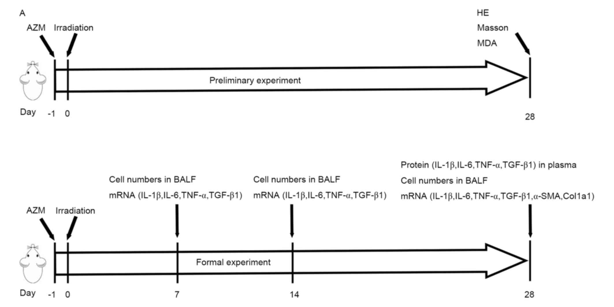 | Figure 1.Schematic diagram of the experimental
protocol and the effect of AZM on histological changes in the
control, RT, AZM-10 and AZM-100 groups on day 28 following
irradiation. (A) The mouse thorax was irradiated with 16 Gy X-ray.
One day prior to radiation, the mice received AZM or vehicle alone
and were administered with the original protocol, as described in
the preliminary study, once daily until the mice were sacrificed
under anesthesia. Lung tissues, plasma and bronchoalveolar lavage
fluid were collected at indicated time points for each experiment.
(B) Representative photomicrographs of hematoxylin and eosin
stained lung sections. Characteristic morphology of each group is
shown at magnifications (a-d) 100× and (e-h) 200x. Scale bar, 100
µm. (C) Representative photomicrographs of Masson stained lung
sections. Characteristic morphology of each group is shown at (a-d)
100× and (e-h) 200x. Examples of collagen deposition lesions (red
arrows) are marked. Scale bar, 100 µm. Control, non-irradiated
control + vehicle; RT group, irradiation + vehicle; AZM-10 group,
irradiation + 10 mg/kg/day AZM; AZM-100 group; irradiation + 100
mg/kg/day AZM. AZM, azithromycin; BALF, bronchoalveolar lavage
fluid; COL1A1, α-1 type I collagen; H&E, hematoxylin and eosin;
MDA, malondialdehyde; TNF, tumor necrosis factor; TGF, transforming
growth factor; IL, interleukin; α-SMA, smooth muscle actin. (D)
Scoring of lung tissue inflammation as assessed by one-way ANOVA.
Data are represented as the mean ± standard error. **P<0.01, RT
group vs. AZM-100 group; n=5 per group. (E) Grading of lung tissue
fibrosis as assessed by one-way ANOVA. Data are represented as the
mean ± standard error. **P<0.01, RT group vs. AZM-100 group; n=5
per group. Control, non-irradiated control + vehicle; RT group,
irradiation + vehicle; AZM-10 group, irradiation + 10 mg/kg/day
AZM; AZM-100 group; irradiation + 100 mg/kg/day AZM. ANOVA, one-way
analysis of variance; AZM, azithromycin. |
AZM treatment reduces the level of
lipid peroxidation
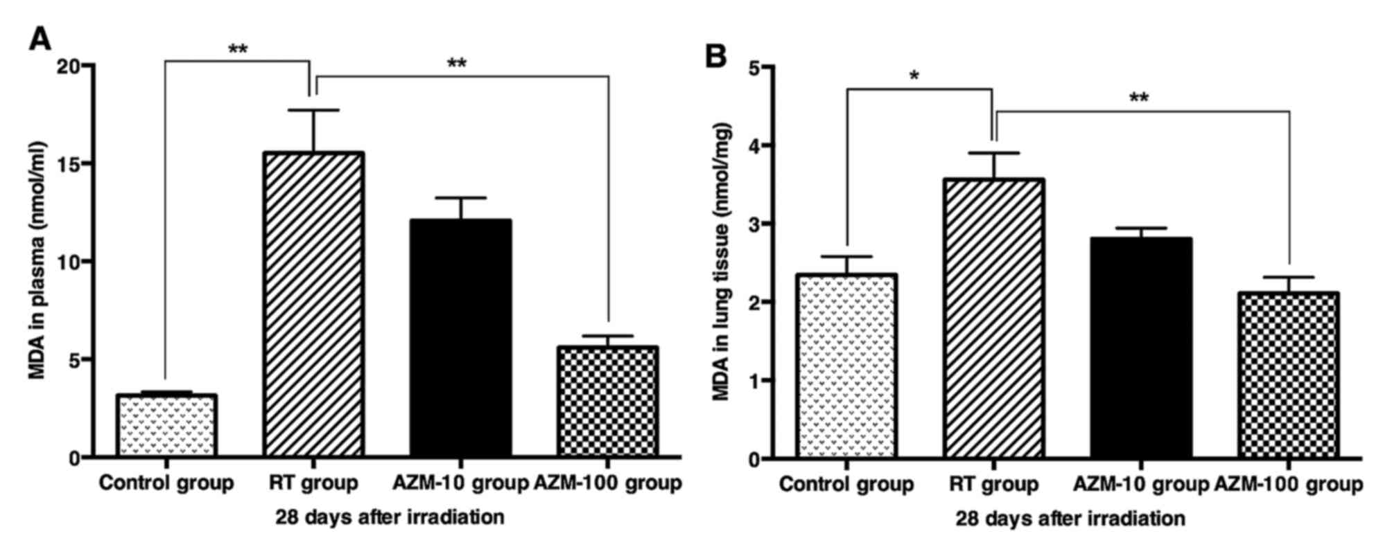
The present authors measured the levels of MDA in
plasma and lung tissue homogenates in order to investigate the
effects of AZM on radiation-induced lipid peroxidation. Irradiation
treatment increased the levels of MDA. However, the levels of MDA
in the AZM-100 group significantly decreased in plasma and lung
tissue (both P<0.01 vs. RT group; Fig.
2A and B). These results indicated that 100 mg/kg/day AZM was
effective in reducing the level of lipid peroxidation. Compared
with the RT group, the levels of MDA in the AZM-10 group were lower
in the plasma and lung tissue. However, no significant differences
were observed in plasma and lung tissue. Therefore, 100 mg/kg/day
was selected as the high AZM dose in subsequent experiments.
AZM administration decreases total
cell counts in BALF
In the formal study, the mice were sacrificed under
anesthesia on day 7, 14 and 28 post-irradiation. The authors
evaluated the effect of AZM on total cell counts in BALF following
irradiation (Fig. 3). In the RT
group, the counts decreased on day 7 compared with the control, and
the levels in the RT group decreased further on day 14 (day 7 and
14 vs. control, P<0.01; Fig. 3).
By day 28, a marked increase was observed compared with the control
(day 28 vs. control). However, in the AZM-100 group, total cell
counts were not significantly different to those in the control
group on day 7 (P>0.05 vs. control. The total cell counts in the
AZM-100 group were increased on day 14 compared with the RT group
(P<0.05 vs. RT group; Fig. 3),
while the influx of total cells in the AZM-100 group was
significantly decreased on day 28 compared with the RT group
(P<0.05; Fig. 3).
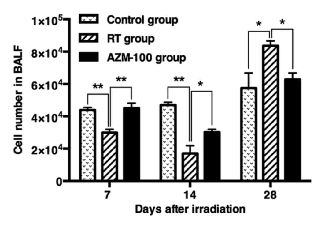 | Figure 3.Effect of AZM on changes in total
cell counts in BALF on day 7, 14 and 28 following irradiation.
Control group vs. RT group and AZM-100 group at the same time
points, by one-way analysis of variance. Data are represented as
the mean ± standard error. Day 7: Control vs. RT group and RT group
vs. AZM-100 group, **P<0.01; day 14: Control vs. RT group,
**P<0.01 and RT group vs. AZM-100 group, *P<0.05; day 28:
Control vs. RT group and RT group vs. AZM-100 group, *P<0.05; 5
samples/group. Control, non-irradiated control + vehicle; RT group,
irradiation + vehicle; AZM-10 group, irradiation + 10 mg/kg/day
AZM; AZM-100 group; irradiation + 100 mg/kg/day AZM. AZM,
azithromycin; BALF, bronchoalveolar lavage fluid. |
AZM treatment reduces the levels of
pro-inflammatory cytokine expression in plasma
The concentrations of pro-inflammatory cytokines in
plasma, including IL-1β, IL-6 and TNF-α, were measured by liquichip
on day 28 following irradiation. Thorax irradiation resulted in the
abundant production of IL-1β, IL-6 and TNF-α, and the plasma levels
of these cytokines significantly increased in RILI mice on day 28
(all P<0.05 vs. control group). By contrast, AZM treatment
significantly decreased the irradiation-induced protein release of
IL-1β, IL-6 and TNF-α in plasma compared with the RT group
(Fig. 4A-C).
AZM treatment reduces pro-inflammatory
cytokine gene expression in lung tissue
To gain further insight into the effect of AZM on
RILI, lung mRNA samples from these mice were measured on day 7, 14
and 28 following irradiation using RT-qPCR. As shown in Fig. 5A and B, irradiation resulted in a
slight increase in the levels of IL-1β and IL-6 on day 7, 14 and 28
compared with the control group (Fig. 5A
and B). By contrast, AZM treatment significantly inhibited this
increase on day 28 (Fig. 5A and B).
On day 14 following irradiation, there was an increase in the
levels of TNF-α compared with the control group, and this increase
was significantly reduced in AZM-treated mice (Fig. 5C).
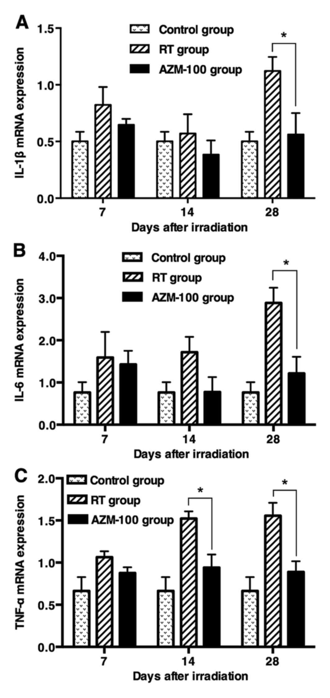 | Figure 5.Analysis of changes in
pro-inflammatory cytokine gene expression in lung tissues by
quantitative reverse transcription polymerase chain reaction on day
7, 14 and 28 following irradiation. (A) IL-1β expression. (B) IL-6
expression. (C) TNF-α expression. Data are represented as the mean
± standard error. In (C) on day 14, RT group vs. AZM-100 group,
*P<0.05; in (A-C) on day 28, RT group vs. AZM-100 group,
*P<0.05; 5 samples/group. Control, non-irradiated control +
vehicle; RT group, irradiation + vehicle; AZM-10 group, irradiation
+ 10 mg/kg/day AZM; AZM-100 group; irradiation + 100 mg/kg/day AZM.
TNF, tumor necrosis factor; IL, interleukin. |
AZM treatment reduces pro-fibrotic
factor expression
The present authors also examined TGF-β1 expression
in plasma and lung tissue using liquichip or western blotting on
day 28 following irradiation. As expected, AZM treatment
significantly decreased irradiation-induced TGF-β1 expression in
plasma and lung tissues compared with the RT group (Fig. 6A-C). In order to further determine the
changes in TGF-β1, the authors measured TGF-β1 mRNA expression in
injured lungs using RT-qPCR on day 7, 14 and 28 following
irradiation. The irradiation-induced increase in TGF-β1 mRNA
expression was significantly decreased in the AZM-100 group on day
7, 14 and 28 (Fig. 6D).
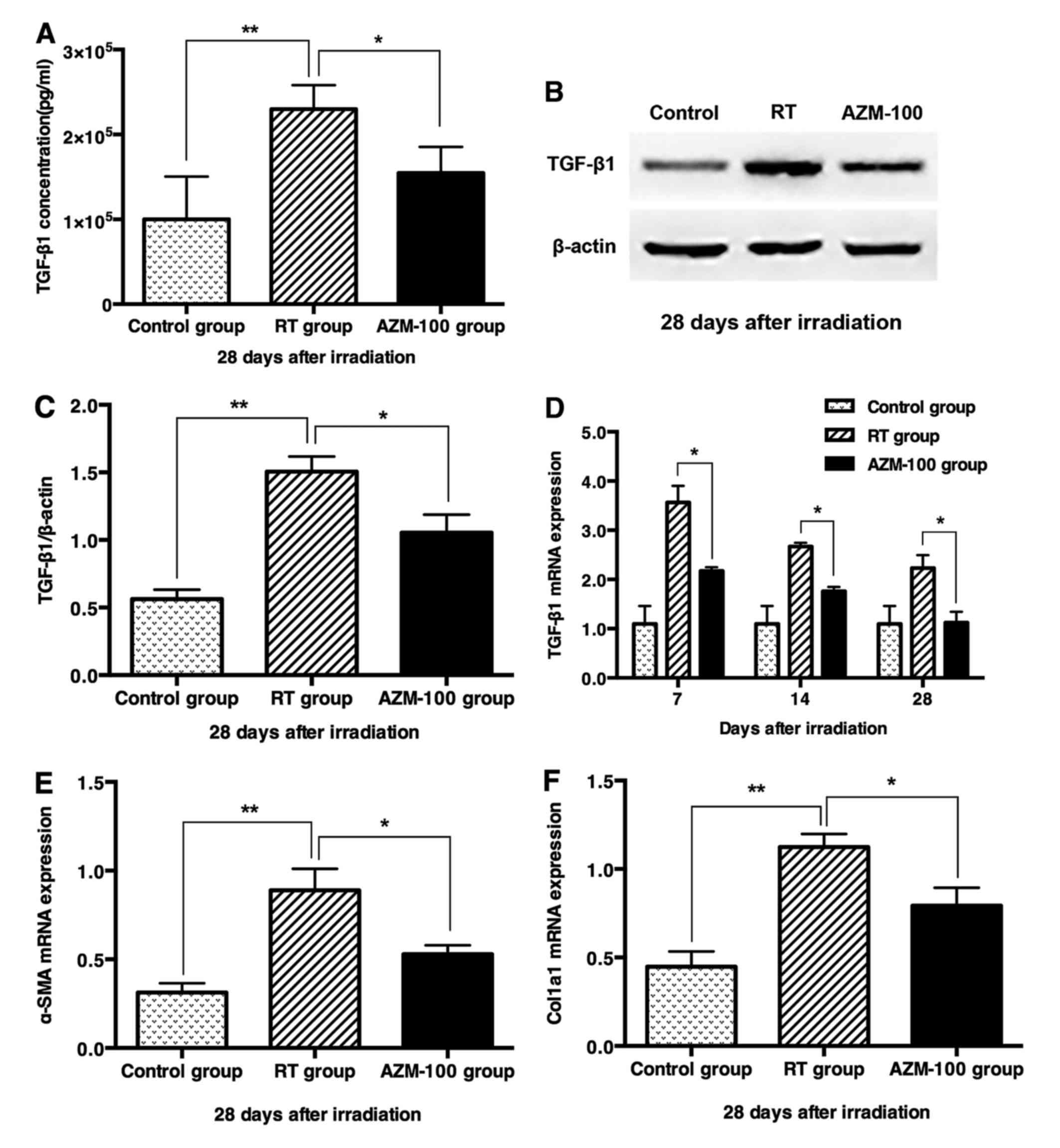 | Figure 6.Effect of AZM on changes in the level
of pro-fibrotic factor in plasma and lung tissue following
irradiation. (A) Changes in TGF-β1 concentration in plasma on day
28 following irradiation. (B) Western blot analysis of TGF-β1 in
lung tissue on day 28 following irradiation. (C) Relative TGF-β1
protein expression in lung tissue. Densitometry values were
normalized to β-actin. (D) Changes in TGF-β1 mRNA expression in
lung tissue on day 7, 14 and 28 following irradiation. (E) α-SMA
mRNA expression in lung tissue on 28 day following irradiation. (F)
COL1A1 mRNA expression in lung tissue on 28 day following
irradiation. Data are represented as the mean ± standard error.
Control vs. RT group, **P<0.01; RT group vs. AZM-100 group,
*P<0.05; 5 samples/group. Control, non-irradiated control +
vehicle; RT group, irradiation + vehicle; AZM-10 group, irradiation
+ 10 mg/kg/day AZM; AZM-100 group; irradiation + 100 mg/kg/day AZM.
AZM, azithromycin; COL1A1, α-1 type I collagen; TGF, transforming
growth factor; IL, interleukin; α-SMA, α-smooth muscle actin. |
The mRNA expression of α-smooth muscle actin (α-SMA)
and α-1 type 1 collagen (COL1A1) was examined in lung tissue on day
28 following irradiation. The irradiation-induced increase in α-SMA
and COL1A1 mRNA expression in injured lung tissue significantly
decreased in the AZM-100 group (Fig. 6E
and F).
Discussion
In the present study, a murine model was used to
investigate the effect of AZM, as a new biological strategy, to
ameliorate acute RILI. The results showed that AZM decreased
radiation-induced early lung injury. Oxidative stress, inflammatory
cell infiltration, cytokine production, and associated gene
expression have pivotal roles in the pathogenesis of RILI (22). Although, the exact mechanisms by which
RILI is mitigated by AZM are not well described, anti-inflammatory
and anti-fibrotic effects may be involved.
Lipid peroxidation is one of the oxidative
conversions of polyunsaturated fatty acids to products such as MDA,
which is an important indicator of oxidative damage (23,24). In
the present study, marked increases in MDA content were observed
between week 1 and 24 following whole-lung irradiation, which
demonstrated oxidative stress due to radiation-induced pneumonitis
and lung fibrosis. Oxidative stress has been previously shown to
ameliorate RILI (25). A previous
study indicated that AZM decreased the levels of MDA in a pig model
of otitis media (26). These findings
were confirmed by the results of the present study. In addition,
radiation is known to induce the expression of NO synthase (NOS)
and results in nitrotyrosine formation in the lungs (27). Evidence to support the harmful role of
NOS in RILI includes a study in which partial attenuation of RILI
was observed following treatment with L-nitro-arginine methyl
ester, a relatively nonspecific NOS inhibitor (28). Notably, AZM also significantly reduced
NOS activity in a rat model of colonic damage (29).
Evidence suggests a central role for the
inflammatory response in the initiation and establishment of RILI.
Local recruitment of inflammatory cells and the production of
inflammatory cytokines exert an important role in mediating,
amplifying and maintaining the RILI process. Intensive
anti-inflammatory treatment mitigates the signs and symptoms of
RILI (30,31).
Many studies have investigated radiation-induced
lung damage using BALF, as it is thought to reflect the lung
inflammatory response (32,33). Previous studies have indicated that
most patients did not show clinical symptoms of radiation
pneumonitis, and lymphocytosis was not very pronounced. However in
symptomatic patients, changes in BALF, including total cellularity
and lymphocytosis [which appeared to be mainly activated cluster of
differentiation (CD)4+ cells], were significantly
greater compared with asymptomatic patients (34). In the present study, the total number
of cells obtained by BALF decreased on day 7 following irradiation
and was very low on day 14. However on day 28, a marked increase
was observed. These dynamic changes are in agreement with previous
studies (35,36). A previous study indicated that the
late increase in BALF cell number was associated with the
development of radiation-induced pulmonary lethality (35,36). As
shown in the present study, administration of AZM inhibited these
dynamic changes. In particular, the number of cells on day 28 was
reduced in the AZM-treated group compared with the control. These
results are supported by previous data which showed that AZM
treatment decreased total cell counts in BALF in a mouse model of
ventilator-associated pneumonia and bleomycin-induced acute lung
injury (37,38). In addition, inflammatory cell
infiltration or exudation into lung parenchyma appears to have to
an important role in the development of RILI. Agents that decrease
inflammatory cell exudation have the potential to alleviate RILI
(39). The present study demonstrated
that AZM markedly reduced inflammatory infiltration in the alveolar
septa, therefore alleviating the extent of RILI (as determined by
H&E staining).
Many studies have indicated that AZM reduces the
inflammatory response by decreasing the levels of pro-inflammatory
cytokine in lipopolysaccharide- and bleomycin-induced acute lung
injury models. These findings are consistent with those in the
present study (40,41). Alveolar macrophages have an important
role in alveolar physiology. Activated macrophages are the main
source of pro-inflammatory cytokines in the early stages of
radiation-induced lung disease (42).
A number of studies have confirmed that the anti-inflammatory
properties of AZM can be attributed, at least partly, to its action
on macrophages (41,43). For example, AZM prevented the
production of pro-inflammatory cytokines by macrophages in the
lipopolysaccharide-induced lung neutrophilia mouse model and
inhibited inflammatory cytokine production by J774A.1 macrophage
cell lines (41,43). Therefore, the authors hypothesize that
AZM regulates inflammatory cytokine production by targeting
macrophages following irradiation.
TGF-β, which is a potent stimulator of collagen
protein synthesis, exerts a critical role in the pathogenesis of
radiation-induced lung fibrosis (44–46). Gene
expression of TGF-β has been demonstrated to increase markedly 1–14
days following irradiation, in parallel with changes in fibroblast
gene expression of collagen I and fibronectin. The administration
of anti-TGF-β1 antibody 1D11 or the TGF-β receptor inhibitor
LY210976 is now an option for ameliorating RILI (46–48). In a
rat model of bleomycin-induced pulmonary fibrosis, the levels of
TGF-β protein and mRNA were reduced following treatment with AZM in
the early stage of pulmonary fibrosis (49). In the present study, it was observed
that AZM treatment significantly decreased expression of TGF-β1,
α-SMA and COL1A1, and grade of fibrosis in Masson stained lung
sections, indicating that AZM may contribute to the anti-fibrotic
effects of post-irradiation.
Thorax irradiation not only affects macrophages in
lung tissue, but also triggers the recruitment of various immune
cells into the lung, including CD4+ T-lymphocytes, which
exert a critical role in the pathogenesis of radiation-induced
pneumonitis preceding lung fibrosis (50). A pronounced increase in
CD4+ lymphocytes was demonstrated 4 weeks following
irradiation. Co-culture of isolated CD4+ T cells from
irradiated lungs with fibroblasts resulted in increased collagen
production (51). Furthermore,
depletion of CD4+ T cells by specific antibodies prior
to partial lung irradiation decreased the degree of
radiation-induced lung fibrosis (50). As mentioned previously, AZM has also
been shown to be beneficial in lung transplantation for the
prevention and treatment of chronic allograft rejection due to its
immunomodulatory properties (52).
AZM treatment significantly decreased CD4+ T cells in
BALF and lung collagen deposition in a murine model of
noninfectious lung injury (53). The
authors of the present study hypothesize that the anti-fibrotic
effects of AZM may be attributed to its immunomodulatory
properties.
The authors observed an improvement in
radiation-induced lung tissue morphology with high-dose (100
mg/kg/day) and low-dose (10 mg/kg/day) AZM in the preliminary
study. As shown in Fig. 1D and E,
compared with the RT group, the inflammation score and the grade of
fibrosis in the AZM-10 group were lower, but not significantly
different. In addition, a lower but not a statistically significant
difference in MDA content was observed in plasma and lung tissue in
the AZM-10 group compared with the RT group. These results indicate
that 100 mg/kg/day is a more appropriate dose for AZM in acute
RILI. Future studies are required to determine the optimal dose of
AZM with the highest efficacy and minimal dose-limiting toxicities
in acute RILI.
RILI refers to a continuous process, which is
triggered following lung RT (3).
Additionally, acute pneumonitis is associated with radiation
fibrosis (39). A limitation in the
present study is the lack of a long-term investigation on the
survival rate of RILI mice treated with AZM. A future study by the
present authors will focus on whether AZM has a similar capacity to
ameliorate late RILI and improve survival in mice.
In conclusion, AZM has therapeutic potential in RILI
management. The present study demonstrated the beneficial role of
AZM in treating RILI, including its anti-inflammatory and
anti-fibrotic effects.
Acknowledgements
The present study was supported by the National
Natural Science Foundation of China (grant nos. 81472196 and
81472808). The abstract was presented at the 58th Annual Meeting of
the American Society for Radiation Oncology on 25–28 September
2016, Boston, USA and published as abstract no. 3439 in the
Proceedings of the American Society for Radiation Oncology Vol. 92
(2). The authors would like to thank
the members of the Laboratory of Stem Cell Biology of Sichuan
University (Chengdu, China) for their assistance in the present
study, and the members of the Department of Radiation Oncology of
West China Hospital (Chengdu, China) for their assistance with the
dosimetry verification and mouse radiation therapy.
References
|
1
|
Baskar R, Lee KA, Yeo R and Yeoh KW:
Cancer and radiation therapy: Current advances and future
directions. Int J Med Sci. 9:193–199. 2012. View Article : Google Scholar : PubMed/NCBI
|
|
2
|
Graves PR, Siddiqui F, Anscher MS and
Movsas B: Radiation pulmonary toxicity: From mechanisms to
management. Semin Radiat Oncol. 20:201–207. 2010. View Article : Google Scholar : PubMed/NCBI
|
|
3
|
Sprung CN, Forrester HB, Siva S and Martin
OA: Immunological markers that predict radiation toxicity. Cancer
Lett. 368:191–197. 2015. View Article : Google Scholar : PubMed/NCBI
|
|
4
|
Marks LB, Bentzen SM, Deasy JO, Kong FM,
Bradley JD, Vogelius IS, El Naqa I, Hubbs JL, Lebesque JV,
Timmerman RD, et al: Radiation dose-volume effects in the lung. Int
J Radiat Oncol Biol Phys. 76 (3 Suppl):S70–S76. 2010. View Article : Google Scholar : PubMed/NCBI
|
|
5
|
Yamashita H, Nakagawa K, Nakamura N,
Koyanagi H, Tago M, Igaki H, Shiraishi K, Sasano N and Ohtomo K:
Exceptionally high incidence of symptomatic grade 2–5 radiation
pneumonitis after stereotactic radiation therapy for lung tumors.
Radiat Oncol. 2:212007. View Article : Google Scholar : PubMed/NCBI
|
|
6
|
Kong FM and Wang S: Nondosimetric risk
factors for radiation-induced lung toxicity. Semin Radiat Oncol.
25:100–109. 2015. View Article : Google Scholar : PubMed/NCBI
|
|
7
|
Togami K, Chono S and Morimoto K:
Distribution characteristics of clarithromycin and azithromycin,
macrolide antimicrobial agents used for treatment of respiratory
infections, in lung epithelial lining fluid and alveolar
macrophages. Biopharm Drug Dispos. 32:389–397. 2011. View Article : Google Scholar : PubMed/NCBI
|
|
8
|
Spagnolo P, Fabbri LM and Bush A:
Long-term macrolide treatment for chronic respiratory disease. Eur
Respir J. 42:239–251. 2013. View Article : Google Scholar : PubMed/NCBI
|
|
9
|
Southern KW, Barker PM, Solis-Moya A and
Patel L: Macrolide antibiotics for cystic fibrosis. Cochrane
Database Syst Rev. 11:CD0022032012.PubMed/NCBI
|
|
10
|
Wong C, Jayaram L, Karalus N, Eaton T,
Tong C, Hockey H, Milne D, Fergusson W, Tuffery C, Sexton P, et al:
Azithromycin for prevention of exacerbations in non-cystic fibrosis
bronchiectasis (EMBRACE): A randomised, double-blind,
placebo-controlled trial. Lancet. 380:660–667. 2012. View Article : Google Scholar : PubMed/NCBI
|
|
11
|
Albert RK, Connett J, Bailey WC, Casaburi
R, Cooper JA Jr, Criner GJ, Curtis JL, Dransfield MT, Han MK,
Lazarus SC, et al: Azithromycin for prevention of exacerbations of
COPD. N Engl J Med. 365:689–698. 2011. View Article : Google Scholar : PubMed/NCBI
|
|
12
|
Vos R, Vanaudenaerde BM, Verleden SE, De
Vleeschauwer SI, Willems-Widyastuti A, Van Raemdonck DE, Schoonis
A, Nawrot TS, Dupont LJ and Verleden GM: A randomised controlled
trial of azithromycin to prevent chronic rejection after lung
transplantation. Eur Respir J. 37:164–172. 2011. View Article : Google Scholar : PubMed/NCBI
|
|
13
|
Tamaoki J, Kadota J and Takizawa H:
Clinical implications of the immunomodulatory effects of
macrolides. Am J Med. 117 Suppl 9A:5S–11S. 2004.PubMed/NCBI
|
|
14
|
Giamarellos-Bourboulis EJ: Macrolides
beyond the conventional antimicrobials: A class of potent
immunomodulators. Int J Antimicrob Agents. 31:12–20. 2008.
View Article : Google Scholar : PubMed/NCBI
|
|
15
|
Li DQ, Zhou N, Zhang L, Ma P and
Pflugfelder SC: Suppressive effects of azithromycin on
zymosan-induced production of proinflammatory mediators by human
corneal epithelial cells. Invest Ophthalmol Vis Sci. 51:5623–5629.
2010. View Article : Google Scholar : PubMed/NCBI
|
|
16
|
Council NR: Guide for the care and use of
laboratory animals. 8th. The National Academies Press; Washington,
DC: 2011, PubMed/NCBI
|
|
17
|
Hoffmann N, Lee B, Hentzer M, Rasmussen
TB, Song Z, Johansen HK, Givskov M and Høiby N: Azithromycin blocks
quorum sensing and alginate polymer formation and increases the
sensitivity to serum and stationary-growth-phase killing of
Pseudomonas aeruginosa and attenuates chronic P. aeruginosa lung
infection in Cftr(−/−) mice. Antimicrob Agents Chemother.
51:3677–3687. 2007. View Article : Google Scholar : PubMed/NCBI
|
|
18
|
Conte JE Jr, Golden J, Duncan S, McKenna
E, Lin E and Zurlinden E: Single-dose intrapulmonary
pharmacokinetics of azithromycin, clarithromycin, ciprofloxacin,
and cefuroxime in volunteer subjects. Antimicrob Agents Chemother.
40:1617–1622. 1996.PubMed/NCBI
|
|
19
|
Heinzelmann F, Jendrossek V, Lauber K,
Nowak K, Eldh T, Boras R, Handrick R, Henkel M, Martin C, Uhlig S,
et al: Irradiation-induced pneumonitis mediated by the
CD95/CD95-ligand system. J Natl Cancer Inst. 98:1248–1251. 2006.
View Article : Google Scholar : PubMed/NCBI
|
|
20
|
Hübner RH, Gitter W, El Mokhtari NE,
Mathiak M, Both M, Bolte H, Freitag-Wolf S and Bewig B:
Standardized quantification of pulmonary fibrosis in histological
samples. Biotechniques. 44:507–517. 2008. View Article : Google Scholar : PubMed/NCBI
|
|
21
|
Schmittgen TD and Livak KJ: Analyzing
real-time PCR data by the comparative C(T) method. Nat Protoc.
3:1101–1108. 2008. View Article : Google Scholar : PubMed/NCBI
|
|
22
|
Fleckenstein K, Gauter-Fleckenstein B,
Jackson IL, Rabbani Z, Anscher M and Vujaskovic Z: Using biological
markers to predict risk of radiation injury. Semin Radiat Oncol.
17:89–98. 2007. View Article : Google Scholar : PubMed/NCBI
|
|
23
|
Kergonou JF, Bernard P, Braquet M and
Rocquet G: Effect of whole-body gamma irradiation on lipid
peroxidation in rat tissues. Biochimie. 63:555–559. 1981.
View Article : Google Scholar : PubMed/NCBI
|
|
24
|
Taysi S, Uslu C, Akcay F and Sutbeyaz MY:
Malondialdehyde and nitric oxide levels in the plasma of patients
with advanced laryngeal cancer. Surg Today. 33:651–654. 2003.
View Article : Google Scholar : PubMed/NCBI
|
|
25
|
Kang SK, Rabbani ZN, Folz RJ, Golson ML,
Huang H, Yu D, Samulski TS, Dewhirst MW, Anscher MS and Vujaskovic
Z: Overexpression of extracellular superoxide dismutase protects
mice from radiation-induced lung injury. Int J Radiat Oncol Biol
Phys. 57:1056–1066. 2003. View Article : Google Scholar : PubMed/NCBI
|
|
26
|
Aktan B, Taysi S, Gümüştekin K, Uçüncü H,
Memişoğullari R, Save K and Bakan N: Effect of macrolide
antibiotics on nitric oxide synthase and xanthine oxidase
activities, and malondialdehyde level in erythrocyte of the guinea
pigs with experimental otitis media with effusion. Pol J Pharmacol.
55:1105–1110. 2003.PubMed/NCBI
|
|
27
|
Giaid A, Lehnert SM, Chehayeb B, Chehayeb
D, Kaplan I and Shenouda G: Inducible nitric oxide synthase and
nitrotyrosine in mice with radiation-induced lung damage. Am J Clin
Oncol. 26:e67–e72. 2003. View Article : Google Scholar : PubMed/NCBI
|
|
28
|
Nozaki Y, Hasegawa Y, Takeuchi A, Fan ZH,
Isobe KI, Nakashima I and Shimokata K: Nitric oxide as an
inflammatory mediator of radiation pneumonitis in rats. Am J
Physiol. 272:L651–L658. 1997.PubMed/NCBI
|
|
29
|
Mahgoub A, El-Medany A, Mustafa A, Arafah
M and Moursi M: Azithromycin and erythromycin ameliorate the extent
of colonic damage induced by acetic acid in rats. Toxicol Appl
Pharmacol. 205:43–52. 2005. View Article : Google Scholar : PubMed/NCBI
|
|
30
|
Ward PA and Hunninghake GW: Lung
inflammation and fibrosis. Am J Respir Crit Care Med.
157:S123–S129. 1998. View Article : Google Scholar : PubMed/NCBI
|
|
31
|
Hong ZY, Song KH, Yoon JH, Cho J and Story
MD: An experimental model-based exploration of cytokines in
ablative radiation-induced lung injury in vivo and in vitro. Lung.
193:409–419. 2015. View Article : Google Scholar : PubMed/NCBI
|
|
32
|
Rosiello RA, Merrill WW, Rockwell S,
Carter D, Cooper JA Jr, Care S and Amento EP: Radiation
pneumonitis. Bronchoalveolar lavage assessment and modulation by a
recombinant cytokine. Am Rev Respir Dis. 148:1671–1676. 1993.
View Article : Google Scholar : PubMed/NCBI
|
|
33
|
Kawana A, Shioya S, Katoh H, Tsuji C,
Tsuda M and Ohta Y: Expression of intercellular adhesion molecule-1
and lymphocyte function-associated antigen-1 on alveolar
macrophages in the acute stage of radiation-induced lung injury in
rats. Radiat Res. 147:431–436. 1997. View
Article : Google Scholar : PubMed/NCBI
|
|
34
|
Morgan GW and Breit SN: Radiation and the
lung: A reevaluation of the mechanisms mediating pulmonary injury.
Int J Radiat Oncol Biol Phys. 31:361–369. 1995. View Article : Google Scholar : PubMed/NCBI
|
|
35
|
Hong JH, Jung SM, Tsao TC, Wu CJ, Lee CY,
Chen FH, Hsu CH, McBride WH and Chiang CS: Bronchoalveolar lavage
and interstitial cells have different roles in radiation-induced
lung injury. Int J Radiat Biol. 79:159–167. 2003. View Article : Google Scholar : PubMed/NCBI
|
|
36
|
Chiang CS, Liu WC, Jung SM, Chen FH, Wu
CR, McBride WH, Lee CC and Hong JH: Compartmental responses after
thoracic irradiation of mice: Strain differences. Int J Radiat
Oncol Biol Phys. 62:862–871. 2005. View Article : Google Scholar : PubMed/NCBI
|
|
37
|
Yamada K, Yanagihara K, Kaku N, Harada Y,
Migiyama Y, Nagaoka K, Morinaga Y, Nakamura S, Imamura Y, Miyazaki
T, et al: Azithromycin attenuates lung inflammation in a mouse
model of ventilator-associated pneumonia by multidrug-resistant
Acinetobacter baumannii. Antimicrob Agents Chemother. 57:3883–3888.
2013. View Article : Google Scholar : PubMed/NCBI
|
|
38
|
Kawashima M, Yatsunami J, Fukuno Y, Nagata
M, Tominaga M and Hayashi S: Inhibitory effects of 14-membered ring
macrolide antibiotics on bleomycin-induced acute lung injury. Lung.
180:73–89. 2002. View Article : Google Scholar : PubMed/NCBI
|
|
39
|
Tsoutsou PG and Koukourakis MI: Radiation
pneumonitis and fibrosis: Mechanisms underlying its pathogenesis
and implications for future research. Int J Radiat Oncol Biol Phys.
66:1281–1293. 2006. View Article : Google Scholar : PubMed/NCBI
|
|
40
|
Wuyts WA, Willems S, Vos R, Vanaudenaerde
BM, De Vleeschauwer SI, Rinaldi M, Vanhooren HM, Geudens N,
Verleden SE, Demedts MG, et al: Azithromycin reduces pulmonary
fibrosis in a bleomycin mouse model. Exp Lung Res. 36:602–614.
2010. View Article : Google Scholar : PubMed/NCBI
|
|
41
|
Bosnar M, Bosnjak B, Cuzic S, Hrvacic B,
Marjanovic N, Glojnaric I, Culic O, Parnham MJ and Haber Erakovic
V: Azithromycin and clarithromycin inhibit
lipopolysaccharide-induced murine pulmonary neutrophilia mainly
through effects on macrophage-derived granulocyte-macrophage
colony-stimulating factor and interleukin-1beta. J Pharmacol Exp
Ther. 331:104–113. 2009. View Article : Google Scholar : PubMed/NCBI
|
|
42
|
Finkelstein JN, Johnston CJ, Baggs R and
Rubin P: Early alterations in extracellular matrix and transforming
growth factor beta gene expression in mouse lung indicative of late
radiation fibrosis. Int J Radiat Oncol Biol Phys. 28:621–631. 1994.
View Article : Google Scholar : PubMed/NCBI
|
|
43
|
Ianaro A, Ialenti A, Maffia P, Sautebin L,
Rombolà L, Carnuccio R, Iuvone T, D'Acquisto F and Di Rosa M:
Anti-inflammatory activity of macrolide antibiotics. J Pharmacol
Exp Ther. 292:156–163. 2000.PubMed/NCBI
|
|
44
|
Martin M, Lefaix J and Delanian S:
TGF-beta1 and radiation fibrosis: A master switch and a specific
therapeutic target? Int J Radiat Oncol Biol Phys. 47:277–290. 2000.
View Article : Google Scholar : PubMed/NCBI
|
|
45
|
Vujaskovic Z and Groen HJ: TGF-beta,
radiation-induced pulmonary injury and lung cancer. Int J Radiat
Biol. 76:511–516. 2000. View Article : Google Scholar : PubMed/NCBI
|
|
46
|
Xue J, Li X, Lu Y, Gan L, Zhou L, Wang Y,
Lan J, Liu S, Sun L, Jia L, et al: Gene-modified mesenchymal stem
cells protect against radiation-induced lung injury. Mol Ther.
21:456–465. 2013. View Article : Google Scholar : PubMed/NCBI
|
|
47
|
Flechsig P, Dadrich M, Bickelhaupt S,
Jenne J, Hauser K, Timke C, Peschke P, Hahn EW, Gröne HJ, Yingling
J, et al: LY2109761 attenuates radiation-induced pulmonary murine
fibrosis via reversal of TGF-β and BMP-associated proinflammatory
and proangiogenic signals. Clin Cancer Res. 18:3616–3627. 2012.
View Article : Google Scholar : PubMed/NCBI
|
|
48
|
Anscher MS, Thrasher B, Rabbani Z, Teicher
B and Vujaskovic Z: Antitransforming growth factor-beta antibody
1D11 ameliorates normal tissue damage caused by high-dose
radiation. Int J Radiat Oncol Biol Phys. 65:876–881. 2006.
View Article : Google Scholar : PubMed/NCBI
|
|
49
|
Chen J, He B, Li Y, Wang G and Zhang W: An
experimental study on the effect of azithromycin treatment in
bleomycin-induced pulmonary fibrosis of rats. Zhonghua Nei Ke Za
Zhi. 38:677–680. 1999.(In Chinese). PubMed/NCBI
|
|
50
|
Westermann W, Schöbl R, Rieber EP and
Frank KH: Th2 cells as effectors in postirradiation pulmonary
damage preceding fibrosis in the rat. Int J Radiat Biol.
75:629–638. 1999. View Article : Google Scholar : PubMed/NCBI
|
|
51
|
Büttner C, Skupin A and Rieber EP:
Transcriptional activation of the type I collagen genes COL1A1 and
COL1A2 in fibroblasts by interleukin-4: Analysis of the functional
collagen promoter sequences. J Cell Physiol. 198:248–258. 2004.
View Article : Google Scholar : PubMed/NCBI
|
|
52
|
Vos R, Vanaudenaerde BM, Verleden SE,
Ruttens D, Vaneylen A, Van Raemdonck DE, Dupont LJ and Verleden GM:
Anti-inflammatory and immunomodulatory properties of azithromycin
involved in treatment and prevention of chronic lung allograft
rejection. Transplantation. 94:101–109. 2012. View Article : Google Scholar : PubMed/NCBI
|
|
53
|
Radhakrishnan SV, Palaniyandi S, Mueller
G, Miklos S, Hager M, Spacenko E, Karlsson FJ, Huber E, Kittan NA
and Hildebrandt GC: Preventive azithromycin treatment reduces
noninfectious lung injury and acute graft-versus-host disease in a
murine model of allogeneic hematopoietic cell transplantation. Biol
Blood Marrow Transplant. 21:30–38. 2015. View Article : Google Scholar : PubMed/NCBI
|


