Introduction
Acute lung injury (ALI) is a serious health disorder
that leads to dysfunction of the respiratory system and has a
mortality rate of >50% (1). The
disease is a clinical challenge for researchers worldwide due to
the high rate of mortality. The factors responsible for inducing
ALI include inhalation of the endotoxins from gram-negative
bacteria and sepsis. Administration of lipopolysaccharide (LPS) has
been used to prepare ALI animal models for use in numerous studies
(2–6).
It has been reported that LPS treatment in rats induces
inflammatory reactions, leading to the development of injury and
ultimately lung dysfunction (7). ALI
in animals is associated with degeneration of lung tissues,
enhancement in the protein concentration and fluid, developing into
edema and breakage of capillary walls (8). LPS administration in rats causes
accumulation and penetration of neutrophils into pulmonary tissues,
which then secrete oxidants leading to lung injury (9,10). These
findings are supported by the fact that treatment of
LPS-administered animals with oxidant scavengers can protect
against lung injury (11).
Natural products have shown potential in the
treatment of numerous diseases and therefore many of these are used
either directly as drugs or modified to develop as chemotherapeutic
agents (12). Danshen (Salvia
miltiorrhiza Bunge) is used in traditional Chinese medicine for
the treatment of diseases related to blood flow (13,14).
Phytochemical analysis of Danshen plant has led to the isolation of
a large number of compounds. The major compounds are phenolics and
diterpenoids, including tanshinone II (15). It is reported that tanshinone II is
the most biologically active compound isolated from Salvia
miltiorrhiza (16). Tanshinone II
treatment has been reported to cause apoptosis in various types of
carcinoma cell lines (16,17). The present study was performed to
investigate the effect of tanshinone II on LPS-induced ALI in rats.
The results demonstrated that tanshinone II exhibits protective
effects against LPS-induced ALI in rats.
Materials and methods
Chemicals and reagents
Tanshinone II (purity 98%), dimethyl sulphoxide
(DMSO), LPS and other common chemicals were obtained from
Sigma-Aldrich (Merck KGaA, Darmstadt, Germany). Stock solution of
tanshinone II was prepared in DMSO and stored until use in
experiments at 10°C.
Animals
Thirty male Sprague-Dawley rats (8-week-old)
weighing 200–220 g were obtained from the Laboratory Animal Center,
Academy of Military Medical Sciences (Beijing, China). The animals
were housed one week before the start of experiment under standard
laboratory conditions at a stable temperature (22–24°C), humidity
(55+5%) and a 12/12-h light/dark cycle with food and water ad
libitum. Animals were maintained as per the guidelines for Care
and Use of Laboratory Animals of the National Institutes of Health
and Academy of Military Medical Sciences (18). The study was approved by the Ethics
Committee of Animal Experiments of the Zhumadian Central Hospital,
Zhumadian, Henan China. Sodium pentobarbital (Bi Biotech India Pvt
Ltd, New Delhi, India) was used to sacrifice the animals for
extraction of lungs.
Treatment strategy
The animals were divided randomly into three groups
of 10 animals each: Control, LPS and tanshinone II groups. Animals
in the LPS and tanshinone II groups were administered 10 mg/kg body
weight LPS intraperitoneally. Tanshinone II at a concentration of
30 µg/kg was injected intraperitoneally to the animals in the
tanshinone II group at 1 h after administration of LPS. Animals in
the normal control group received an equal volume of normal saline
alone. On day 8 after administration of tanshinone II, the animals
were sacrificed to extract the lungs and collect abdominal aorta
blood samples. The weight of the lungs was recorded to calculate
the lung wet/dry (W/D) ratio in the animals.
Lung W/D ratio
The lung W/D ratio was determined using a previously
reported protocol (11). Briefly, the
lungs were extracted, followed by separation of the right lung. The
right lung was weighed immediately after extraction to determine
its wet weight followed by drying for 5 min under microwaves at low
power (200 W) to measure the dry weight. The W/D ratio was
calculated as follows: W/D=weight wet/weight dry.
Bronchoalveolar lavage
Left lung of the animals was washed with normal
saline three times (5 min each time) and the washing fluid was
collected for determination of protein concentration. An ELISA
protein kit (IL-6, SEA079Hu; and TNF-α, E90133Hu; Uscn Life
Sciences, Inc., Wuhan, China) was used to determine the
concentration of proteins according to manufacturer's protocol.
Analysis of myeloperoxidase (MPO) and
malondialdehyde (MDA) activities in lung homogenates
Lung tissues were lysed using RIPA buffer (Cell
Signaling Technology, Inc., Danvers, MA, USA) and a DC protein
assay (Bio-Rad Laboratories, Inc., Hercules, CA, USA) was used for
the determination of protein concentration in the homogenates. The
cellular lysates were centrifuged at 15,000 rpm for 15 min at 4°C,
and the supernatants were harvested and mixed with loading buffer.
MPO and MDA activity was determined using MPO ELISA kit (cat. no.
DRE 30329; Xinfan Biomart Co., Ltd., Shanghai, China) and MDA kit
(cat. no. ab118970; Abcam; Cambridge, UK) respectively, according
to the manufacturer's protocols.
Tumor necrosis factor (TNF)-α and
interleukin (IL)-6 levels in blood samples
Blood samples collected from the aortic artery in
the abdominal region of the rats were anti-coagulated using EDTA.
The blood was then centrifuged for 15 min at 4,000 × g at 0°C to
collect the plasma. The levels of TNF-α (cat. no. RAB0476) and IL-6
(cat. no. RAB0311; Sigma-Aldrich; Merck KGaA) in the plasma were
determined using ELISA kits.
RNA interference
The lung tissue samples were transfected with siRNA
sequences, as described previously (19). The siRNA sequences,
5′-ACUUUGCUGUAACCCUGUA(dTdT)-3′ (sense) and
5′-UACAGGGUUACAGCAAAGU(dTdT)-3′ (antisense) were used to silence
Sirt1 expression.
Western blot analysis
Lung tissues were lysed by treatment with RIPA
buffer (Cell Signaling Technology, Inc.) for 15 min at 0°C and
lysates were centrifuged at 14,000 × g for 20 min at 4°C. The
concentration of proteins in tissue lysates was detected using the
DC™ protein assay (Bio-Rad, Laboratories, Inc., Hercules, CA, USA).
The protein samples (10 µg) were separated by SDS-PAGE using 7.5%
polyacrylamide gels and transferred onto PVDF membranes (Millipore
Corp., Bedford, MA, USA). The membranes were blocked for 1 h at
room temperature in 5% non-fat dry milk in TBS. The membranes were
then subjected to immunodetection with a 1:1,000 dilution of
respective primary antibodies against MMP9 (cat. no. 3852), iNOS
(cat. no. 2982), IL-1β (cat. no. 5204), IL-6 (cat. no. 5216) and
Sirt1 (cat. no. 2310; dilution 1:500, Cell Signaling Technology,
Inc.) overnight at 4°C. Following washing, the membranes were
incubated with a 1:20,000 dilution of horseradish peroxidase
(HRP)-conjugated goat anti-rabbit or anti-mouse secondary antibody
(cat. no. 27647; Cell Signaling Technology, Inc.) for 1 h at room
temperature. Antigen-antibody complexes were visualized with
chemiluminescence HRP substrate (EMD Millipore, Billerica, MA, USA)
and LAS-1000 image analyzer (Fuji Film, Tokyo, Japan).
Histochemical examination
A portion of the right lung was exposed to buffered
formaldehyde (3%) for 1 h at room temperature and then subjected to
paraffin embedding. The paraffin-embedded lung tissues were cut
into 2-µm sections, followed by staining with hematoxylin and eosin
at room temperature for 4 h. The paraffin-embedded lung sections
were deparaffinized by boiling in xylene followed by rehydration
through a graded ethanol series (Japan Alcohol Trading Co., Ltd.,
Tokyo, Japan). The sections were treated for 15 min with 3%
hydrogen peroxide solution for quenching the activity of endogenous
peroxidase. The sections were examined under a light microscope and
assessed for alterations in lung morphology.
Statistical analysis
Data are expressed as the mean ± standard deviation.
Comparisons among the groups was performed using one-way analysis
of variance with the Bonferroni post-hoc test. Statistical analyses
were performed using SPSS v17 software (SPSS, Inc., Chicago, IL,
USA). P<0.05 was considered to indicate a statistically
significant difference.
Results
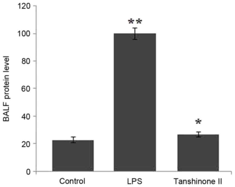
Tanshinone II inhibits expression of
proteins present in bronchoalveolar lavage fluid (BALF)
Analysis of the BALF revealed a significant increase
in the protein level in LPS-administered rats compared with the
control group (Fig. 1; P<0.02).
Tanshinone II treatment exhibited a significant (P<0.05)
inhibitory effect on the level of proteins in the BALF of
LPS-administered rats. The concentration of proteins in
LPS-administered rats was reduced to the level of the control group
at 30 µg/kg concentration of tanshinone II (Fig. 1).
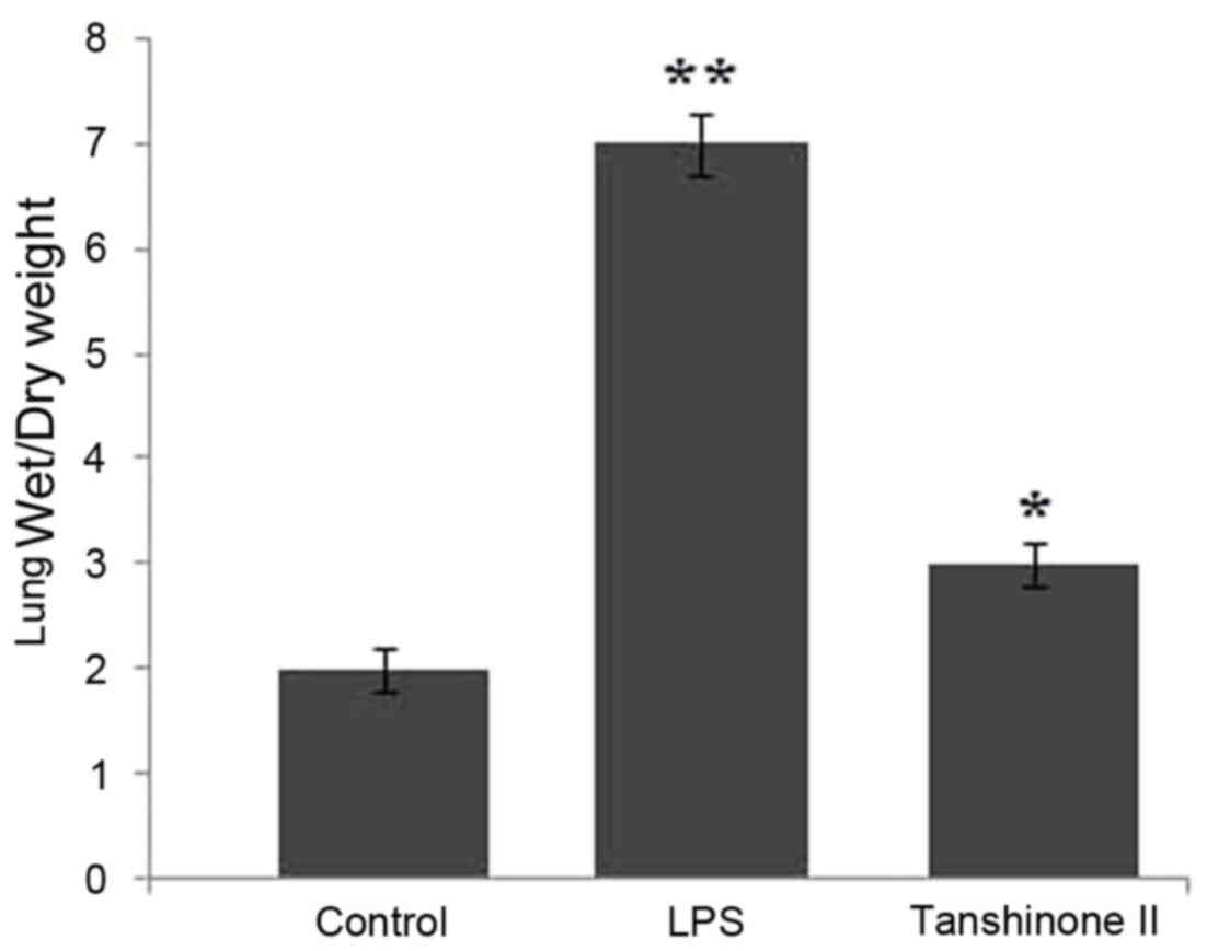
Effects of tanshinone II on lung W/D
ratio
The lung W/D ratio in the control, LPS and
tanshinone II groups were compared. The data revealed that LPS
administration significantly increased the lung W/D ratio compared
with the rats in the control group (P<0.05; Fig. 2). Treatment of the LPS-administered
rats with 30 µg/kg concentration of tanshinone II significantly
(P<0.02) inhibited the increase in lung W/D ratio (Fig. 2). The ratio of wet and dry weight in
the rats of tanshinone II treatment and control groups was nearly
similar.
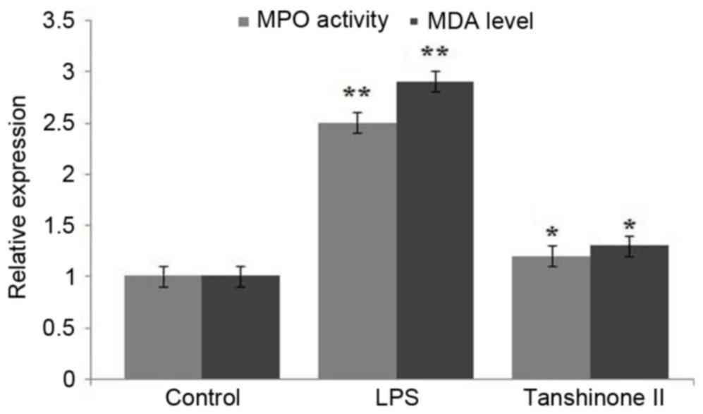
Tanshinone II inhibited MPO activity
and MDA level in LPS-administered rats
LPS administration significantly enhanced the level
of MPO activity in rats compared with those in the control group
(Fig. 3; P<0.03). Tanshinone II
treatment of the LPS-administered rats caused a significant
(P<0.02) inhibition of LPS-induced increase in activity of MPO
(Fig. 3). Analysis of MDA activity
indicated a significantly higher level in LPS-administered rats
compared with the control group (Fig.
3; P<0.03). However, treatment of the LPS-administered rats
with 30 µg/kg concentration of tanshinone II caused a significant
(P<0.05) decrease in MDA and reduced its level to that observed
in the control group (Fig. 3).
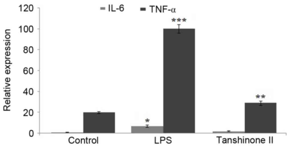
Inhibition of TNF-α and IL-6 in blood
samples of LPS-administered rats by tanshinone II
Blood samples collected from the rats were analyzed
for the concentration of TNF-α and IL-6. The results revealed a
significant increase in the level of TNF-α (P<0.02) and IL-6
(P<0.05) on LPS administration compared with the control
(Fig. 4). However, treatment of the
LPS-administered rats with tanshinone II markedly reduced the level
of TNF-α and IL-6 compared with the LPS group (Fig. 4).
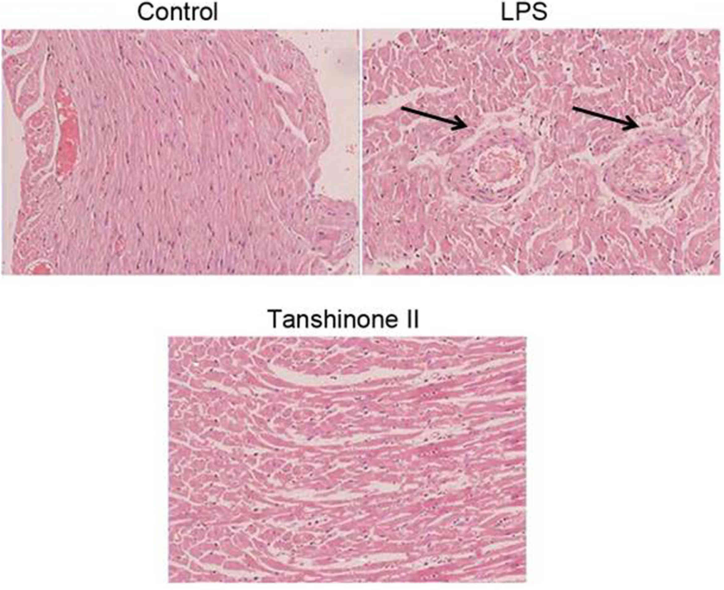
Tanshinone II exhibits a protective
effect against LPS-induced pulmonary alterations
Light microscopy examination of the pulmonary
tissues revealed accumulation of neutrophils and degradation of
cells in LPS-administered animals (Fig.
5). Pulmonary tissues were intact without any appearance of
degradation in the LPS-administered rats treated with tanshinone
II. No evident symptoms of LPS administration were observed in
tanshinone II treated rats (Fig.
5).
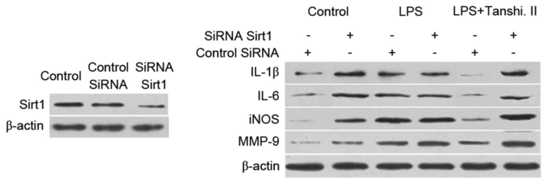
Tanshinone II prevents inflammation by
targeting Sirt1 expression
Administration of small interfering RNA (siRNA) to
inhibit the expression of Sirt1 in rats was performed. A marked
reduction in the expression of gene corresponding to Sirt1 was
observed after 48 h of siRNA administration (Fig. 6). Inhibition of matrix
metalloproteinase (MMP)-9, inducible nitric oxide synthase (iNOS),
IL-1β and IL-6 expression was attenuated after tanshinone II
treatment was attenuated on administration of rats with siRNA
corresponding to Sirt1 (Fig. 6).
Discussion
ALI is a serious health problem detected frequently
in developing countries and the rate of associated mortality is
very high (20). The characteristic
features of ALI are enhanced concentration of proteins, increased
lung W/D ratio, and accumulation and penetration of neutrophils in
pulmonary tissues (21). In the
present study, the effect of tanshinone II on ALI in an LPS-induced
lung injury rat model was analyzed. Administration of LPS induces
cellular inflammatory processes and subsequent pulmonary injury in
animals (22,23). The present results demonstrated that
LPS administration enhanced the concentration of proteins in
pulmonary fluid of rats. Tanshinone II exhibited an inhibitory
effect on the increase of pulmonary protein content in LPS-induced
rats. Comparison of the lung W/D ratio in the LPS and control group
indicated that LPS administration caused pulmonary edema in the
rats. Treatment of the rats with tanshinone II inhibited
LPS-induced lung edema, which was evidenced by similar lung W/D
ratios in the tanshinone II and control groups. Neutrophils secrete
an enzyme, MMP-9, which has been demonstrated to induce degradation
of tissues by producing oxidative species (24). The present results revealed that
tanshinone II treatment of LPS-induced rats reduced the expression
of MMP-9 in the blood samples. These findings were further
confirmed by histopathological examination of pulmonary tissues in
tanshinone II-treated rats. Accumulation of neutrophils and tissue
degradation was reduced in the LPS-induced rats treated with
tanshinone II. The level of MDA in the blood samples of LPS-induced
rats was also reduced by treatment with tanshinone II, indicating
inhibition of peroxidation of membrane lipids (25).
TNF-α and ILs serve critical functions in
proinflammatory activity and have been identified to increase the
rate of mortality in animals (26).
The present results demonstrated that tanshinone II treatment in
the LPS-induced rats inhibited the expression of TNF-α and IL-6 in
blood samples. The level of these cytokines was significantly
promoted in the rats on administration of LPS. Inflammatory
processes or LPS administration induces expression of Sirt1 in
cells (27,28). In the present study, administration of
siRNA against Sirt1 resulted in a significant reduction in the
protein expression of Sirt1. Tanshinone II treatment inhibited the
expression of MMP-9, iNOS, IL-1β and IL-6, and this effect was
attenuated by administration of siRNA against Sirt1. These results
suggest that tanshinone II inhibits LPS-induced lung injury through
targeting Sirt1.
In summary, the present study demonstrates that
tanshinone II inhibits LPS-induced lung injury in the rats by
inhibiting TNF-α and IL-6 expression through targeting Sirt1. It
also inhibits the LPS-induced increase in activity of MPO and level
of MDA. Therefore, tanshinone II may be of therapeutic importance
for the treatment of ALI. However, further studies need to be
performed to understand the mechanism of action of tanshinone II
clearly.
References
|
1
|
Herridge MS, Cheung AM, Tansey CM,
Matte-Martyn A, Diaz-Granados N, Al-Saidi F, Cooper AB, Guest CB,
Mazer CD, Mehta S, et al: One-year outcomes in survivors of the
acute respiratory distress syndrome. N Engl J Med. 348:683–693.
2003. View Article : Google Scholar : PubMed/NCBI
|
|
2
|
Chung YJ, Jarvis B and Pestka J:
Modulation of lipopolysaccharide-induced proinflammatory cytokine
production by satratoxins and other macrocyclic trichothecenes in
the murine macrophage. J Toxicol Environ Health A. 66:379–391.
2003. View Article : Google Scholar : PubMed/NCBI
|
|
3
|
Brandolini L, Asti C, Ruggieri V,
Intilangelo A, Pellegrini L, Chiusaroli R, Caselli GF and Bertini
R: Lipopolysaccharide-induced lung injury in mice. II. Evaluation
of functional damage in isolated parenchyma strips. Pulm Pharmacol
Ther. 13:71–78. 2000. View Article : Google Scholar : PubMed/NCBI
|
|
4
|
Bucher M and Taeger K: Endothelin-receptor
gene-expression in rat endotoxemia. Intensive Care Med. 28:642–647.
2002. View Article : Google Scholar : PubMed/NCBI
|
|
5
|
Emery DA, Nagaraja KV, Sivanandan V, Lee
BW, Zhang CL and Newman JA: Endotoxin lipopolysaccharide from
Escherichia coli and its effects on the phagocytic function of
systemic and pulmonary macrophages in turkeys. Avian Dis.
35:901–909. 1991. View
Article : Google Scholar : PubMed/NCBI
|
|
6
|
Esbenshade AM, Newman JH, Lams PM, Jolles
H and Brigham KL: Respiratory failure after endotoxin infusion in
sheep: Lung mechanics and lung fluid balance. J Appl Physiol Respir
Environ Exerc Physiol. 53:967–976. 1982.PubMed/NCBI
|
|
7
|
Rojas M, Woods CR, Mora AL, Xu J and
Brigham KL: Endotoxin-induced lung injury in mice: Structural,
functional and biochemical responses. Am J Physiol Lung Cell Mol
Physiol. 288:L333–L341. 2005. View Article : Google Scholar : PubMed/NCBI
|
|
8
|
Matuschak GM and Lechner AJ: Acute lung
injury and the acute respiratory distress syndrome: Pathophysiology
and treatment. Mo Med. 107:252–258. 2010.PubMed/NCBI
|
|
9
|
Cross CE, Forte T, Stocker R, Louie S,
Yamamoto Y, Ames BN and Frei B: Oxidative stress and abnormal
cholesterol metabolism in patients with adult respiratory distress
syndrome. J Lab Clin Med. 115:396–404. 1990.PubMed/NCBI
|
|
10
|
Sabarirajan J, Vijayaraj P and Nachiappan
V: Induction of acute respiratory distress syndrome in rats by
lipopolysaccharide and its effect on oxidative stress and
antioxidant status in lung. Indian J Biochem Biophys. 47:278–284.
2010.PubMed/NCBI
|
|
11
|
Blackwell TS, Blackwell TR, Holden EP,
Christman BW and Christman JW: In vivo antioxidant treatment
suppresses nuclear factor-kappa B activation and neutrophilic lung
inflammation. J Immunol. 157:1630–1637. 1996.PubMed/NCBI
|
|
12
|
Gordaliza M: Natural products as leads to
anticancer drugs. Clin Transl Oncol. 9:767–776. 2007. View Article : Google Scholar : PubMed/NCBI
|
|
13
|
Sheen WS, Tsai IL, Teng CM and Chen IS:
Nor-neolignan and phenyl propanoid from Zanthoxylum ailanthoides.
Phytochemistry. 36:213–215. 1994. View Article : Google Scholar
|
|
14
|
Yang Z, Hon MH, Chui KY, Xu HM, Lee CM,
Cui YX, Wong HNC, Poon CD and Fung BM: Naturally occurring
benzofuran: Isolation, structure elucidation and total synthesis of
5-(3-hydroxypropyl)-7-methoxy-2-(3′-methoxy-4′hydroxyphenyl)-3-benzo
[b]furancarbaldehyde, a novel adenosine A1 receptor ligand isolated
from salvia miltiorrhiza bunge (danshen). Tetrahedron Lett.
32:2061–2064. 1991. View Article : Google Scholar
|
|
15
|
Zhou L, Zuo Z and Chow MS: Danshen: An
overview of its chemistry, pharmacology, pharmacokinetics, and
clinical use. J Clin Pharmacol. 45:1345–1359. 2005. View Article : Google Scholar : PubMed/NCBI
|
|
16
|
Yuan SL, Wei YQ, Wang XJ, Xiao F, Li SF
and Zhang J: Growth inhibition and apoptosis induction of
tanshinone II-A on human hepatocellular carcinoma cells. World J
Gastroenterol. 10:2024–2028. 2004. View Article : Google Scholar : PubMed/NCBI
|
|
17
|
Sung HJ, Choi SM, Yoon Y and An KS:
Tanshinone IIA, an ingredient of Salvia miltiorrhiza BUNGE, induces
apoptosis in human leukemia cell lines through the activation of
caspase-3. Exp Mol Med. 31:174–178. 1999. View Article : Google Scholar : PubMed/NCBI
|
|
18
|
Hua Y, Li W, Wan-Liang S, Lei W, Zai-Liang
Y, Yuan L, Ke Z, Ying W and Wei-Jing Z: The green tea extract
epigallocatechin-3-gallate inhibits irradiation-induced pulmonary
fibrosis in adult rats. Int J Mol Med. 34:92–102. 2014. View Article : Google Scholar : PubMed/NCBI
|
|
19
|
Ford J, Jiang M and Milner J:
Cancer-specific functions of SIRT1 enable human epithelial cancer
cell growth and survival. Cancer Res. 65:10457–10463. 2005.
View Article : Google Scholar : PubMed/NCBI
|
|
20
|
Dushianthan A, Grocott MP, Postle AD and
Cusack R: Acute respiratory distress syndrome and acute lung
injury. Postgrad Med J. 87:612–622. 2011. View Article : Google Scholar : PubMed/NCBI
|
|
21
|
Gattinoni L, Bombino M, Pelosi P, Lissoni
A, Pesenti A, Fumagalli R and Tagliabue M: Lung structure and
function in different stages of severe adult respiratory distress
syndrome. JAMA. 271:1772–1779. 1994. View Article : Google Scholar : PubMed/NCBI
|
|
22
|
Mu E, Ding R, An X, Li X, Chen S and Ma X:
Heparin attenuates lipopolysaccharide-induced acute lung injury by
inhibiting nitric oxide synthase and TGF-β/Smad signaling pathway.
Thromb Res. 129:479–485. 2012. View Article : Google Scholar : PubMed/NCBI
|
|
23
|
Ni YF, Tian F, Lu ZF, Yang GD, Fu HY, Wang
J, Yan XL, Zhao YC, Wang YJ and Jiang T: Protective effect of
nicotine on lipopolysaccharide-induced acute lung injury in mice.
Respiration. 81:39–46. 2011. View Article : Google Scholar : PubMed/NCBI
|
|
24
|
Ma Z, Ji W, Fu Q and Ma S: Formononetin
inhibited the inflam-mation of LPS-induced acute lung injury in
mice associated with induction of PPAR gamma expression.
Inflammation. 36:1560–1566. 2013. View Article : Google Scholar : PubMed/NCBI
|
|
25
|
Torun AN, Kulaksizoglu S, Kulaksizoglu M,
Pamuk BO, Isbilen E and Tutuncu NB: Serum total antioxidant status
and lipid peroxidation marker malondialdehyde levels in overt and
subclinical hypothyroidism. Clin Endocrinol (Oxf). 70:469–474.
2009. View Article : Google Scholar : PubMed/NCBI
|
|
26
|
Van Lent PL, van de Loo FA, Holthuysen AE,
van den Bersselaar LA and Vermeer H: Major role for interleukin-1
but not tumor necrosis factor in early cartilage damage in immune
complex arthritis in mice. J Rheumatol. 22:2250–2058.
1995.PubMed/NCBI
|
|
27
|
Lee SJ and Kim MM: Resveratrol with
antioxidant activity inhibits matrix metalloproteinase via
modulation of SIRT1 in human fibrosarcoma cells. Life Sci.
88:465–472. 2011. View Article : Google Scholar : PubMed/NCBI
|
|
28
|
Niederer F, Ospelt C, Brentano F, Hottiger
MO, Gay RE, Gay S, Detmar M and Kyburz D: SIRT1 overexpression in
the rheumatoid arthritis synovium contributes to proinflammatory
cytokine production and apoptosis resistance. Ann Rheum Dis.
70:1866–1873. 2011. View Article : Google Scholar : PubMed/NCBI
|