Introduction
Lung cancer was a rare disease at the beginning of
the 20th century; however, nowadays, it is the leading cause of
cancer-related deaths worldwide, with 17.2% morbidity in male and
11.6% in female smokers compared with 1.3 and 1.4% in non-smokers,
respectively (1–3). Lung cancer can be divided into two
categories: Non-small cell lung cancer (NSCLC) and small cell lung
cancer (SCLC), of which the former one includes adenocarcinoma,
squamous carcinoma, adenosquamous carcinoma and large cell lung
cancer. Although efforts to develop novel therapies for NSCLC were
made in the last decades, including chemotherapy and molecular
targeted therapy, the five-yearsurvival rate remains <5% for
patients with metastatic NSCLC (4).
Perennial black cohosh is a common plant which is
used by Native Americans to treat a variety of conditions,
including arthritis, rheumatism muscle pain and dysmenorrhoea for
centuries (5). It has also been a
popular alternative for female menopausal symptoms (6). Recent studies indicate that black cohosh
has minimal effects on tumor development. However, the extract of
black cohosh has potential anticancer properties (7). The extract of black cohosh contains two
major classes of secondary metabolites, triterpene glycosides and
phenylpropanoids (8,9). Of the >42 triterpene glycosides
present in black cohosh, Actein has emerged to exert potent tumor
inhibitory effects in human breast cancer (10). Although actein was demonstrated to
exhibit other functions, such as prevention of oxidative damage to
osteoblasts and regulation of lipid disorders, a major role of
actein consists of its anticancer activity, particularly in breast
cancer (11), where it was first
reported that actein and a fraction of black cohosh inhibit breast
cancer cells growth in vitro. Actein and fractions of black
cohosh potentiate the antiproliferative effects of chemotherapy
agents on human breast cancer cells, and its growth inhibitory
effects are associated with activation of the stress response
pathway and induction of calcium release (11). Of note, actein potentiatesdigitoxin's
inhibitory effect on Na+-K+-ATPase activity
and MDA-MB-453 breast cancer cell growth, indicating the potential
of synergistic combination for cancer therapy (12).
An increasing number of studies have focused on the
effects of actein in tumorigenesis. It has been reported that
actein inhibits cell growth rates and promotes cell apoptosis in
human osteosarcoma cells (13). In
addition, actein increases the activity of glyoxalase I and levels
of reduced glutathione by activating the transcriptional factor
nuclear factor 2-related factor 2 (14). However, the specific role of actein in
NSCLC remains largely unknown. The present study examined the
effects of actein treatment on the cell survival rate, cell
proliferation and migration of NSCLC cell lines. The results may
provide novel information for the diagnosis and treatment of NSCLC
in the clinic.
Materials and methods
Cell culture and reagents
NSCLC cell lines A549, H1975, SPC-A-1, H125 and 95D
were commercially purchased from American Type Culture Collection
(ATCC; Manassas, VA, USA) and cultured in the recommended medium
supplied with 10% fetal bovine serum (FBS) (both from (Gibco;
Thermo Fisher Scientific, Inc., Waltham, MA, USA) in a 5%
CO2 atmosphere. Actein was purchased form Sigma-Aldrich
(Merck KGaA, Darmstadt, Germany), prepared as a stock solution in
methanol at a concentration of 120 µM and diluted to desired doses
based on experimental designs. Inhibitory rate was defined as (OD
value in control group - OD value in experimental group)/(OD value
in control group - OD value in blank control group) × 100%.
Cell viability assay
Cell viability was assessed using an MTT assay. A
total of 1×10⁴ NSCLC cells were seeded in 96-well plates
and treated with different concentrations of actein in triplicate.
Following treatment (at different time intervals), 10 µl of MTT (5
µg/ml) was mixed with the medium in each well and incubated for
another 3 h at 37°C in the dark. Formazan crystals were dissolved
in 100 µl DMSO and the optical absorbance of each well was measured
at 570 nM with a TECAN plate-reader.
Colony formation assay
A549 and 95D cells in 6-well plates were treated
with different concentrations of actein (0, 5, 10, 20, 40 and 80
µM), and following 24 h of treatment, cells were seeded into
12-well plates (100 cells/well) in triplicate. The medium was
changed every other day and following incubation for 10 days, the
colonies were fixed with pre-iced methanol and stained with crystal
violet. Colonies containing more than 50 cells were selected and
counted as survivors under a Nikon microscope at a ×200
magnification. The following formula was used to calculate the rate
of colony formation: Colony formation rate = (number of
colonies/number of seeded cells) × 100%.
Transwell assay
Cell migration and invasion were determined by
transwell assays. First, cells were seeded in 6-well plates and
treated with different doses of actein for 24 h. Then, cells were
resuspended in recommended medium without FBS and 1×10⁴
A549 or 95D cells were placed in the upper chambers (Corning Inc.,
Corning, NY, USA) with a volume of 100 µl per well. A total of 600
µl of medium with 10% FBS was added in the bottom chambers. After
incubation for 12 h, cells were washed with PBS for three times,
fixed with methanol for 10 min, and stained with crystal violet for
5 min. Cell migration was quantified by counting cells migrated
across the filter towards the lower surface of the chamber. Five
random fields were calculated under a Nikon microscope. For
invasion assays, the upper surface of the chambers was pre-coated
with Matrigel (BD Biosciences, Franklin Lakes, NJ, USA) for 6 h in
a 37°C incubator prior to the assays. All experiments were repeated
at least three times in triplicate.
Wound-healing assay
Wound-healing assays were performed by creating
identical wound areas for the anchorage-dependent cells A549 and
95D with a 10 µl sterile pipette tip. Cells were seeded into 6-well
plates and incubated with different concentrations of actein for 24
h. Next, cells were washed with PBS and a cross was scraped in the
center of each well. Both A549 and 95D cells were washed with PBS
and fresh serum-free medium was immediately added. Following 24 h
growth, cells were observed and photographed under a Nikon
microscope at a ×200, magnification for each group (6 fields were
counted/well and the experiment was repeated 3 times).
Cell apoptosis assay
Cell apoptosis was examined with Hoechst 33258
(Beyotime Institute of Biotechnology, Nanjing, China) according to
the manufacturer's instructions. A total of 5×10⁵ A549
and 95D cells were seeded into 12-well plates in triplicate and
treated with different concentrations of actein (0, 5, 10, 20, 40
and 80 µM) for 48 h. Afterwards, Hoechst 33258 was mixed with the
medium in each well and incubated for 15 min at 37°C. Fluorescence
images were captured randomly (6 fields were counted per well and
the experiment was repeated 3 times) with an inverted fluorescence
microscope (Nikon Corporation, Tokyo, Japan). The percentage of
apoptotic neurons were calculated with the following formula:
Apoptotic rate = (apoptotic cells/total cells) × 100%.
Caspase activity assay
The activity of caspase-3, −8 and −9 was determined
by caspase activity kits (Beyotime Institute of Biotechnology),
according to the manufacturer's instructions. Briefly, A549 and 95D
cells were treated with actein (0, 20 and 40 µM) for 24 h and cell
lysates were collected. Assays were performed in 96-well plates by
incubating proteins of cell lysates and reaction buffers containing
substrates for caspase-3, −8 and −9, respectively. Following
co-incubation for 4 h, samples were measured with a TECAN
plate-reader at an absorbance of 405 nm. All experiments were
repeated at least three times in triplicate.
Western blot analysis
A549 cells were treated with different doses of
actein (0, 20 and 40 µM) for 24 h. Total proteins were extracted
with NP-40 lysis buffer (Beyotime Institute of Biotechnology) and
quantified by the standard BCA method (Thermo Fisher Scientific,
Inc.). Equal amount of proteins from each sample (50 µg) was
separated with 12% SDS-PAGE and electroblotted onto nitrocellulose
membranes. Following blocking with TBST/5% milk for 1 h at room
temperature, the membranes were incubated with primary antibodies
at 4°C overnight. Primary antibodies against BCL2 apoptosis
regulator (Bcl-2; 1:2,000, cat. no. ab32124) and BCL2 associated X
protein (Bax; 1:2,000, cat. no. ab32503) were both purchased from
Abcam (Cambridge, UK). Primary antibodies against caspase-9
(1:2,000; cat. no. sc4704), caspase-3 (1:2,000; cat. no. sc1224),
cytochrome c (cyto c; 1:2,000, cat. no. sc8385) and
GAPDH (1:1,000; cat. no. sc365062) and secondary antibodies were
purchased from Santa Cruz Biotechnology, Inc. (1:1,500, cat. no.
sc-2354; Dallas, TX, USA). The protein bands labeled with the
antibodies were visualized using the SuperSignal West Pico
Chemiluminescent Substrate Trial kit (Pierce Protein Biology;
Thermo Fisher Scientific, Inc.). Images were obtained using the
ChemiDoc XRS system with Quantity One software (Bio-Rad
Laboratories, Inc., Hercules, CA, USA). Protein expression was
analyzed using BandScan version 5.0 software (Glyko, Inc., Novato,
CA, USA).
Statistical analysis
All data were presented as the mean ± standard
deviation. Each experiment was repeated at least three times in
triplicate, except if otherwise stated. Statistical evaluation was
performed using Student's t-test or one-way analysis of variance
followed by Student-Newman-Keuls test with SAS (version 9.2; SAS
Institute Inc., Cary, NC, USA). P<0.05 was considered to
indicate a statistically significant difference.
Results
Actein inhibits cell viability in
NSCLC cells
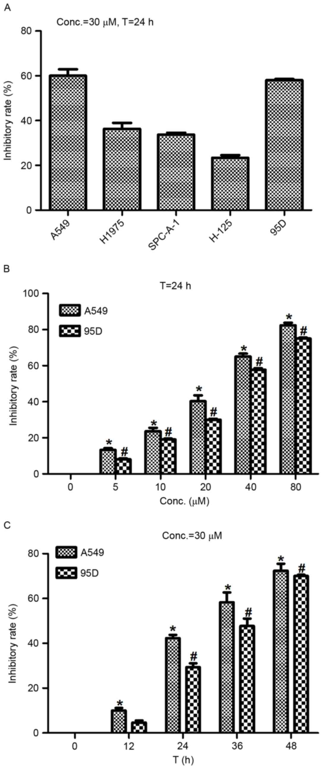
To test the effects of actein administration on
NSCLC cell proliferation, NSCLC cell lines A549, H1975, SPC-A-1,
H-125 and 95D were cultured and treated with actein (30 µM) 24 h
prior to cell viability assays. The results demonstrated that cell
viability rates in all these five cell lines were reduced following
actein administration, especially in A549 cells and 95D cells
(Fig. 1A). Therefore, the A549 and
95D cells were selected for subsequent experiments. Next, the 2
cell lines were treated with different doses of actein at diverse
time intervals. Fig. 1B and C
illustrate that actein significantly suppressed cell viability in a
dose- and time-dependent manner, compared with control cells. The
lethal dose 50 value (LD50) was approximate 20 and 30
µM, respectively, for A549 and 95D cells 24 h following actein
administration (Fig. 1B). The
inhibitory rate was progressively increased with extended treatment
time for both cell lines stimulated with 30 µM actein (Fig. 1C). These data revealed that actein
decreased the cell viability of NSCLC cell lines in a dose- and
time-dependent manner.
Actein suppresses cell colony
formation in NSCLC cells
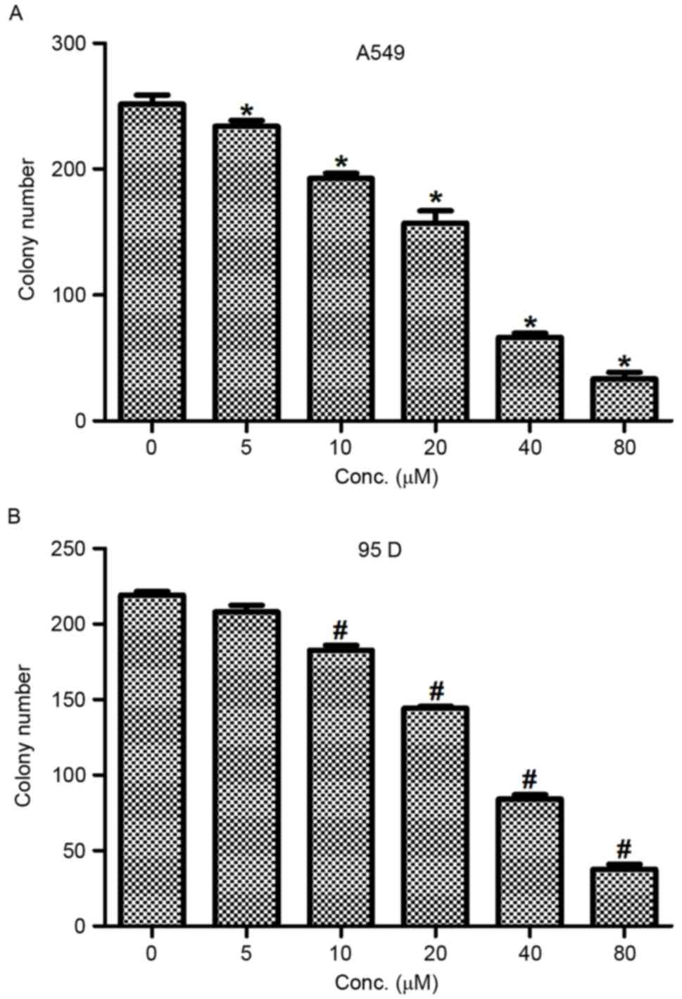
To further assess the inhibitory effects of actein
in NSCLC, a colony formation assay was conducted in A549 and 95D
cells. Cells were treated with different doses of actein (0, 5, 10,
20, 40 and 80 µM). In control untreated A549 cells, ~250 colonies
were visualized; however, only 160 and 35 colonies were visualized
when A549 cells were treated with 20 and 80 µM actein, respectively
(Fig. 2A). In 95D cells, the colony
numbers were also dramatically decreased from 230 in the control
group to 35 in the 80 µM actein treated group (Fig. 2B). These data demonstrated that actein
inhibited colony formation, jointly leading to the conclusion that
actein treatment suppressed cell proliferation in NSCLC cell
lines.
Actein inhibits cell migration and
invasion in NSCLC cells
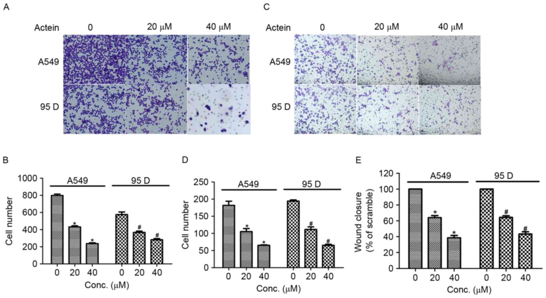
Next, transwell assays were performed to explore the
effects of actein in cell migration and invasion in human NSCLC
cells. As illustrated in Fig. 3A and
B, the number of cells migrated to the lower surface of the
chamber was decreased by 50 and 75% when A549 cells were treated
with 20 and 40 µM actein respectively, compared with control.
Significant decreases in cell migration were also observed in 95D
cells upon actein administration. Cell invasion ability was also
measured by using Transwell chambers that was pre-coated with
Matrigel for 6 h. The results demonstrated that the number of cells
invaded through the membrane was significantly decreased in A549
cells treated with actein, in a dose-dependent manner (Fig. 3C and D). Similarly, the invasion
abilities of 95D cells treated with 20 or 40 µM actein were
decreased compared with control (Fig. 3C
and D). Furthermore, wound-healing assays were performed to
further confirm the effects of actein on NSCLC cell migration. A549
and 95D cells were treated with 20 or 40 µM actein prior to
scraping a cross in each well. Fig.
3E illustrates that the rates of wound closure were
significantly decreased for both actein-treated cell lines compared
with untreated controls. These effects were also dose-dependent.
The present data suggested that actein suppressed cell migration
and invasion in vitro.
Actein increases NSCLC cell apoptosis
in a dose-dependent manner
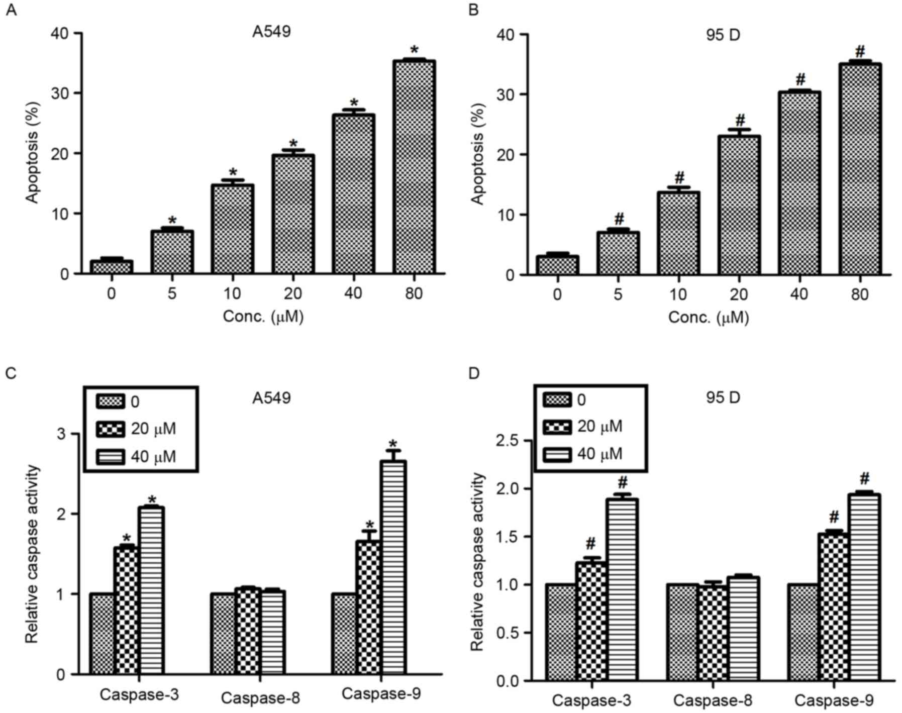
Actein was reported to exhibit a significant
regulatory role in cell apoptosis in melanoma cells (15). Thus, the effects of actein in NSCLC
cell apoptosis were examined in the present study with Hoechst
33258 staining analysis. Following exposure to various
concentrations (0, 5, 10, 20, 40 and 80 µM) of actein, the cell
nuclei of A549 and 95D cells were observed to be crushed and
fragmented with Hoechst 33258 staining. The results demonstrated
that cell apoptosis rates were increased by 5, 12, 18, 23 and 30%
in A549 cells treated with 5, 10, 20, 40 and 80 µM actein
respectively (Fig. 4A). Similar
results were observed in 95D cells. Apoptosis rates were increased
from 3% in control cells to 35% in the 80 µM actein-treated cells
(Fig. 4B). Next, the relative caspase
activity was explored in cells that were treated with actein (0, 20
and 40 µM). It was demonstrated that the activities of caspase-3
and caspase-9 were significantly increased by actein treatment in a
dose-dependent manner, while the activity of caspase-8 remained
unchanged in both NSCLC cell lines (Fig.
4C and D).
Finally, the expression of apoptosis-related
proteins in A549 cells was examined by western blot analysis. As
illustrated in Fig. 5, when A549
cells were treated with various doses of actein (0, 20 and 40 µM),
the protein expression levels of Bcl-2 were decreased as the
concentration of actein increased. In addition, the protein
expression levels of mitochondrial Bax, cleaved caspase-3, −9 and
cytoplasmic cyto c were significantly upregulated following
actein treatment in a dose-dependent manner (Fig. 5). Together with Fig. 4, the present results suggested that
actein promoted NSCLC cell apoptosis in a dose-dependent
manner.
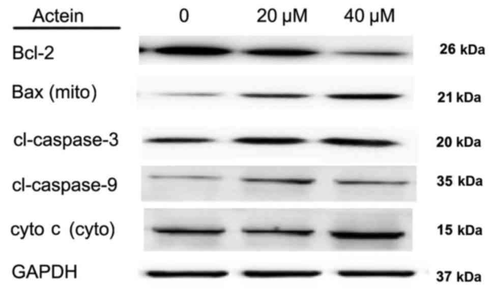 | Figure 5.Actein treatment alters expression of
apoptosis-related proteins in A549 cells. A549 cells were treated
with different doses of actein (0, 20 and 40 µM) and total proteins
in each group were subjected to western blot analysis. Protein
levels of Bcl-2 were decreased, while levels of Bax (mito),
cl-caspase-3, −9, cyto c (cyto) were increased following
actein treatment in a dose-dependent manner. Bcl-2, BCL2 apoptosis
regulator; Bax, BCL2 associated X; cyto, cytoplasmic; mito,
mitochondrial; cl, cleaved; cyto c, cytochrome c. |
Discussion
NSCLC is a life-threatening cancer with an obvious
etiology and various pathogenic factors. In the future, it is
likely to become the world's leading cause of cancer-related deaths
despite worldwide efforts to control the etiology of the disease.
The identification of novel genetic biomarkers and effective
therapeutic agents has been a huge step forward to understanding
the tumorigenesis, diagnosis and treatment of NSCLC (16–18).
However, further studies are required in order to increase the
five-year survival rate for patients suffering from NSCLC.
The present study demonstrated the inhibitive
effects of actein on NSCLC cell proliferation and migration in
vitro. Actein suppressed cell viability in a dose- and
time-dependent manner. Furthermore, the colony formation assay
confirmed that, upon actein administration, NSCLC cell lines A549
and 95D displayed reduced abilities to form colonies, which
suggested an inhibited proliferation property. Transwell assays
revealed that actein treatment suppressed cell migration and
invasion in NSCLC cells. Since cell proliferation and migration are
hallmarks of cancer, these data suggest that actein might function
as a potential therapeutic agent against NSCLC through its strong
growth inhibitory effects. Furthermore, lower and higher
concentrations of actein were explored in the present preliminary
study. It was demonstrated that the cell proliferative rate and
invasive capacity of both A549 and 95D cells were suppressed
following actein treatment, even in a lower concentration (30 µM).
Therefore, the inhibitory effects of actein on cell proliferation
and invasion were synchronous. The detailed mechanism underlying
the inhibitory effects of actein remains unknown and this will be
the focus of future studies.
The induction of apoptosis can be divided into two
categories: Intrinsic and extrinsic pathways. The initiation of the
former is associated with the pro-apoptotic factors Bax and BCL2
associated agonist of cell death (Bad), and it results in increased
permeability of the mitochondria membrane, loss of membrane
potential and release of cyto c into the cytosol (19). Cyto c serves a regulatory role
since it precedes morphological changes associated with apoptosis.
Cyto c binds to apoptotic protease activating factor-1 and
then pro-caspase-9 to form a protein complex called apoptosome, the
role of which is to cleave pro-caspase to its active form of
caspase-9 and then in turn activate caspase-3 (20). In the present study, the apoptosis
rate following actein administration was explored and the results
demonstrated thatactein could positively promote cell apoptosis in
NSCLC cells. Furthermore, the expression levels of mitochondrial
Bax, caspase-3, −9 and cyto c were all upregulated in
actein-treated A549 cells in a dose-dependent manner, while actein
inhibited the protein levels of Bcl-2 and cytoplasmic Bax. Bcl-2
and Bax both belong to the BCL2 family, the former of which is
anti-apoptotic while the latter one is proapoptotic. When apoptosis
occurs, the proapoptotic homodimers are essential to make the
mitochondrial membrane permeable for the release of caspase
activators including cyto c (21), resulting in decreased expression of
cytoplasmic Bax and increased mitochondrial Bax during apoptosis.
Since induction of apoptosis is always associated with
proliferation inhibition, it is hypothesized that actein may
inhibit cell proliferation and migration through the apoptosis
pathway. However, the detailed mechanisms and signaling processes
by which actein affects the apoptosis pathways remain to be
elucidated.
In conclusion, the present study revealed the growth
inhibitory effectof actein in NSCLC cells, which was related with
cell apoptosis. Theseresults might provide new clues for the
treatment of NSCLC in the clinic. Actein may serve as a potential
therapeutic agent against NSCLC.
References
|
1
|
Villeneuve PJ and Mao Y: Lifetime
probability of developing lung cancer, by smoking status, Canada.
Can J Public Health. 85:385–388. 1994.PubMed/NCBI
|
|
2
|
Torre LA, Bray F, Siegel RL, Ferlay J,
Lortet-Tieulent J and Jemal A: Global cancer statistics, 2012. CA
Cancer J Clin. 65:87–108. 2015. View Article : Google Scholar : PubMed/NCBI
|
|
3
|
Jemal A, Bray F, Center MM, Ferlay J, Ward
E and Forman D: Global cancer statistics. CA Cancer J Clin.
61:69–90. 2011. View Article : Google Scholar : PubMed/NCBI
|
|
4
|
Chen W, Zheng R, Zeng H and Zhang S:
Epidemiology of lung cancer in China. Thorac Cancer. 6:209–215.
2015. View Article : Google Scholar : PubMed/NCBI
|
|
5
|
McKenna DJ, Jones K, Humphrey S and Hughes
K: Black cohosh: Efficacy, safety, and use in clinical and
preclinical applications. Altern Ther Health Med. 7:93–100.
2001.PubMed/NCBI
|
|
6
|
Einbond LS, Su T, Wu HA, Friedman R, Wang
X, Ramirez A, Kronenberg F and Weinstein IB: The growth inhibitory
effect of actein on human breast cancer cells is associated with
activation of stress response pathways. Int J Cancer.
121:2073–2083. 2007. View Article : Google Scholar : PubMed/NCBI
|
|
7
|
Lee YS and Choi EM: Actein isolated from
black cohosh promotes the function of osteoblastic MC3T3-E1 cells.
J Med Food. 17:414–423. 2014. View Article : Google Scholar : PubMed/NCBI
|
|
8
|
Einbond LS, Soffritti M, Esposti DD, Park
T, Cruz E, Su T, Wu HA, Wang X, Zhang YJ, Ham J, et al: Actein
activates stress- and statin-associated responses and is
bioavailable in Sprague-Dawley rats. Fundam Clin Pharmacol.
23:311–321. 2009. View Article : Google Scholar : PubMed/NCBI
|
|
9
|
Einbond LS, Shimizu M, Xiao D, Nuntanakorn
P, Lim JT, Suzui M, Seter C, Pertel T, Kennelly EJ, Kronenberg F
and Weinstein IB: Growth inhibitory activity of extracts and
purified components of black cohosh on human breast cancer cells.
Breast Cancer Res Treat. 83:221–231. 2004. View Article : Google Scholar : PubMed/NCBI
|
|
10
|
Einbond LS, Shimizu M, Nuntanakorn P,
Seter C, Cheng R, Jiang B, Kronenberg F, Kennelly EJ and Weinstein
IB: Actein and a fraction of black cohosh potentiate
antiproliferative effects of chemotherapy agents on human breast
cancer cells. Planta Med. 72:1200–1206. 2006. View Article : Google Scholar : PubMed/NCBI
|
|
11
|
Einbond LS, Mighty J, Redenti S and Wu HA:
Actein induces calcium release in human breast cancer cells.
Fitoterapia. 91:28–38. 2013. View Article : Google Scholar : PubMed/NCBI
|
|
12
|
Einbond LS, Shimizu M, Ma H, Wu HA,
Goldsberry S, Sicular S, Panjikaran M, Genovese G and Cruz E:
Actein inhibits the Na+-K+-ATPase and
enhances the growth inhibitory effect of digitoxin on human breast
cancer cells. Biochem Biophys Res Commun. 375:608–613. 2008.
View Article : Google Scholar : PubMed/NCBI
|
|
13
|
Chen Z, Wu J and Guo Q: Actein inhibits
cell proliferation and migration in human osteosarcoma. Med Sci
Monit. 22:1609–1616. 2016. View Article : Google Scholar : PubMed/NCBI
|
|
14
|
Suh KS, Chon S and Choi EM: Actein
protects against methylglyoxal-induced oxidative damage in
osteoblastic MC3T3-E1 cells. J Sci Food Agric. 97:207–214. 2017.
View Article : Google Scholar : PubMed/NCBI
|
|
15
|
Du J, Lu X, Long Z, Zhang Z, Zhu X, Yang Y
and Xu J: In vitro and in vivo anticancer activity of aconitine on
melanoma cell line B16. Molecules. 18:757–767. 2013. View Article : Google Scholar : PubMed/NCBI
|
|
16
|
Lee PN and Hamling J: The relation between
smokeless tobacco and cancer in Northern Europe and North America.
A commentary on differences between the conclusions reached by two
recent reviews. BMC Cancer. 9:2562009. View Article : Google Scholar : PubMed/NCBI
|
|
17
|
Hackshaw AK, Law MR and Wald NJ: The
accumulated evidence on lung cancer and environmental tobacco
smoke. BMJ. 315:980–988. 1997. View Article : Google Scholar : PubMed/NCBI
|
|
18
|
King PT: Inflammation in chronic
obstructive pulmonary disease and its role in cardiovascular
disease and lung cancer. Clin Transl Med. 4:682015. View Article : Google Scholar : PubMed/NCBI
|
|
19
|
Spencer SL and Sorger PK: Measuring and
modeling apoptosis in single cells. Cell. 144:926–939. 2011.
View Article : Google Scholar : PubMed/NCBI
|
|
20
|
Dejean LM, Martinez-Caballero S and
Kinnally KW: Is MAC the knife that cuts cytochrome c from
mitochondria during apoptosis? Cell Death Differ. 13:1387–1395.
2006. View Article : Google Scholar : PubMed/NCBI
|
|
21
|
Murphy KM, Ranganathan V, Farnsworth ML,
Kavallaris M and Lock RB: Bcl-2 inhibits Bax translocation from
cytosol to mitochondria during drug-induced apoptosis of human
tumor cells. Cell Death Differ. 7:102–111. 2000. View Article : Google Scholar : PubMed/NCBI
|