Introduction
Esophageal cancer is the eighth most common cancer
and the seventh leading cause of cancer-related death in men
worldwide (1). It is composed of two
major types, i.e. esophageal squamous cell carcinoma (ESCC) and
esophageal adenocarcinoma. ESCC is the predominant type of
esophageal cancer in Asian countries (2,3).
Tumor resection is the frontline treatment for ESCC
patients. However, its benefit is limited to patients in early
stage disease and those with resectable tumors. For patients with
locally advanced tumors, they are alternatively treated with
neoadjuvant chemoradiation for tumor down-staging before tumor
resection. Based on the reports from our group and other research
teams, such neoadjuvant treatment can enhance the chance of cure by
tumor resection and therefore can profoundly increase patient
outcomes (4–6). Despite that, patients exhibit a range of
different responses towards chemoradiation from complete response,
partial response to non-response. Till now, no clinical parameters
or biomarkers are used to predict patient chemoradiation response
and to identify non-responders to exclude them from treatment.
Dismally, patients who do not respond to chemoradiation have to
suffer from unnecessary and adverse side-effects of the treatment,
such as drug-related toxicity and post-treatment complications. To
address this clinical limitation, identification of biomarkers that
can predict chemoradiation response can provide a guideline for
selecting patients for chemoradiation, such that responders will be
offered chemoradiation and non-responders will be excluded from
such treatment.
MicroRNAs (miRNAs) are single-stranded, non-coding
small RNA of 19 to 25 nucleotides. With their main functions on
regulating gene expressions, they are capable of regulating
diversified cellular functions and are involved in a range of
physiological processes of growth and development (7). However, their deregulation can be
commonly found in various diseased conditions including cancer of
various types (8). MiRNAs involve in
tumorigenesis by functioning both as tumor suppressors and
oncogenes. A number of miRNAs have been found to have deregulated
expressions in tumors when compared to adjacent non-tumor tissues.
These observations have implicated for the potential uses of miRNAs
for cancer prognosis and therapy (7,9).
Apart from the uses of miRNAs for cancer prognosis
and therapy, recent studies have started to investigate the
capability of miRNAs to predict patient responses towards different
anti-cancer treatments including chemotherapy and targeting therapy
for various cancers including ESCC (10–14).
Collectively, these findings have consolidated the use of miRNAs
for treatment response prediction. Due to the secretory nature of
miRNAs and their small sizes making them resistant to degradation
(15,16), current efforts have been focused on
validating the use of circulatory miRNAs as non-invasive biomarkers
for treatment response prediction for cancer patients (8,16,17).
The purpose of this study was to identify serum
miRNAs that may serve as non-invasive biomarkers for treatment
response prediction for ESCC patients. MiRNA array was employed in
the analysis of serum samples from good responders vs. poor
responders towards chemoradiation to identify candidate miRNAs with
most differential difference in the serum levels between good
responders and poor responders. The candidate miRNAs were further
validated and analyzed for their potential use as predictive
biomarkers for chemoradiation response of ESCC patients.
Materials and methods
Patient specimens
From 2000 to 2010, 47 locally advanced ESCC patients
managed in Queen Mary Hospital, Hong Kong were included in this
study. Age range of this patient cohort was 44 to 82 years old and
the sex ratio was 44 (M) to 3 (F). All patients were treated with
neoadjuvant cisplatin- and 5-fluorouracil-based chemoradiation
followed by tumor resection. Chemoradiation was given concurrently.
The chemotherapy regimen composed of cisplatin at 100
mg/m2 on day 1 and then day 22 and 5-fluorouracil at 500
mg/m2 per day from day 1 to day 5 and day 22 to day 26.
Radiotherapy was given at a dosage of 40Gy at 2Gy per fraction
(18). Serum specimens were prepared
from blood samples collected from patients before treatment with
chemoradiation. Patient chemoradiation responses were determined
pathologically based on the percentage of viable tumor cells in the
resected tissues collected from tumor resection (18). In this study, patients with 0% viable
tumor cells were classified as good chemoradiation responders,
while those with at least 50% viable tumor cells were regarded as
poor responders. Informed consent was obtained for each patient
recruited in this study. AJCC 6th edition was used for cancer
staging. The use of clinical specimens for research was approved by
the Institutional Review Board of The University of Hong
Kong/Hospital Authority Hong Kong West Cluster (HKU/HA HKW
IRB).
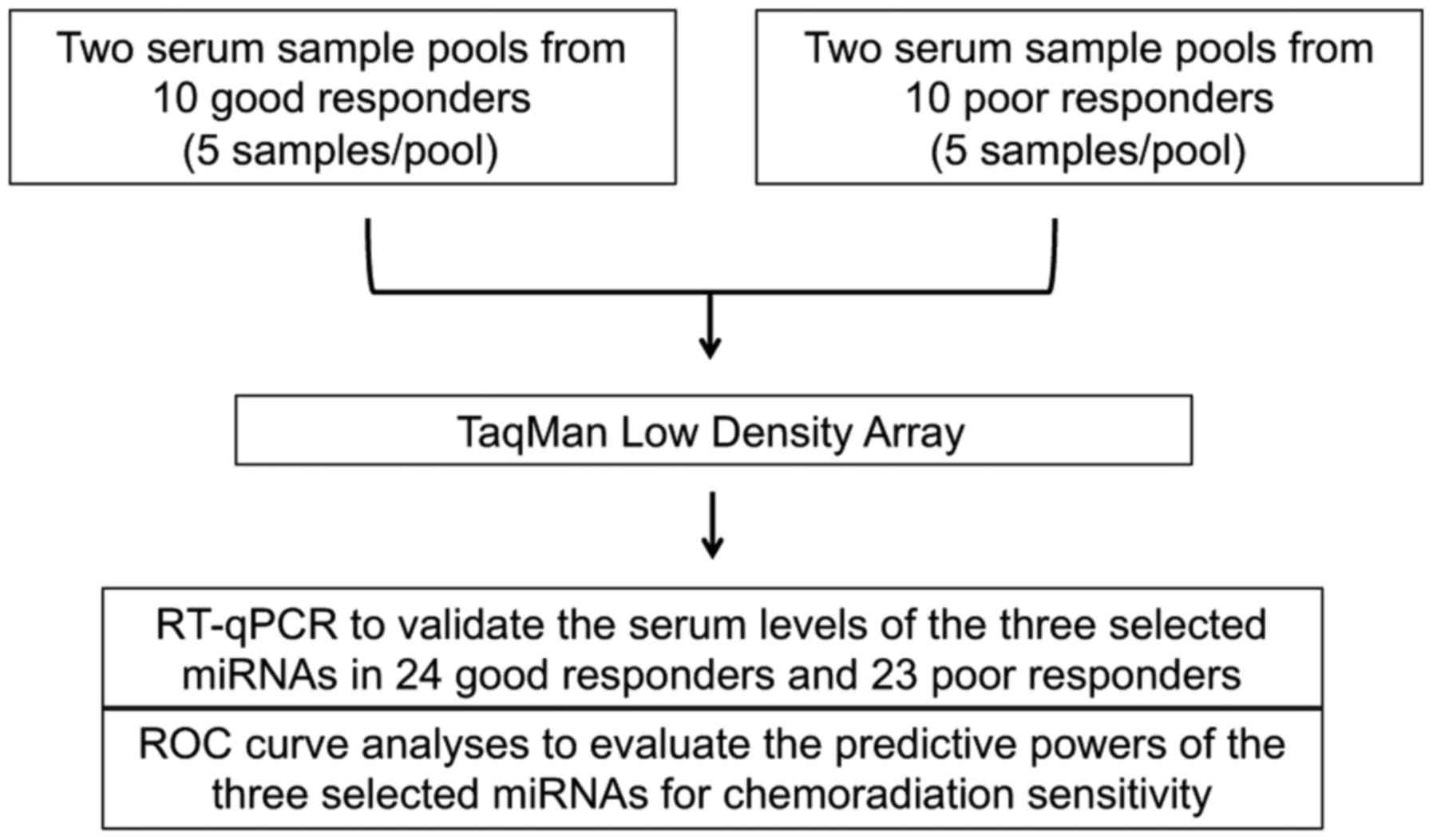
MiRNA array
Small RNAs from serum samples (10 samples from good
responders and 10 samples from poor responders) were extracted
using miRNeasy Serum/Plasma kit (Qiagen Inc., Hilden, Germany). RNA
concentration and quality were determined using a NanoDrop ND-1000
spectrophotometer (NanoDrop Technologies, Wilmington, DE, USA). For
both good responders and poor responders, pooled serum samples were
prepared by mixing extracted RNAs from 5 samples, and that resulted
in 2 serum sample pools for good responders and poor responders.
The pooled RNA samples were then reverse transcribed to cDNA using
TaqMan MicroRNA Reverse Transcription kit and Megaplex RT Primers
(Applied Biosystems, Foster City, CA, USA) with the following
thermal condition: 40 cycles of 16°C for 2 min; 42°C for 1 min;
50°C for 1 sec and 85°C for 5 min. TaqMan Low Density Array (TaqMan
Array Human MicroRNA A+B Cards Set v3.0; Applied Biosystems) was
employed to generate miRNA profiles from 754 different human miRNAs
and U6, which was selected as an endogenous control. The reactions
were performed in a 7900HT Fast Real-Time PCR System (Applied
Biosystems), following the manufacturer protocol. Differentially
expressed miRNAs were identified by comparing the miRNA profiles
obtained from good responders and poor responders, and that miRNAs
with at least 2.5-fold elevation in good responders vs. poor
responders were subjected to further validation.
Reverse transcription-quantitative
polymerase chain reaction (RT-qPCR)
RNAs from serum specimens were prepared using
miRNeasy Serum/Plasma kit (Qiagen). Concentration and quality of
the extracted RNAs were determined as described above. To validate
the miRNA array data, a TaqMan probe-based RT-qPCR system with the
use of specific TaqMan MicroRNA Assays (Applied Biosystems) and
TaqMan Fast Universal PCR Master Mix (Applied Biosystems) was
performed on 24 good responders and 23 poor responders (including
those used in the test cohort) (19–21). The
reactions in duplicates were run on a 7900HT Fast Real-Time PCR
System (Applied Biosystems) under the following thermal cycling
condition: 2 min at 50°C; 10 min at 95°C and 45 cycles of 15 sec at
95°C and 1 min at 60°C. U6 was used as an endogenous control.
Relative expressions for the examined miRNAs were calculated using
the ∆∆Cq method (22).
Statistical analyses
GraphPad Prism 6.0 (GraphPad Software, La Jolla, CA,
USA) was used for statistical analyses. Quantitative data are
described as the means with standard deviations (SD). Students
t-test was used to compare expression level difference of each
miRNA (expressed as ∆∆Cq value) between studied groups. To assess
the capability of the studied miRNA for discriminating good
responders and poor responders towards chemoradiation, receiver
operating characteristic (ROC) curve and the area under the ROC
curve (AUC) with 95% confidence interval (CI) were generated
(19,23). Survival was calculated by the
Kaplan-Meier method from the date of operation to the time of death
or last follow-up date. Log-rank test was used to compare survival
difference between groups. Clinical categorical data were analyzed
using chi-squared test together with Pearson correlation. P<0.05
was considered to indicate a statistically significant
difference.
Results
Identification of candidate miRNAs for
predicting patient chemoradiation responses
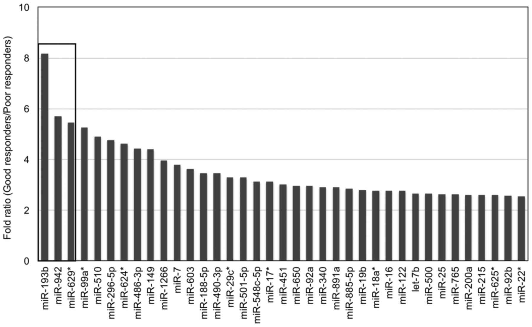
MiRNA array was performed on pooled serum samples
from good responders and poor responders (Fig. 1). Of the 754 miRNAs examined, 37
miRNAs had at least 2.5-fold elevation in sera from good responders
when compared to poor responders, while 23 of them (highlighted in
grey) displayed consistent data between the two serum sample pools
from the same group (Table I). Three
serum miRNAs (miR-193b, miR-942 and miR-629*) with the most
difference on the fold ratio between good responders and poor
responders were selected as candidates for further validation
(Fig. 2).
 | Table I.Serum miRNAs with at least 2.5-fold
elevation in good responders (GR) versus poor responders (PR). |
Table I.
Serum miRNAs with at least 2.5-fold
elevation in good responders (GR) versus poor responders (PR).
|
| 1st array | 2nd array | 1st array | 2nd array |
|
|
|
|---|
|
|
|
|
|
|
|
|
|
|---|
| miRNAs | GR1 | GR2 | PR1 | PR2 | Relative level
(GR) | Relative level
(PR) | Fold ratio
(GR/PR) |
|---|
|
miR-193ba | 34.83 | 35.17 | nd | nd | 0.3004 | 0.0368 | 8.1681 |
|
miR-942a | 34.03 | 36.31 | nd | nd | 0.2544 | 0.0445 | 5.7160 |
|
miR-629*a | 35.03 | 35.44 | nd | nd | 0.2432 | 0.0445 | 5.4642 |
| miR-99a* | 34.94 | nd | nd | 36.61 | 0.3132 | 0.0595 | 5.2598 |
| miR-510 | nd | 35.04 | 36.6 | nd | 0.2922 | 0.0597 | 4.8906 |
| miR-296-5p | 34.8 | 34.87 | 36.12 | nd | 0.3368 | 0.0706 | 4.7733 |
|
miR-624*a | 34.97 | 35.98 | nd | nd | 0.2059 | 0.0445 | 4.6268 |
|
miR-486-3pa | 34.17 | 34.78 | 36.32 | 37.56 | 0.4118 | 0.0928 | 4.4383 |
| miR-149 | 33.89 | 35.37 | 35.48 | nd | 0.3882 | 0.0881 | 4.4076 |
|
miR-1266a | 35.29 | 36.1 | nd | nd | 0.1768 | 0.0445 | 3.9724 |
| miR-7a | 34.81 | 35.14 | 36.49 | 37.25 | 0.3057 | 0.0805 | 3.7974 |
| miR-603 | nd | 33.65 | nd | nd | 0.1615 | 0.0445 | 3.6301 |
| miR-188-5p | nd | 34.47 | nd | nd | 0.1276 | 0.0368 | 3.4702 |
| miR-490-3p | 34.17 | 30.59 | 32.61 | 35.67 | 1.8468 | 0.5340 | 3.4581 |
|
miR-29c*a | 35.89 | 36.04 | nd | 38 | 0.1466 | 0.0445 | 3.2944 |
| miR-501-5p | 33.77 | 35.79 | nd | 34.93 | 0.3499 | 0.1066 | 3.2830 |
|
miR-548c-5pa | 35.01 | 37.07 | nd | nd | 0.1392 | 0.0445 | 3.1275 |
|
miR-17*a | 34.5 | 37.59 | nd | nd | 0.1387 | 0.0445 | 3.1167 |
|
miR-451a | 20.23 | 21.1 | 22.48 | 21.98 | 6208.3751 | 2055.1101 | 3.0209 |
|
miR-650a | 35.01 | 37.22 | nd | nd | 0.1321 | 0.0445 | 2.9690 |
|
miR-92aa | 24.97 | 25.08 | 26.61 | 26.5 | 302.3341 | 102.5369 | 2.9485 |
|
miR-340a | 37.07 | 35.9 | nd | nd | 0.1073 | 0.0368 | 2.9180 |
| miR-891a | nd | 34.98 | nd | nd | 0.1069 | 0.0368 | 2.9079 |
| miR-885-5p | 36.11 | 35.02 | 36.08 | nd | 0.2031 | 0.0715 | 2.8382 |
|
miR-19ba | 25.61 | 26.35 | 27.29 | 27.57 | 155.9565 | 55.9087 | 2.7895 |
|
miR-18a*a | 32.67 | 33.65 | 34.95 | 34.25 | 1.0755 | 0.3882 | 2.7702 |
| miR-16a | 23.72 | 24.28 | 25.55 | 25.33 | 615.2377 | 222.0899 | 2.7702 |
|
miR-122a | 30.57 | 29.55 | 31.26 | 31.74 | 9.2215 | 3.3288 | 2.7702 |
| let-7ba | 28.18 | 28.66 | 29.64 | 29.96 | 28.7402 | 10.8153 | 2.6574 |
|
miR-500a | 34.39 | 35.29 | 36.38 | 36.06 | 0.3356 | 0.1263 | 2.6574 |
| miR-25a | 28.08 | 28.8 | 29.98 | 29.64 | 28.3446 | 10.7406 | 2.6390 |
| miR-765 | 36.39 | 35.97 | nd | 37.09 | 0.1326 | 0.0504 | 2.6299 |
| miR-200a | nd | 35.79 | nd | 37.14 | 0.1737 | 0.0668 | 2.6027 |
|
miR-215a | 32.69 | 33.68 | 33.91 | 35.16 | 1.0570 | 0.4061 | 2.6027 |
| miR-625* | 35.45 | 35.07 | 35.21 | nd | 0.2509 | 0.0967 | 2.5937 |
| miR-92b | 35.49 | 36.07 | nd | 36.23 | 0.1749 | 0.0679 | 2.5758 |
|
miR-22*a | 34.05 | 33.68 | 34.72 | 35.66 | 0.6598 | 0.2579 | 2.5580 |
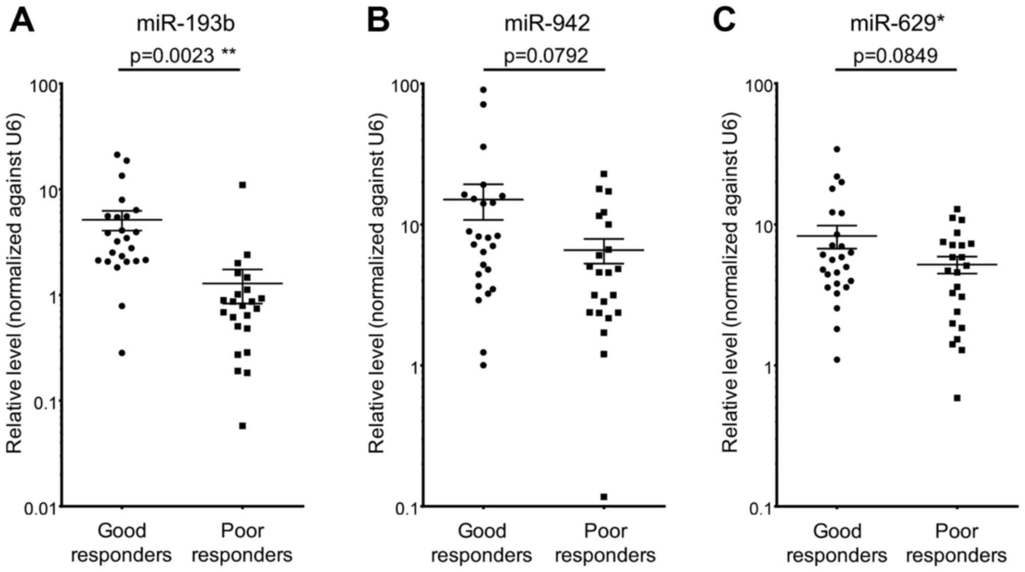
Validation of candidate miRNAs by
RT-qPCR
To validate the miRNA array data, RT-qPCR on serum
miR-193b, miR-942 and miR-629* was performed on 24 good responders
and 23 poor responders. The RT-qPCR results were consistent with
those obtained by miRNA array, such that all three examined miRNAs
demonstrated higher levels in sera from good responders when
compared to poor responders (Fig. 3).
However, only miR-193b achieved a statistically significant result
(P<0.05) (Fig. 3A). These results
suggested that high serum level of miR-193b could potentially be
served as a biomarker to predict patient chemoradiation
responses.
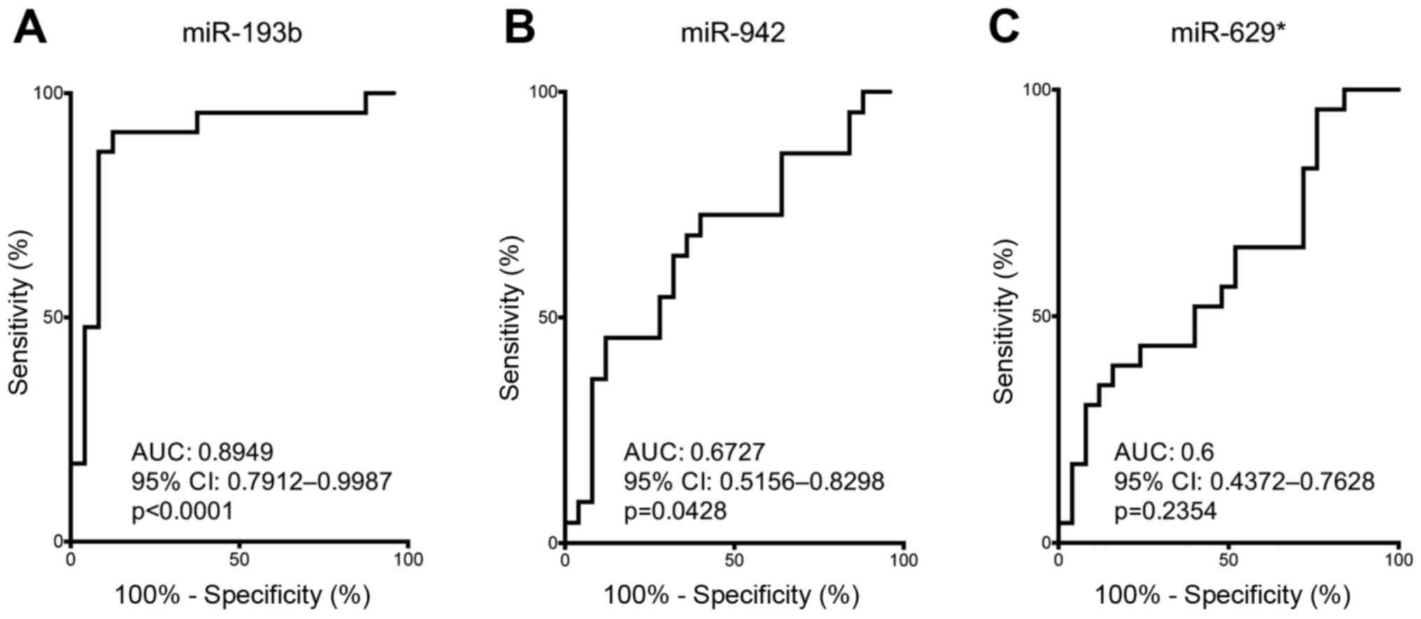
Serum miR-193b serves as a
non-invasive predictive biomarker for patient chemoradiation
response and survival outcome
To examine the predictive power of serum miR-193b,
miR-942 and miR-629* on distinguishing good responders and poor
responders towards chemoradiation, ROC curves analysis was
performed to obtain the AUC values and 95% CI. ROC curve data of
serum miR-193b revealed a strong predictive power to distinguish
good responders and poor responders (AUC: 0.8949, 95% CI:
0.7912–0.9987, P<0.0001) (Fig.
4A). Although ROC curve analysis supporting the use of serum
miR-942 for distinguishing good responders and poor responders
(AUC: 0.6727, 95% CI: 0.5156–0.8298, P=0.0428) (Fig. 4B), serum miR-942 failed to demonstrate
significant difference between its level in good responders and
poor responders (Fig. 3B). On the
other hand, serum miR-629* failed to show sufficient discriminative
power to distinguish good responders from poor responders (Fig. 4C). Based on the differential
difference of serum miR-193b level in good responders vs. poor
responders and the good discriminative power of serum miR-193b on
distinguishing good responders from poor responders, our data have
supported the potential use of miR-193b for predicting
chemoradiation response.
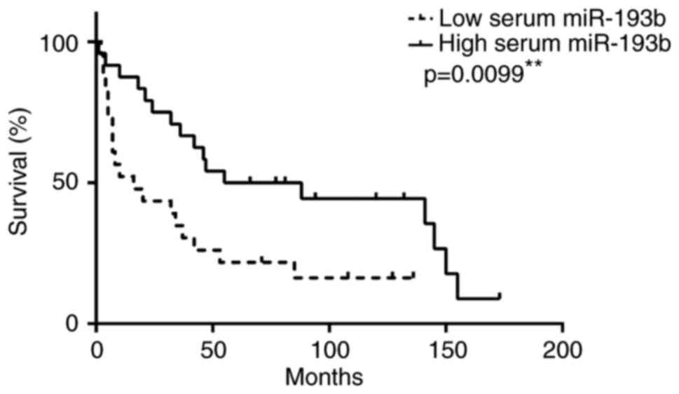
As miR-193b is a potential predictor for
chemoradiation response in ESCC patients, we also performed
survival and clinical analyses by using the median value as a
cut-off value to divide the patients into two groups with high and
low serum level of miR-193b. Patients with high serum miR-193b
level associated better survival (median survival=71.5 months) than
those with low serum miR-193b level (median survival=16 months)
(P=0.0099) (Fig. 5). High serum
miR-193b level was inversely correlated with post-therapy
pathological ypT stage (P<0.001), ypN stage (P=0.045), ypM stage
(P=0.035) and overall ypTNM stage (P<0.001) (Table II). Moreover, high serum miR-193b
level was associated with R0 category (no residual tumor after
surgery) (P<0.001) and with pathological complete response
(ypCR) (P<0.001) (Table II).
Collectively, our data have suggested that high serum level of
miR-193b can potentially serve as a non-invasive predictive
biomarker for patient chemoradiation response and survival
outcome.
| Table II.Clinical correlation analyses of
serum miR-193b level in ESCC patients treated with neoadjuvant
chemoradiation. |
Table II.
Clinical correlation analyses of
serum miR-193b level in ESCC patients treated with neoadjuvant
chemoradiation.
|
|
| Serum miR-193b
level |
|
|---|
|
|
|
|
|
|---|
| Clinicopathological
parameters | Patient number | High | Low | P-value |
|---|
| Age (years) |
|
|
|
|
|
≤64.5 | 23 | 10 | 13 | 0.308 |
|
>64.5 | 24 | 14 | 10 |
|
| Gender |
|
|
|
|
|
Male | 44 | 24 | 20 | 0.067 |
|
Female | 3 | 0 | 3 |
|
| R category |
|
|
|
|
| R0 | 34 | 24 | 10 |
<0.001 |
|
R1/R2 | 13 | 0 | 13 |
|
| Pathological
stage |
|
|
|
|
| ypT
stage |
|
|
|
|
|
ypT0 | 24 | 21 | 3 |
<0.001 |
|
ypT1/2 | 4 | 0 | 4 |
|
|
ypT3/4 | 19 | 3 | 16 |
|
| ypN
stage |
|
|
|
|
|
ypN0 | 33 | 20 | 13 | 0.045 |
|
ypN1 | 14 | 4 | 10 |
|
| ypM
stage |
|
|
|
|
|
ypM0 | 40 | 23 | 17 | 0.035 |
|
ypM1 | 7 | 1 | 6 |
|
|
ypTNM |
|
|
|
|
|
ypCRa | 24 | 21 | 3 |
<0.001 |
| y-stage
I/II | 9 | 1 | 8 |
|
| y-stage
III/IV | 14 | 2 | 12 |
|
Discussion
Neoadjuvant chemoradiation followed by tumor
resection has become the standard treatment for locally advanced
ESCC (24–26). However, patients respond differently
towards such neoadjuvant treatment. On average, only 20–30% of
patients demonstrate complete chemoradiation response. Poor
responders, on the other hand, suffer unnecessary
chemoradiation-related toxicity and complications without benefits.
In view of this clinical situation, identification of reliable
biomarker that can predict patient response before chemoradiation
is important for selecting optimal treatment for patients.
For ESCC, previous studies have proposed the use of
several biomarkers identified from ESCC tumors to predict patient
chemoradiation response. A study has demonstrated the association
of excision repair cross-complementing 1 (ERCC1) with cisplatin
response in ESCC by revealing tumoral ERCC1 levels in patients with
partial responses to chemoradiation were significantly lower than
those in non-responsive patients. Besides, ESCC cell lines with
lower ERCC1 level showed greater sensitivity to clinically relevant
concentrations of cisplatin and its related drug oxaliplatin when
compared to cell lines with higher ERCC1 level (27). In addition to this single molecule
study, a number of studies have taken into accounts the use of more
than one molecule for predicting chemoradiation responses. Using
cDNA microarray to analyze pre-chemoradiation tumor biopsies from
ESCC patients has identified a 32-gene classifier that can be used
to predict patient responses towards chemoradiation (28). Another study by Wen et al has
carried out gene expression analysis on pre-treatment ESCC biopsies
from patients who have received neoadjuvant chemoradiation followed
by surgery. Based on the gene expression analysis, they have
developed a prediction model composing of three genes (MMP1,
LIMCH1 and C1orf226) that can be used to predict
pathological response towards chemoradiation with high accuracy
(29). Apart from the above use of
protein-coding genes for predicting chemoradiation responses of
ESCC patients, research has also been focused on exploring the
potential use of non-coding RNAs for this purpose. MiRNAs are
non-coding RNAs with diversified roles on tumor progression and
development, implicating their plausible uses for cancer
classification and prognostication (7,30). By
analyzing the pre-treatment tumor biopsies, a recent miRNA study
has identified the combined use of four differentially expressed
miRNAs (miR-145-5p, miR-152, miR-193b-3p and miR-376a-3p) for
chemoradiation response prediction for ESCC patients (14).
When compared to the biomarkers derived from ESCC
tumors, blood-based biomarkers are better predictors for
chemotherapy/chemoradiation responses of patients as the procedure
for blood collection is minimally invasive. Most studies on finding
blood-based biomarkers for chemotherapy/chemoradiation response
prediction put the focus on miRNAs because of their stability in
human circulation (15,16). Kurashige et al have reported
the use of serum miR-21 for indicating chemotherapy response by
showing a significant reduction of its serum level in ESCC patients
who are responsive to chemotherapy, but not in non-responders,
after treatment (31). On the other
hand, Tanaka et al have demonstrated serum level of miR-200c
can be used for predicting chemotherapy responses for ESCC patients
receiving chemotherapy (32). In the
current study, we identified serum miR-193b by miRNA array, and
validated it as a reliable predictor for chemoradiation responses
for ESCC patients using TaqMan probe-based qPCR. Specifically, high
serum level of miR-193b can indicate ESCC patients who are
responsive to chemoradiation. Moreover, high serum level of
miR-193b was significantly associated with better survival and
inversely correlated with post-therapy pathological stages. Despite
the advantageous feature of miRNAs, some studies have alternatively
investigated the possible use of other circulatory protein or mRNA
for predicting chemoradiation responses. Maher et al have
utilized proteomics to analyze pre-treatment sera from esophageal
cancer patients and have revealed significant elevation on the
serum levels of complement C4a and C3a in poor responders towards
chemoradiation (33). Moreover, a
more recent study has also demonstrated the usefulness of
circulating FAM84B mRNA and protein for predicting
pathological chemoradiation responses (34). The above studies have supported the
idea of using circulating biomarkers for predicting chemoradiation
responses of ESCC patients.
In this study, we have reported high serum miR-193b
level in ESCC patients responsive to chemoradiation, implicating
the link between miR-193b and chemoradiation sensitivity.
Similarly, Nyhan et al have reported the positive
association between miR-193b and increased chemosensitivity of
esophageal cancer cells, such that overexpression of miR-193b in
chemoresistant esophageal cancer cells sensitized them towards
chemotherapeutic drug treatment (35). Apart from the direct link between
miR-193b with chemotherapy/chemoradiation sensitivity, this miRNA
is proven to involve in other treatment-related sensitivities. In
melanoma, over-expression of miR-193b in ABT-737-resistant tumor
cells can restore their sensitivity towards ABT-737 (BH3 mimetic)
treatment (36). In another scenario,
over-expression of miR-193b in liver cancer cells can sensitize
them towards treatment with molecular targeting drug sorafenib
(37). Together, these prior studies
including the current one depict the importance of miR-193b on
determining the treatment sensitivity of tumor cells and implicate
the potential of restoring miR-193b for treatment
sensitization.
In summary, the present study has demonstrated high
level of serum miR-193b as a promising biomarker for predicting
chemoradiation responses and post-therapy survival of ESCC
patients. Further validation on a larger and independent patient
cohort is required for full assessment of its predictive power. Our
findings can benefit the treatment selection process for locally
advanced ESCC patients by predicting their sensitivities towards
chemoradiation, such that only good responders will be offered this
treatment.
References
|
1
|
Jemal A, Bray F, Center MM, Ferlay J, Ward
E and Forman D: Global cancer statistics. CA Cancer J Clin.
61:69–90. 2011. View Article : Google Scholar : PubMed/NCBI
|
|
2
|
Enzinger PC and Mayer RJ: Esophageal
cancer. N Engl J Med. 349:2241–2252. 2003. View Article : Google Scholar : PubMed/NCBI
|
|
3
|
Hiyama T, Yoshihara M, Tanaka S and
Chayama K: Genetic polymorphisms and esophageal cancer risk. Int J
Cancer. 121:1643–1658. 2007. View Article : Google Scholar : PubMed/NCBI
|
|
4
|
Tong DK, Law S, Kwong DL, Wei WI, Ng RW
and Wong KH: Current management of cervical esophageal cancer.
World J Surg. 35:600–607. 2011. View Article : Google Scholar : PubMed/NCBI
|
|
5
|
Law S, Kwong DL, Kwok KF, Wong KH, Chu KM,
Sham JS and Wong J: Improvement in treatment results and long-term
survival of patients with esophageal cancer: Impact of
chemoradiation and change in treatment strategy. Ann Surg.
238:339–348. 2003.PubMed/NCBI
|
|
6
|
Allum WH, Blazeby JM, Griffin SM,
Cunningham D, Jankowski JA and Wong R; Association of Upper
Gastrointestinal Surgeons of Great Britain and Ireland, the British
Society of Gastroenterology and the British Association of Surgical
Oncology, : Guidelines for the management of oesophageal and
gastric cancer. Gut. 60:1449–1472. 2011. View Article : Google Scholar : PubMed/NCBI
|
|
7
|
Di Leva G, Garofalo M and Croce CM:
MicroRNAs in cancer. Annu Rev Pathol. 9:287–314. 2014. View Article : Google Scholar : PubMed/NCBI
|
|
8
|
Mostert B, Sieuwerts AM, Martens JW and
Sleijfer S: Diagnostic applications of cell-free and circulating
tumor cell-associated miRNAs in cancer patients. Expert Rev Mol
Diagn. 11:259–275. 2011.PubMed/NCBI
|
|
9
|
Garofalo M, Leva GD and Croce CM:
MicroRNAs as anti-cancer therapy. Curr Pharm Des. 20:5328–5335.
2014. View Article : Google Scholar : PubMed/NCBI
|
|
10
|
Tumilson CA, Lea RW, Alder JE and Shaw L:
Circulating microRNA biomarkers for glioma and predicting response
to therapy. Mol Neurobiol. 50:545–558. 2014. View Article : Google Scholar : PubMed/NCBI
|
|
11
|
Schwarzenbach H: The potential of
circulating nucleic acids as components of companion diagnostics
for predicting and monitoring chemotherapy response. Expert Rev Mol
Diagn. 15:267–275. 2015. View Article : Google Scholar : PubMed/NCBI
|
|
12
|
Chen X, Xu Y, Liao X, Liao R, Zhang L, Niu
K, Li T, Li D, Chen Z, Duan Y and Sun J: Plasma miRNAs in
predicting radiosensitivity in non-small cell lung cancer. Tumour
Biol. 37:11927–11936. 2016. View Article : Google Scholar : PubMed/NCBI
|
|
13
|
DAngelo E, Fassan M, Maretto I,
Pucciarelli S, Zanon C, Digito M, Rugge M, Nitti D and Agostini M:
Serum miR-125b is a non-invasive predictive biomarker of the
pre-operative chemoradiotherapy responsiveness in patients with
rectal adenocarcinoma. Oncotarget. 7:28647–28657. 2016.PubMed/NCBI
|
|
14
|
Wen J, Luo K, Liu H, Liu S, Lin G, Hu Y,
Zhang X, Wang G, Chen Y, Chen Z, et al: MiRNA expression analysis
of pretreatment biopsies predicts the pathological response of
esophageal squamous cell carcinomas to neoadjuvant
chemoradiotherapy. Ann Surg. 263:942–948. 2016. View Article : Google Scholar : PubMed/NCBI
|
|
15
|
Hoy AM and Buck AH: Extracellular small
RNAs: What, where, why? Biochem Soc Trans. 40:886–890. 2012.
View Article : Google Scholar : PubMed/NCBI
|
|
16
|
Jarry J, Schadendorf D, Greenwood C, Spatz
A and van Kempen LC: The validity of circulating microRNAs in
oncology: Five years of challenges and contradictions. Mol Oncol.
8:819–829. 2014. View Article : Google Scholar : PubMed/NCBI
|
|
17
|
Cortez MA, Bueso-Ramos C, Ferdin J,
Lopez-Berestein G, Sood AK and Calin GA: MicroRNAs in body
fluids-the mix of hormones and biomarkers. Nat Rev Clin Oncol.
8:467–477. 2011. View Article : Google Scholar : PubMed/NCBI
|
|
18
|
Tong DK, Law S, Kwong DL, Chan KW, Lam AK
and Wong KH: Histological regression of squamous esophageal
carcinoma assessed by percentage of residual viable cells after
neoadjuvant chemoradiation is an important prognostic factor. Ann
Surg Oncol. 17:2184–2192. 2010. View Article : Google Scholar : PubMed/NCBI
|
|
19
|
Liu AM, Yao TJ, Wang W, Wong KF, Lee NP,
Fan ST, Poon RT, Gao C and Luk JM: Circulating miR-15b and miR-130b
in serum as potential markers for detecting hepatocellular
carcinoma: A retrospective cohort study. BMJ Open. 2:e0008252012.
View Article : Google Scholar : PubMed/NCBI
|
|
20
|
Liu AM, Xu Z, Shek FH, Wong KF, Lee NP,
Poon RT, Chen J and Luk JM: miR-122 targets pyruvate kinase M2 and
affects metabolism of hepatocellular carcinoma. PLoS One.
9:e868722014. View Article : Google Scholar : PubMed/NCBI
|
|
21
|
Tsang FH, Au V, Lu WJ, Shek FH, Liu AM,
Luk JM, Fan ST, Poon RT and Lee NP: Prognostic marker microRNA-125b
inhibits tumorigenic properties of hepatocellular carcinoma cells
via suppressing tumorigenic molecule eIF5A2. Dig Dis Sci.
59:2477–2487. 2014. View Article : Google Scholar : PubMed/NCBI
|
|
22
|
Livak KJ and Schmittgen TD: Analysis of
relative gene expression data using real-time quantitative PCR and
the 2(-Delta Delta C(T)) Method. Methods. 25:402–408. 2001.
View Article : Google Scholar : PubMed/NCBI
|
|
23
|
Sun S, Poon RT, Lee NP, Yeung C, Chan KL,
Ng IO, Day PJ and Luk JM: Proteomics of hepatocellular carcinoma:
Serum vimentin as a surrogate marker for small tumors (<or=2
cm). J Proteome Res. 9:1923–1930. 2010. View Article : Google Scholar : PubMed/NCBI
|
|
24
|
Berger AC, Farma J, Scott WJ, Freedman G,
Weiner L, Cheng JD, Wang H and Goldberg M: Complete response to
neoadjuvant chemoradiotherapy in esophageal carcinoma is associated
with significantly improved survival. J Clin Oncol. 23:4330–4337.
2005. View Article : Google Scholar : PubMed/NCBI
|
|
25
|
Stahl M, Stuschke M, Lehmann N, Meyer HJ,
Walz MK, Seeber S, Klump B, Budach W, Teichmann R, Schmitt M, et
al: Chemoradiation with and without surgery in patients with
locally advanced squamous cell carcinoma of the esophagus. J Clin
Oncol. 23:2310–2317. 2005. View Article : Google Scholar : PubMed/NCBI
|
|
26
|
Bonnetain F, Bouché O, Michel P, Mariette
C, Conroy T, Pezet D, Roullet B, Seitz JF, Paillot B, Arveux P, et
al: A comparative longitudinal quality of life study using the
Spitzer quality of life index in a randomized multicenter phase III
trial (FFCD 9102): Chemoradiation followed by surgery compared with
chemoradiation alone in locally advanced squamous resectable
thoracic esophageal cancer. Ann Oncol. 17:827–834. 2006. View Article : Google Scholar : PubMed/NCBI
|
|
27
|
Tanaka K, Mohri Y, Ohi M, Yokoe T, Koike
Y, Morimoto Y, Miki C, Tonouchi H and Kusunoki M: Excision-repair
cross-complementing 1 predicts response to cisplatin-based
neoadjuvant chemoradiotherapy in patients with esophageal squamous
cell carcinoma. Mol Med Rep. 2:903–909. 2009. View Article : Google Scholar : PubMed/NCBI
|
|
28
|
Duong C, Greenawalt DM, Kowalczyk A,
Ciavarella ML, Raskutti G, Murray WK, Phillips WA and Thomas RJ:
Pretreatment gene expression profiles can be used to predict
response to neoadjuvant chemoradiotherapy in esophageal cancer. Ann
Surg Oncol. 14:3602–3609. 2007. View Article : Google Scholar : PubMed/NCBI
|
|
29
|
Wen J, Yang H, Liu MZ, Luo KJ, Liu H, Hu
Y, Zhang X, Lai RC, Lin T, Wang HY and Fu JH: Gene expression
analysis of pretreatment biopsies predicts the pathological
response of esophageal squamous cell carcinomas to
neo-chemoradiotherapy. Ann Oncol. 25:1769–1774. 2014. View Article : Google Scholar : PubMed/NCBI
|
|
30
|
Hemmatzadeh M, Mohammadi H, Jadidi-Niaragh
F, Asghari F and Yousefi M: The role of oncomirs in the
pathogenesis and treatment of breast cancer. Biomed Pharmacother.
78:129–139. 2016. View Article : Google Scholar : PubMed/NCBI
|
|
31
|
Kurashige J, Kamohara H, Watanabe M,
Tanaka Y, Kinoshita K, Saito S, Hiyoshi Y, Iwatsuki M, Baba Y and
Baba H: Serum microRNA-21 is a novel biomarker in patients with
esophageal squamous cell carcinoma. J Surg Oncol. 106:188–192.
2012. View Article : Google Scholar : PubMed/NCBI
|
|
32
|
Tanaka K, Miyata H, Yamasaki M, Sugimura
K, Takahashi T, Kurokawa Y, Nakajima K, Takiguchi S, Mori M and
Doki Y: Circulating miR-200c levels significantly predict response
to chemotherapy and prognosis of patients undergoing neoadjuvant
chemotherapy for esophageal cancer. Ann Surg Oncol. 20 Suppl
3:S607–S615. 2013. View Article : Google Scholar : PubMed/NCBI
|
|
33
|
Maher SG, McDowell DT, Collins BC, Muldoon
C, Gallagher WM and Reynolds JV: Serum proteomic profiling reveals
that pretreatment complement protein levels are predictive of
esophageal cancer patient response to neoadjuvant chemoradiation.
Ann Surg. 254:809–817. 2011. View Article : Google Scholar : PubMed/NCBI
|
|
34
|
Hsu FM, Cheng JC, Chang YL, Lee JM, Koong
AC and Chuang EY: Circulating mRNA profiling in esophageal squamous
cell carcinoma identifies FAM84B as a biomarker in predicting
pathological response to neoadjuvant chemoradiation. Sci Rep.
5:102912015. View Article : Google Scholar : PubMed/NCBI
|
|
35
|
Nyhan MJ, ODonovan TR, Boersma AW, Wiemer
EA and McKenna SL: MiR-193b promotes autophagy and non-apoptotic
cell death in oesophageal cancer cells. BMC Cancer. 16:1012016.
View Article : Google Scholar : PubMed/NCBI
|
|
36
|
Chen J, Zhang X, Lentz C, Abi-Daoud M,
Paré GC, Yang X, Feilotter HE and Tron VA: miR-193b regulates Mcl-1
in melanoma. Am J Pathol. 179:2162–2168. 2011. View Article : Google Scholar : PubMed/NCBI
|
|
37
|
Mao K, Zhang J, He C, Xu K, Liu J, Sun J,
Wu G, Tan C, Zeng Y, Wang J and Xiao Z: Restoration of miR-193b
sensitizes Hepatitis B virus-associated hepatocellular carcinoma to
sorafenib. Cancer Lett. 352:245–252. 2014. View Article : Google Scholar : PubMed/NCBI
|