Introduction
The Gannan area of China (southern Jiangxi) is
praised as the ‘rare earth kingdom’, processing >30% of the
country's medium and heavy rare earth reserves and 35% of the
ion-type rare earth reserves; therefore, the region occupies a
pivotal position in the global rare earth industry (1,2). However,
due to the year-round exploitation of a large amount of rare earth
elements (REE), rare earth contamination is a serious concern. REE
have special physical and chemical properties, including the
ability to enter the human body through the skin, respiratory tract
and gastrointestinal tract (3).
Following transportation by the blood, REEs may be retained and
accumulated in all tissues and organs, leading to an uneven
distribution throughout the body (4,5).
The role of REE in the occurrence and development of
tumors has received notable attention from the scientific community
(6,7);
however, studies of their association with nasopharyngeal carcinoma
(NPC) remain rare. The present study aimed to investigate the
associations of REE and Epstein-Barr virus (EBV) with NPC, to
identify their roles in the occurrence and development of NPC.
Inductively coupled plasma-tandem mass spectrometry (ICP-MS/MS) was
used to measure the concentrations of REE in NPC tissues, and
reverse transcription-quantitative polymerase chain reaction
(RT-qPCR) was used to detect EBV; subsequently, their correlations
with clinical parameters were determined.
Materials and methods
Ethics statement
The Ethical Committee of the Tumor Hospital of
Ganzhou Review Board (Ganzhou, China) approved the study protocol
(approval no. 20130102), and the study was conducted in accordance
with the principles of the Declaration of Helsinki regarding
research involving human subjects. Each of the patients provided
written informed consent to participate once the nature of the
study was explained to them.
Patient inclusion and exclusion
criteria
The inclusion criteria were as follows: Nodular or
cauliflower-type NPC diagnosed with an electronic
nasopharyngoscope; NPC confirmed by pathological examination;
patients were undergoing their first treatment; no history of other
malignant tumors; a Karnofsky performance score of ≥80 points; an
expected survival period of >6 months; and patients who
volunteered and signed an informed consent form. The exclusion
criteria were as follows: Other types of NPC (diagnosed by
electronic nasopharyngoscope); recurrent NPC; and the presence of
other malignant tumors.
Clinical information
There were 30 patients with NC who met the inclusion
criteria and were enrolled in the study between January 2013 and
June 2014. The cohort included 17 males and 13 females, 24–78 years
of age (mean, 47 years). The pathological findings from the biopsy
analyses revealed 1 case of keratinizing squamous cell carcinoma,
28 cases of non-keratinizing squamous cell carcinoma and 1 case of
undifferentiated carcinoma combined with neuroendocrine
differentiation (Table I).
 | Table I.General clinical information of the
patients enrolled in the present study. |
Table I.
General clinical information of the
patients enrolled in the present study.
| Categories | Study subjects
(n=30) |
|---|
| Age range
(years) | 24–78 |
| Median age | 47.0 |
| Sex |
|
|
Male | 17 |
|
Female | 13 |
|
Male:female ratio | 1.31:1 |
| Stage |
|
| 0 | 0 |
| I | 0 |
| II | 5 |
|
III | 14 |
|
IVa | 10 |
|
IVb | 1 |
|
IVc | 0 |
Pre-treatment of tissue specimens
In total, NPC tissue specimens were obtained from 30
patients who had originated from the rare earth-rich region of
Gannan. It was particularly important that patients originated from
this region as this population exhibits an increased incidence of
NPC. Endoscopy-guided biopsy was completed in the Department of
Endoscopy between January 2013 and June 2014, Ganzhou Tumor
Hospital (Jiangxi, China) to remove NPC samples. NPC tissues were
collected and stored in a −80°C refrigerator. For detection assays,
the samples were lyophilized at 35°C for 1 h, and then ground using
an agate mortar to form a powder. Samples were then placed in a
polyethylene bottle and stored in a vacuum freeze drying machine
(cat. no. FD-1A-50, Shanghai Bilon Instrument Co., Ltd, Shanghai,
China) at −35°C and were vacuumed at 15pa for 1 h, or until dry.
Ultrapure nitric acid (cat. no. 140730-1, China Xilong Chemical
Co., Ltd., Guangdong, China) and perchloric acid (cat. no.
20140412; Tianjin Kemiou Chemical Reagent Co., Ltd., Tianjin,
China) reagents were used. For water, 18 MΩ ultrapure water was
prepared using a Water Purification System (Milli-Q®
Advantage A10; Merck KGaA, Darmstadt, Germany). For each powdered
sample, 10 mg was precisely weighed and dissolved in 1 ml of mixed
acid (68% HNO3; 70% HClO4; 10:1). The
prepared mixed acid was slowly heated in a water-bath (65°C) for
digestion until the solution became a light yellow transparent
liquid, indicating that the powder was fully dissolved. The volume
was then increased to 2 ml with distilled water, and this solution
was used for measuring the REE concentrations. A blank control was
prepared using the aforementioned solution and processed under the
same conditions. The mass ratio was calculated according to the
measured blank and sample concentrations, and the dry weight of the
samples.
Sample measurement
Measurement of the concentrations of 15 REE,
including yttrium (Y), lanthanum (La) and lutetium (Lu), was
performed by the National Tungsten & Rare-Earth Product Quality
Supervision Testing Center, using an Agilent 8000 ICP-MS/MS
(Agilent Technologies Inc., Santa Clara, CA, USA). The detection
limit was 0.002–0.025 ng/ml. Indium and caesium were used as
internal standard elements for compensation to inhibit the
sensitivity drift caused by the matrix and interface effects. The
instrument automatically calibrated interference caused by oxides.
The standard recovery rates of the 15 REE were between
93.8–108.8%.
EBV measurement
RT-qPCR was performed in an ABI 7500 machine (Thermo
Fisher Scientific, Inc., Waltham, MA, USA) with a nucleic acid
amplification (PCR) fluorescent quantitative detection kit for EB
virus (Da An Gene Co, Guangdong, China). DNA extraction was
performed using DNAzol™ reagent (cat. no. 10503027; Thermo Fisher
Scientific Inc.) according to the manufacturer's instructions. The
amplified target was from the Epstein-Barr virus nuclear antigen
(EBNA-1) fragment of EBV. The upstream primer, downstream primer
and probes were included in the Da An Gene Co., Ltd., reagent kit.
The 5′-end of the probe was labeled with the fluorophore
carboxyfluorescein (FAM), while the 3′-end of the probe was labeled
with the fluorescence-quenching molecule
carboxytetramethylrhodamine. The samples were divided into 4 groups
according to the concentration of EBV: Negative, ≤500 IU/ml; weakly
positive, 501–20,000 IU/ml; positive, 20,001–1,000,000 IU/ml; and
strongly positive, ≥1,000,001 IU/ml (8,9).
Statistical methods
SPSS version 22.0 (IBM Corp., Armonk, NY, USA)
statistical software package was used for data processing. Standard
deviations (SD) of the mean values, geometric mean values and
median values of the representative NPC tissue samples were
calculated. The parameter with the lowest SD was selected as the
representative indicator for the present study. The mean values and
overall deviations for all REE concentrations in NPC tissues were
calculated and analyzed using the compare means function in the
SPSS software package. The normality test was performed using the
Kolmogorov-Smirnov test and the Shapiro-Wilk test. When P~1, it was
considered to indicate an improved fit for normal distribution. The
Kruskal-Wallis method was performed to examine whether the
concentrations of REE in NPC tissues were significantly different
at distinct clinical stages. Correlations were analyzed using the
Spearman's rank correlation analysis, with P=0.05 considered to
indicate a statistically significant difference.
The distributions of light, medium and heavy, and/or
light vs. heavy, REE and the ratios between the light and heavy
element concentrations were calculated. In the calculation of
light, medium and heavy REE concentrations, the light REE
concentration referred to the total concentration of all elements
from La to neodymium; the medium REE concentration referred to the
total concentration of all elements from samarium to holmium; and
the heavy REE concentration referred to the total concentration
from erbium to Lu. For the calculation of light vs. heavy REE
concentrations, the light REE concentration referred to the total
concentration of elements from La to europium; and the heavy REE
concentration referred to the total concentration from gadolinium
to Lu. The concentration of Y was not included in the present study
as a REE. After the REE were normalized using the recommended mean
values for chondrite, their distributions in cancer tissues were
compared.
Results
Concentrations of REE in NPC
tissues
The mean, geometric mean and median concentrations
of REE in NPC tissues were calculated, along with the corresponding
SD, which revealed that the SD was lowest for the mean (Table II).
| Table II.Comparison of mean values, geometric
mean values, median values, and SD of REE in NPC (ng/g). |
Table II.
Comparison of mean values, geometric
mean values, median values, and SD of REE in NPC (ng/g).
|
|
| Mean | Geometric mean | Median |
|---|
|
|
|
|
|
|
|---|
| Elements in NPC
tissues | Mass | Mean | SD | Geometric mean | Variance calculated
using geometric mean | Median | Variance calculated
using median |
|---|
| Yttrium | 89 | 22.3880 | 7.8422 | 21.2478 | 7.9275 | 18.6600 | 8.7108 |
| Lanthanum | 139 | 32.4425 | 9.9812 | 31.1723 | 10.0644 | 29.5700 | 10.3997 |
| Cerium | 140 | 56.0563 | 18.8704 | 53.5996 | 19.0353 | 51.4100 | 19.4535 |
| Praseodymium | 141 | 19.5743 | 12.6486 | 17.8561 | 12.7687 | 15.2100 | 13.4048 |
| Neodymium | 146 | 72.2203 | 44.5399 | 65.8566 | 45.0076 | 56.8100 | 47.2171 |
| Samarium | 147 | 5.5647 | 3.8381 | 4.9980 | 3.8812 | 4.3500 | 4.0320 |
| Europium | 151 | 5.6570 | 2.1905 | 5.2273 | 2.2334 | 5.7700 | 2.1936 |
| Gadolinium | 157 | 96.1065 | 288.2334 | 23.6737 | 297.4995 | 14.6452 | 299.9054 |
| Terbium | 159 | 5.0213 | 5.0379 | <0.0001 | 7.1734 | 4.0850 | 5.1270 |
| Dysprosium | 162 | 13.2283 | 6.5979 | 12.3694 | 6.6555 | 11.5600 | 6.8126 |
| Holmium | 165 | 3.0120 | 2.4210 | 2.4631 | 2.4846 | 1.9750 | 2.6408 |
| Erbium | 166 | 2.4853 | 0.9046 | 2.3437 | 0.9160 | 2.1650 | 0.9616 |
| Thulium | 169 | 0.2047 | 0.1323 | <0.0001 | 0.2473 | 0.1900 | 0.1333 |
| Ytterbium | 172 | 4.6663 | 5.9985 | 3.2258 | 6.1749 | 2.4900 | 6.3940 |
| Lutetium | 175 | 0.8323 | 0.3629 | 0.7681 | 0.3688 | 0.7150 | 0.3822 |
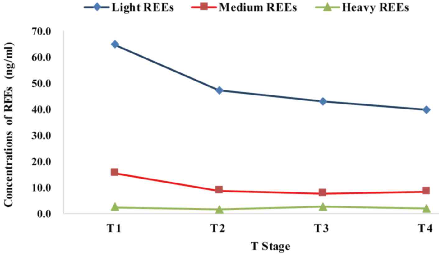
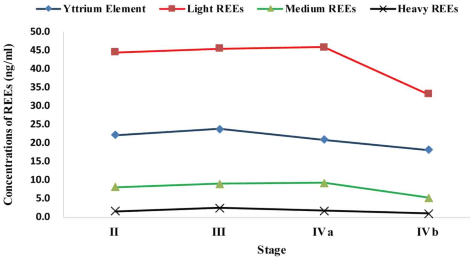
Trends in REE concentrations at the
tumor (T) stage of NPC
The mean concentrations of REE at the T stage of NPC
are demonstrated in Table III,
grouped according to light, medium and heavy elements. Light and
medium REE each exhibited a decreasing trend, while heavy elements
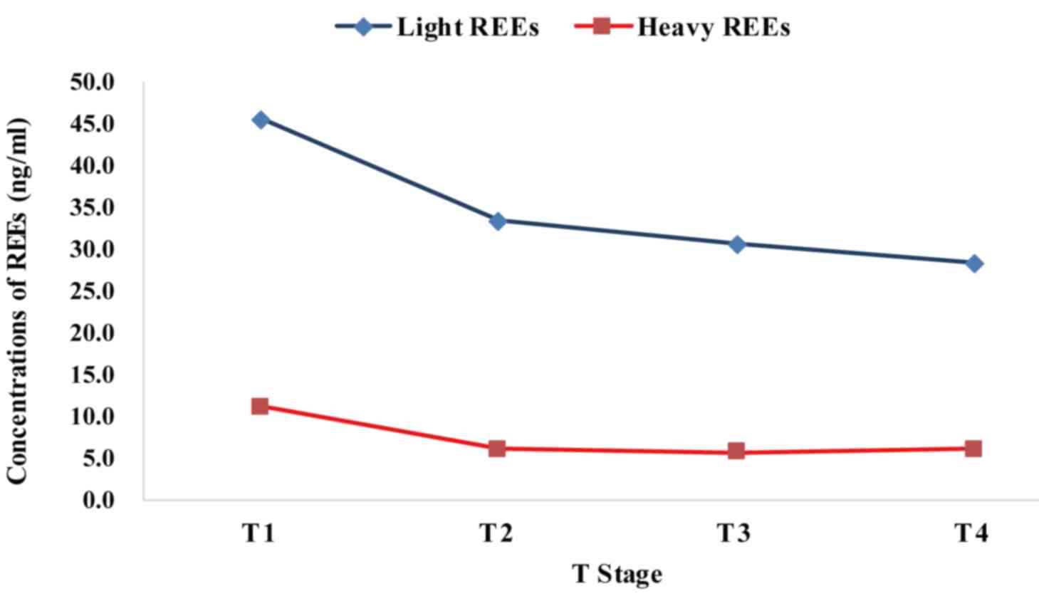
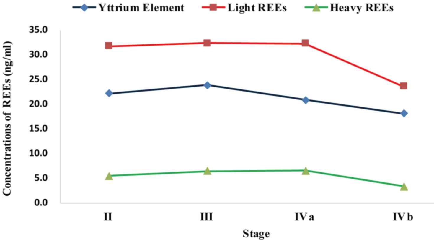
demonstrated a periodically changing trend (Fig. 1). When divided into only light and
heavy REE, both groups showed gradually decreasing trends (Fig. 2).
| Table III.Mean concentrations of light, medium
and heavy REE, or light vs. heavy REE at the T stage of NPC
(ng/g). |
Table III.
Mean concentrations of light, medium
and heavy REE, or light vs. heavy REE at the T stage of NPC
(ng/g).
|
| Method 1 | Method 2 |
|---|
|
|
|
|
|---|
| T stage | Light REE | Medium REE | Heavy REE | Light REE | Heavy REE |
|---|
| T1 | 64.72 | 15.46 | 2.36 | 45.48 | 11.03 |
| T2 | 47.27 | 8.74 | 1.49 | 33.29 | 5.97 |
| T3 | 42.87 | 7.75 | 2.57 | 30.55 | 5.59 |
| T4 | 39.79 | 8.52 | 1.93 | 28.24 | 6.02 |
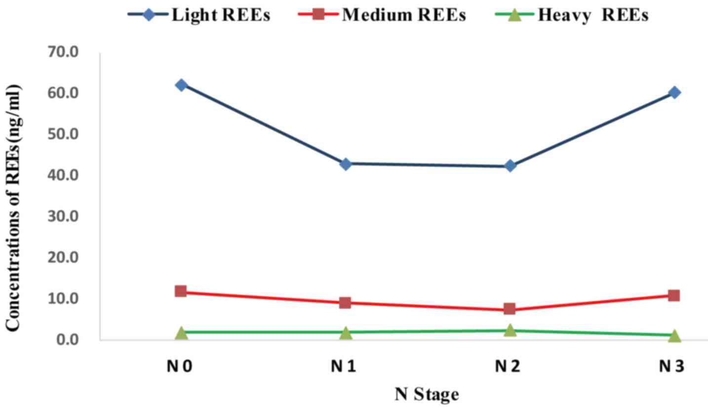
Trends in REE concentrations at the
node (N) stage of NPC
The mean concentrations of REE at the N stage of NPC
are listed in Table IV. When grouped
as light, medium, and heavy elements, no groups exhibited
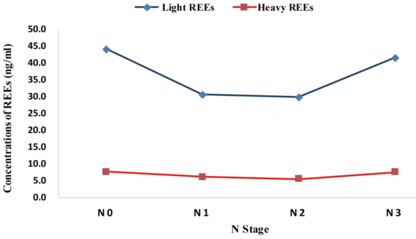
significant trends (Fig. 3). However,
when grouped according to light and heavy elements, each group
exhibited a decreasing trend from stage N0 to N1, remained steady
from stage N1 to N2, and increased from stage N2 to N3 (Fig. 4).
| Table IV.Mean concentrations of light, medium,
and heavy or light vs. heavy REE at the N stage of NPC (ng/g). |
Table IV.
Mean concentrations of light, medium,
and heavy or light vs. heavy REE at the N stage of NPC (ng/g).
|
| Method 1 | Method 2 |
|---|
|
|
|
|
|---|
| N stage | Light REE | Medium REE | Heavy REE | Light REE | Heavy REE |
|---|
| N0 | 62.18 | 11.76 | 1.91 | 44.18 | 7.73 |
| N1 | 42.86 | 9.12 | 1.94 | 30.65 | 6.23 |
| N2 | 42.40 | 7.48 | 2.42 | 29.89 | 5.58 |
| N3 | 60.22 | 10.82 | 1.27 | 41.60 | 7.66 |
Trends in REE concentrations at each
clinical stage of NPC
The mean concentrations of REE at each clinical
stage of NPC are presented in Table
V. When REE were classified as light, medium and heavy, the
concentrations of light and medium REE gradually increased from
stage II to IVa, and then started to decrease. Decreasing trends in
Y and heavy REE began earlier; from stage IVa to IVb, the absolute
value of the concentration of Y was decreased by 13.30%, and the
concentrations of light, medium and heavy REEs was decreased by
27.78, 42.62 and 41.32%, respectively. The change in the
concentrations of light REE was the largest, while the decrease in
medium REE concentrations was the greatest, followed by that of
heavy REE; the change in Y was the smallest. Overall, light, middle
and heavy REE changed by 27.78, 42.62 and 59.25%, respectively,
between their peaks and lowest points at stage IVb. The changes in
the concentrations of light REE and Y were relatively minor
compared with the greater shifts in heavy REE, which were observed
with disease progression. Medium REE also indicated prominent
changes, but not until the late stages of NPC, while the changes in
heavy REE were already notable at moderate disease stages (Fig. 5).
| Table V.Mean concentrations of light, medium
and heavy REE, or light vs. heavy REE at each clinical stage of NPC
(ng/g). |
Table V.
Mean concentrations of light, medium
and heavy REE, or light vs. heavy REE at each clinical stage of NPC
(ng/g).
|
|
| Method 1 | Method 2 |
|---|
|
|
|
|
|
|---|
| TNM stage | Y | Light REE | Medium REE | Heavy REE | Light REE | Heavy REE |
|---|
| II | 22.1400 | 44.5306 | 8.1771 | 1.6255 | 31.7084 | 5.4296 |
| III | 23.8560 | 45.5145 | 8.9385 | 2.4962 | 32.3540 | 6.4212 |
| IVa | 20.8853 | 45.9175 | 9.2184 | 1.7335 | 32.2465 | 6.5207 |
| IVb | 18.1078 | 33.1638 | 5.2899 | 1.0173 | 23.6012 | 3.3571 |
When grouped as light vs. heavy REE, the
concentrations of light REE in the tissues of the patients with NPC
were largely consistent across different stages, with a slight
increase from stage II to III, no major change from stage III to
IVa and a strong decrease from stage IVa to IVb (26.81%). Heavy REE
exhibited an even higher decrease of 48.52% from stage IVa to IVb
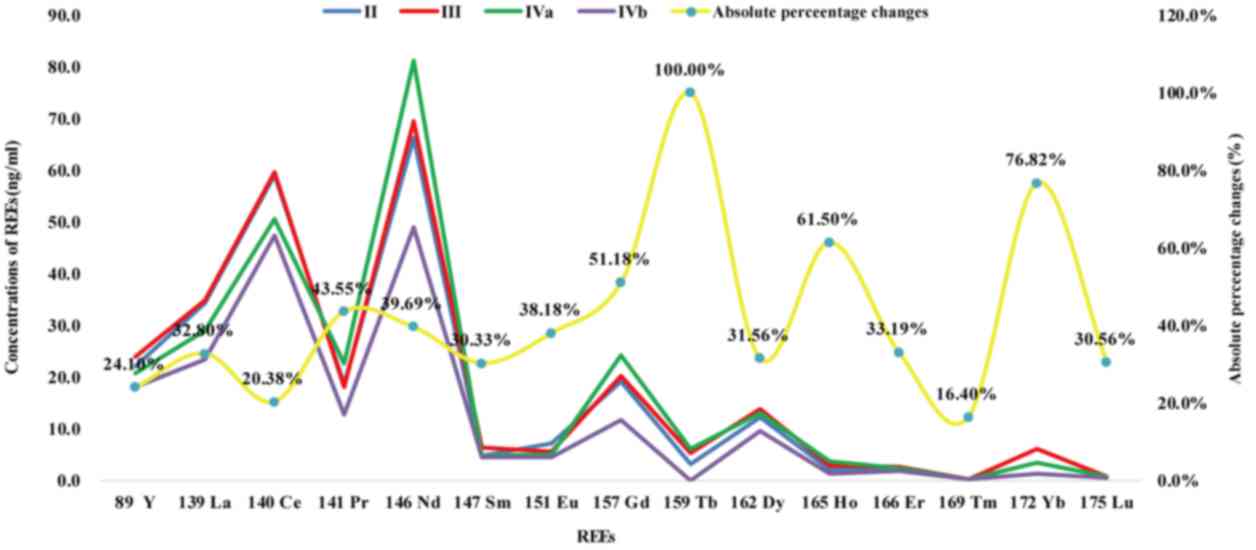
(Fig. 6). The variation in the
absolute values of light REE was the highest, though substantial
variation was also observed for medium and heavy REE. The elements
with the greatest change were terbium (Tb), Ho and ytterbium (Yb)
(Fig. 7).
Correlations of REE with sex, age and
EBV in NPC
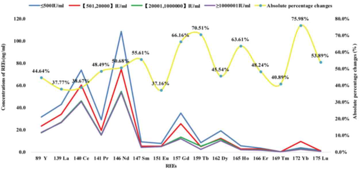
Non-parametric tests demonstrated that the
concentrations of REE in patients with NPC were not associated with
sex (r=0.301, P=0.106) or age (r=−0.011, P=0.955). However, there
was a strong negative correlation between the concentrations of REE
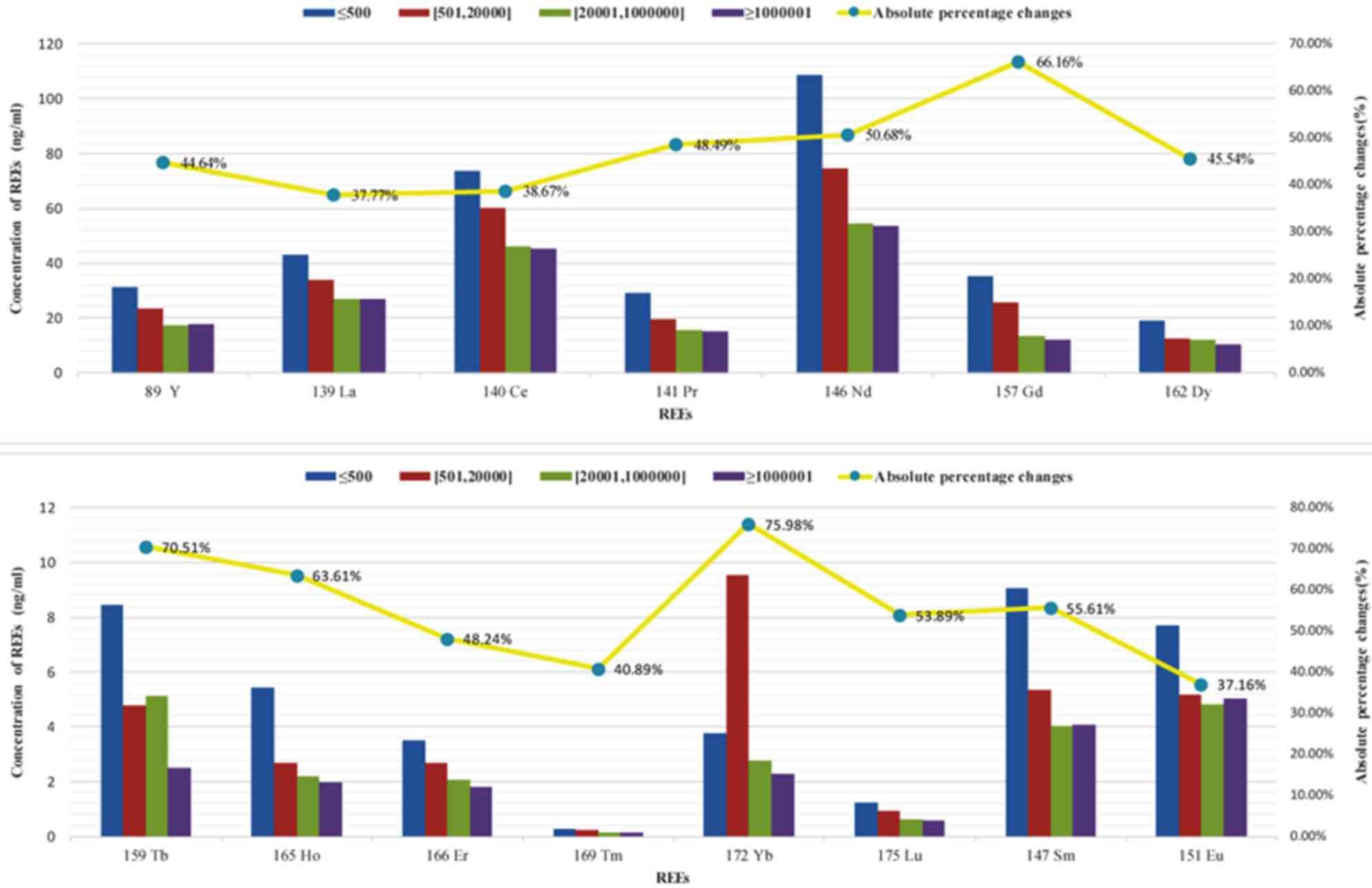
and the EBV copy number (r=−0.744, P<0.001; Table VI). With increasing levels of EBV,
the total concentration of REE demonstrated a decreasing trend, and
the majority of individual REE gradually decreased in
concentration. Judging by the percentage of variation, medium and
heavy elements indicated the greatest changes. Thus, the
association of EBV with REE changes in patients with NPC was more
apparent for the medium and heavy elements (Figs. 8 and 9).
| Table VI.Associations between REE, sex, age
and EBV in NPC. |
Table VI.
Associations between REE, sex, age
and EBV in NPC.
| Type | n | r | P-value |
|---|
| Sex |
|
|
|
|
Male | 17 |
|
|
|
Female | 13 | 0.301 | 0.106 |
| Age (years) |
|
|
|
|
23–37 | 2 |
|
|
|
37–51 | 16 |
|
|
|
51–65 | 7 |
|
|
|
65–79 | 5 | −0.011 | 0.955 |
| EBV (IU/ml) |
|
|
|
|
≤500 | 7 |
|
|
|
501–20,000 | 8 |
|
|
|
20,001–1,000,000 | 6 |
|
|
|
≥1,000,000 | 9 | −0.744 | <0.001 |
Discussion
The potentially toxic effects of REE on human health
are a focus of numerous recent studies (10,11). REE
have various biological activities, affecting cell proliferation
and growth through changes in ATPase activity on the cell surface,
intracellular and extracellular ion exchange, cell division and DNA
synthesis (12–15). Their functions in tumors are
particularly prominent.
REE are closely associated with the occurrence and
development of tumors. In 1971, Schroeder and Mitchener (2) confirmed that
Y(NO3)3 water containing heavy REE had
carcinogenic effects in rats. REE can cause lipid peroxidation
injury, which can induce DNA base damage and strand breakage as
well as the production of numerous types of fluorescent substances
(16,17). The end product, malondialdehyde, can
form DNA adducts, which further causes mutations and tumorigenesis
(10,18). In addition, the process of lipid
peroxidation produces a variety of active free radicals, which have
mutagenic effects (19). However,
some studies have also revealed that REE can have antitumor
effects. Giri et al (20)
demonstrated that the intraperitoneal injection of 0.1 mg/kg cerium
oxide nanoparticles into mice implanted with A2780 ovarian cancer
cells significantly inhibited the growth of the implanted tumor. Su
et al (21) indicated that
treatment with lanthanum citrate for 48 h induced anoikis of HeLa
cells, thus inhibiting tumor growth. The present study determined
that the concentrations of certain REE, including Gd, Tb and Yb,
changed notably with the progression of NPC, while others,
including Y, La and cerium, exhibited only small changes. The
concentrations of REE increased from stage II to IVa and then
greatly decreased from IVa to IVb. Notably, the concentrations of
medium and heavy REE decreased with disease progression.
EBV is closely associated with the occurrence and
development of NPC (22,23). The initial report by Mutirangura et
al (24) in 1998 revealed that
EBV DNA copy number was significantly higher in the peripheral
blood of patients with NPC than in individuals without the disease;
since then, a large amount of evidence has demonstrated that higher
plasma levels of EBV DNA are associated with more advanced clinical
stages and more extensive lymph node metastases (25–27).
Therefore, plasma EBV DNA not only serves as a useful marker for
the screening and early diagnosis of NPC but also has significance
in clinical staging, prognosis determination, and the monitoring of
recurrence and distant metastasis in patients with NPC (28–30). This
present study supports prior studies that indicated that EBV copy
number increased with advancements in clinical stage. In addition
to this increase in the EBV copy number, the concentrations of all
REE demonstrated a decreasing trend with progression of the
disease. The majority of individual REE also gradually decreased,
and their concentrations indicated a strong negative correlation
with EBV levels. The greatest proportional changes were observed
with the medium and heavy REE. The association of EBV with the
changes in REE in patients with NPC was clearest between the medium
and heavy elements. Therefore, it is speculated that changes in the
concentrations of medium and heavy REE may affect the occurrence
and development of NPC.
Chemical carcinogens can enhance EBV activation,
change gene expression in NPC patients and induce tumors (31). EBV infection, chemical carcinogens and
the interaction between these factors could all serve important
roles in the development of NPC (32,33). A
previous study (34) proposed that
REE have dual, dose-dependent effects on cellular physiology, with
high doses exerting inhibitory effects on cell growth and low doses
promoting growth. However, this pattern varies across the different
types of REE, inverting completely in certain cases, having more
prominent effects in tumors. The present study found that compared
with the mean concentration and distribution values of REE in the
chondrite; NPC tissues demonstrated a relative loss of heavy REE
and an enrichment of light REE. Throughout the development and
progression of NPC, heavy REE varied the most, with reductions in
concentration ≤41.32–48.52%. Elucidation of the mechanism
underlying this loss of heavy REE and the enrichment of light REEs
in NPC will require further animal experiments and clinical
studies.
The present study did not include a non-NPC control
group. A survey was performed on the family members of patients
with NPC and also members of the public prior to performing the
study and determined that healthy family members and the public
would not agree to have their nasopharyngeal tissues collected.
Collection of normal nasopharyngeal tissue samples involves
significant trauma and pain and carries a risk of massive bleeding.
The Ethics Committee also rejected the proposal for setting up a
control group following careful consideration. The results in the
current study may also have certain biases. First, the overall
sample size was small. During the calculation of the mean
concentrations of all REE in NPC tissues and the overall
deviations, samples were repeatedly used, and the 95% confidence
interval method was used to expand the number of samples for
prediction of the overall mean value. Second, non-tumor tissues may
have inadvertently been included when the diagnosed nodular or
cauliflower-type NPC tissues were collected using an electronic
nasopharyngoscope for REE detection. Third, to reduce bleeding,
non-tumor tissues and deep-layer tissues were not collected. As a
result, the collected tissues were small, and specimen processing
may have introduced errors. In addition, despite being able to
determine EBV levels in serum, due to the limitation of the NPC
tissue specimens, EBV levels could not be determined in tumor
tissues. We did not determine EBV levels in the tumor tissues.
Although the present study did not completely
elucidate the associations between the occurrence and development
of NPC, and changes in the distribution and concentration of REE in
NPC tissues, the results provide insight for further studies on
this topic, including studies on the correlation of NPC with REE
and EBV.
In conclusion, the concentrations of REE in NPC
tissues were not associated with sex or age, but gradually
increased between stages II and IVa prior to significantly
decreasing following stage IVa. By contrast, the EBV copy number
increased with advanced clinical stages. Changes in the
concentrations of REE in NPC were more prominent for medium and
heavy elements. Shifts in the concentrations of heavy REE may
affect the occurrence and development of NPC.
Acknowledgements
The present study was supported by the Natural
Science Foundation of Jiangxi Province (grant no.
20122BAB205048).
Glossary
Abbreviations
Abbreviations:
|
NPC
|
nasopharyngeal carcinoma
|
|
REE
|
rare earth elements
|
|
EBV
|
Epstein-Barr virus
|
|
ICP-MS/MS
|
inductively coupled plasma-tandem mass
spectrometry
|
|
RT-qPCR
|
reverse transcription-quantitative
polymerase chain reaction
|
|
SD
|
standard deviation
|
|
T
|
tumor
|
|
N
|
node
|
|
Y
|
yttrium
|
|
La
|
lanthanum
|
|
Lu
|
lutetium
|
|
Yb
|
ytterbium
|
|
Tb
|
terbium
|
References
|
1
|
Liu HP: A discussion of the development of
rare earth industry of Ganzhou. Nonferrous Metals Science and
Engineering. 101–104. 2012.(In Chinese).
|
|
2
|
Xu ST: Study on the problems and
countermeasures of Ganzhou sustainable development of rare earth
industry. J Jiangxi Univ Sci Technol. 47–51. 2014.(In Chinese).
|
|
3
|
Chen Z, Liu Y, Cheng W, Zhang L, Li H and
Wang Y: The environmental toxicology of rare earth elements
(147Pm, 141Ce, 147Nd) and their
safety elvaluation in environment. China Nuclear Sci Technol Rep.
33:2001.(In Chinese).
|
|
4
|
Ohnishi K, Usuda K, Nakayama S, Sugiura Y,
Kitamura Y, Kurita A, Tsuda Y, Kimura M and Kono K: Distribution,
elimination, and renal effects of single oral doses of europium in
rats. Biol Trace Elem Res. 143:1054–1063. 2011. View Article : Google Scholar : PubMed/NCBI
|
|
5
|
Guo ZY, Zhang YS and Zhang HJ: Pathway,
distribution, metabolism and toxicity of exogenous rare earth in
animals. Chin Rare Earths. 61–64. 1997.(In Chinese).
|
|
6
|
Schroeder HA and Mitchener M: Scandium,
chromium(VI), gallium, yttrium, rhodium, palladium, indium in mice:
Effects on growth and life span. J Nutr. 101:1431–1437. 1971.
View Article : Google Scholar : PubMed/NCBI
|
|
7
|
Smith JB and Smith L: Initiation of DNA
synthesis in quiescent Swiss 3T3 and 3T6 cells by lanthanum. Biosci
Rep. 4:777–782. 1984. View Article : Google Scholar : PubMed/NCBI
|
|
8
|
Lo YM, Chan LY, Lo KW, Leung SF, Zhang J,
Chan AT, Lee JC, Hjelm NM, Johnson PJ and Huang DP: Quantitative
analysis of cell-free Epstein-Barr virus DNA in plasma of patients
with nasopharyngeal carcinoma. Cancer Res. 59:1188–1191.
1999.PubMed/NCBI
|
|
9
|
Shotelersuk K, Khorprasert C, Sakdikul S,
Pornthanakasem W, Voravud N and Mutirangura A: Epstein-Barr virus
DNA in serum/plasma as a tumor marker for nasopharyngeal cancer.
Clin Cancer Res. 6:1046–1051. 2000.PubMed/NCBI
|
|
10
|
Sojka B, Kuricova M, Liskova A, Bartusova
M, Banski M, Misiewicz J, Dusinska M, Horvathova M, Jahnova E,
Ilavska S, et al: Hydrophobic sodium fluoride-based nanocrystals
doped with lanthanide ions: Assessment of in vitro toxicity to
human blood lymphocytes and phagocytes. J Appl Toxicol.
34:1220–1225. 2014. View
Article : Google Scholar : PubMed/NCBI
|
|
11
|
Pagano G, Guida M, Tommasi F and Oral R:
Health effects and toxicity mechanisms of rare earth
elements-Knowledge gaps and research prospects. Ecotoxicol Environ
Saf. 115:40–48. 2015. View Article : Google Scholar : PubMed/NCBI
|
|
12
|
Cao R, Huang XH and Zhou Q: Research on
Molecular Biomarker of Rare Earths Ions Ecotoxicity. System Sci
Comprehensive Studies Agriculture. 2007.
|
|
13
|
Fernando KC and Barritt GJ:
Characterisation of the divalent cation channels of the hepatocyte
plasma membrane receptor-activated Ca2+ inflow system using
lanthanide ions. Biochim Biophys Acta. 1268:97–106. 1995.
View Article : Google Scholar : PubMed/NCBI
|
|
14
|
Matsumoto Y and Komiyama M: DNA hydrolysis
by rare-earth metal ions. Nucleic Acids Symp Ser. 1–34. 1992.
|
|
15
|
Leuratti C, Singh R, Lagneau C, Farmer PB,
Plastaras JP, Marnett LJ and Shuker DE: Determination of
malondialdehyde-induced DNA damage in human tissues using an
immunoslot blot assay. Carcinogenesis. 19:1919–1924. 1998.
View Article : Google Scholar : PubMed/NCBI
|
|
16
|
Leuratti C, Singh R, Lagneau C, Farmer PB,
Plastaras JP, Marnett LJ and Shuker DE: Determination of
malondialdehyde-induced DNA damage in human tissues using an
immunoslot blot assay. Carcinogenesis. 19:1919–1924. 1998.
View Article : Google Scholar : PubMed/NCBI
|
|
17
|
Mittal S and Pandey AK: Cerium oxide
nanoparticles induced toxicity in human lung cells: Role of ROS
mediated DNA damage and apoptosis. Biomed Res Int. 2014:8919342014.
View Article : Google Scholar : PubMed/NCBI
|
|
18
|
Huang PJ, Vazin M, Matuszek Ż and Liu J: A
new heavy lanthanide-dependent DNAzyme displaying strong metal
cooperativity and unrescuable phosphorothioate effect. Nucleic
Acids Res. 43:461–469. 2015. View Article : Google Scholar : PubMed/NCBI
|
|
19
|
Lin WT, Huang PJ, Pautler R and Liu J: The
group trend of lanthanides binding to DNA and DNAzymes with a
complex but symmetric pattern. Chem Commun (Camb). 50:11859–11862.
2014. View Article : Google Scholar : PubMed/NCBI
|
|
20
|
Giri S, Karakoti A, Graham RP, Maguire JL,
Reilly CM, Seal S, Rattan R and Shridhar V: Nanoceria: A Rare-earth
nanoparticle as a novel Anti-angiogenic therapeutic agent in
ovarian cancer. PLoS One. 8:e545782013. View Article : Google Scholar : PubMed/NCBI
|
|
21
|
Su X, Zheng X and Ni J: Lanthanum citrate
induces anoikis of Hela cells. Cancer Lett. 285:200–209. 2009.
View Article : Google Scholar : PubMed/NCBI
|
|
22
|
Perri F, Della Vittoria Scarpati G,
Giuliano M, D'Aniello C, Gnoni A, Cavaliere C, Licchetta A and
Pisconti S: Epstein-Barr virus infection and nasopharyngeal
carcinoma: The other side of the coin. Anticancer Drugs.
26:1017–1025. 2015. View Article : Google Scholar : PubMed/NCBI
|
|
23
|
Lim CS, Goh SL, Kariapper L, Krishnan G,
Lim YY and Ng CC: Inclusion bodies of recombinant Epstein-Barr
virus capsid antigen p18 as potential immobilized antigens in
enzyme immunoassays for detection of nasopharyngeal carcinoma. Clin
Chim Acta. 448:206–210. 2015. View Article : Google Scholar : PubMed/NCBI
|
|
24
|
Mutirangura A, Pornthanakasem W,
Theamboonlers A, Sriuranpong V, Lertsanguansinchi P, Yenrudi S,
Voravud N, Supiyaphun P and Poovorawan Y: Epstein-Barr viral DNA in
serum of patients with nasopharyngeal carcinoma. Clin Cancer Res.
4:665–669. 1998.PubMed/NCBI
|
|
25
|
Xu J, Wan XB, Huang XF, Chan KC, Hong MH,
Wang LH, Long ZJ, Liu Q, Yan M, Lo YM, et al: Serologic antienzyme
rate of Epstein-Barr virus DNase-specific neutralizing antibody
segregates TNM classification in nasopharyngeal carcinoma. J Clin
Oncol. 28:5202–5209. 2010. View Article : Google Scholar : PubMed/NCBI
|
|
26
|
Li Y, Wang K, Yin SK, Zheng HL and Min DL:
Expression of Epstein-Barr virus antibodies EA-IgG, Rta-IgG, and
VCA-IgA in nasopharyngeal carcinoma and their use in a combined
diagnostic assay. Genet Mol Res. 15:2016.
|
|
27
|
Tiwawech D, Srivatanakul P, Karaluk A and
Ishida T: Significance of plasma IgA and IgG antibodies to
Epstein-Barr virus early and viral capsid antigens in Thai
nasopharyngeal carcinoma. Asian Pac J Cancer Prev. 4:113–118.
2003.PubMed/NCBI
|
|
28
|
Gourzones C, Gelin A, Bombik I, Klibi J,
Vérillaud B, Guigay J, Lang P, Témam S, Schneider V, Amiel C, et
al: Extra-cellular release and blood diffusion of BART viral
micro-RNAs produced by EBV-infected nasopharyngeal carcinoma cells.
Virol J. 7:2712010. View Article : Google Scholar : PubMed/NCBI
|
|
29
|
Yip TT, Ngan RK, Fong AH and Law SC:
Application of circulating plasma/serum EBV DNA in the clinical
management of nasopharyngeal carcinoma. Oral Oncol. 50:527–538.
2014. View Article : Google Scholar : PubMed/NCBI
|
|
30
|
Leung SF, Chan KC, Ma BB, Hui EP, Mo F,
Chow KC, Leung L, Chu KW, Zee B, Lo YM and Chan AT: Plasma
Epstein-Barr viral DNA load at midpoint of radiotherapy course
predicts outcome in advanced-stage nasopharyngeal carcinoma. Ann
Oncol. 25:1204–1208. 2014. View Article : Google Scholar : PubMed/NCBI
|
|
31
|
Tan CH: Epstein-Barr virus, chemical
carcinogens and nasopharyngeal carcinoma. Practical J Cancer.
58–59. 1992.(In Chinese).
|
|
32
|
Fang CY, Huang SY, Wu CC, Hsu HY, Chou SP,
Tsai CH, Chang Y, Takada K and Chen JY: The synergistic effect of
chemical carcinogens enhances Epstein-Barr virus reactivation and
tumor progression of nasopharyngeal carcinoma cells. PLoS One.
7:e448102012. View Article : Google Scholar : PubMed/NCBI
|
|
33
|
Fang CY, Lee CH, Wu CC, Chang YT, Yu SL,
Chou SP, Huang PT, Chen CL, Hou JW, Chang Y, et al: Recurrent
chemical reactivations of EBV promotes genome instability and
enhances tumor progression of nasopharyngeal carcinoma cells. Int J
Cancer. 124:2016–2025. 2009. View Article : Google Scholar : PubMed/NCBI
|
|
34
|
Bhakta-Guha D and Efferth T: Hormesis:
Decoding two sides of the same coin. Pharmaceuticals (Basel).
8:865–883. 2015. View Article : Google Scholar : PubMed/NCBI
|