Introduction
Prostate cancer is the most commonly diagnosed
malignancy in men in western countries (1). As the second most common cause for
cancer-associated mortality among men, prostate cancer caused
~27,540 deaths in the United States (2015) (2). In addition, the incidence and mortality
for prostate cancer have been evidently increasing in Asia,
including in China, in recent years (3).
Although novel therapies for prostate cancer with
proven survival benefits have been developed relatively recently
(4,5),
the overall increase in survival rate has been negligible. A major
clinical challenge in prostate cancer is the insufficient power of
the currently available diagnostic tests. Since the introduction of
serum prostate specific antigen (PSA) screening >30 years ago,
prostate cancer diagnosis and management have been guided by this
biomarker; it remains the most commonly used tumor marker for
prostate cancer diagnosis, postoperative monitoring and prognosis
evaluation. PSA is a protein secreted by the epithelial cells of
the prostate; an increase in serum PSA is often identified in
prostate cancer patients (6,7). However, PSA is susceptible to various
confounding factors, including benign prostatic hyperplasia,
prostatitis and urethral surgery, which may influence the clinical
reliability (8,9). The deficiencies of PSA, including the
lack of specificity and sensitivity, may lead to false-positive or
false-negative results. In view of this, identifying a biomarker
with an improved diagnostic and prognostic potential for prostate
cancer assessment may be of great significance.
Refinements to PSA measurements have been proposed,
including early PSA, benign PSA, free PSA, dynamic PSA parameters
(including PSA velocity and PSA doubling time), PSA density (PSA to
prostate volume ratio) and age-specific PSA level (1). Although these alternative applications
of PSA may improve the diagnosis accuracy of prostate cancer to a
certain extent, their relatively complicated implementation and the
same confounding issues as usually affect tPSA continue to limit
their application (10).
The proteasome is a multicatalytic proteinase
complex responsible for the degradation of the majority of
intracellular proteins, including the proteins required for cell
cycle regulation and apoptosis (11).
The 26S proteasome is comprised of two 19S regulatory subunits and
a 20S core. In the proteasome degradation system, the target
protein is recognized by the 19S subunits following ubiquitination,
and can then access the 20S core for further degradation. The 20S
subunit is a multicatalytic threonine protease with three types of
enzymatic activity, described as chymotrypsin-like, trypsin-like
and caspase-like activities. As part of the ubiquitin-proteasome
pathway, the proteasome plays a vital role in the degradation of
proteins from a broad range of cellular pathways, and contributes
to the pathology of a number of human diseases, including cancer,
in which regulatory proteins may be stabilized due to decreased
degradation, or lost due to accelerated degradation (12). The proteasome degrades a range of
endogenous proteins associated with cancer, including transcription
factors, cyclins, Bcl-2-associated X (Bax), p53, p27 and inhibitor
of NFκB-α (IκB-α), and it has become an important potential target
for cancer therapy.
The majority of studies regarding this topic have
concentrated on the proteasome inhibitor for tumor treatment;
research on the proteasome for disease diagnosis is insufficient.
Stoebner et al (13) reported
that in 20 tumor patients (including those with breast, gastric,
kidney, colon, testicular, liver and lung cancer), the proteasome
20S serum level was significantly elevated compared with controls,
indicating serum proteasome could be applied in tumor diagnostics.
Our previous study demonstrated that a proteasome inhibitor could
affect the proliferation and apoptosis of prostate cancer cells by
inhibiting chymotrypsin-like activity, thus influencing the
expression of the target proteins IκB-α, Bax and p27 (14). Therefore, in the present study, the
chymotrypsin-like, trypsin-like and caspase-like proteasomal
activity in cultured LnCaP cells and tumor-bearing nude mice was
assessed, in addition to the expression of the proteasomal
substrates IκB-α, Bax and p27, in order to analyze the feasibility
of proteasomal activity as a candidate biomarker for prostate
cancer.
Materials and methods
Materials
LNCaP human prostate cells were provided by Ryder
Guanzhou Lian Kang Biological Technology Co., Ltd. (Guangzhou,
China). A Prostate Epithelial Cell Medium BulletKit™ was purchased
from Lonza Group, Ltd. (Basel, Switzerland). The keratinocyte-serum
free medium was purchased from Gibco (Thermo Fisher Scientific,
Inc., Waltham, MA, USA) and PBS was from Hyclone (GE Healthcare
Life Sciences, Logan, UT, USA). RPMI-1640, penicillin and
streptomycin were purchased from Invitrogen (Thermo Fisher
Scientific, Inc.). Fetal bovine serum (FBS) was purchased from
Hyclone (GE Healthcare Life Sciences). Mouse monoclonal antibodies
against Bax (cat. no. sc-23959) and p27 (cat. no. sc-1641), and
rabbit polyclonal antibodies against inhibitor of nuclear factor
(NF)-κB-α (IκB-α; cat. no. sc-203) and GAPDH (FL-335; cat. no.
sc-25778) were both from Santa Cruz Biotechnology, Inc. (Dallas,
TX, USA). Suc-LLVY-AMC, Z-LLE-NA, and BZVGR-AMC was all from
Sigma-Aldrich; Merck KGaA (Darmstadt, Germany).
Cell culture
LNCaP cells were grown in RPMI-1640 supplemented
with 10% FBS, 100 U/ml of penicillin and 100 µg/ml of streptomycin,
and were maintained at 37°C and 5% CO2 for 1 week.
Prostate epithelial cells were obtained from prostate tissue by
conventional tissue culture methods: A human prostate tissue
specimen was obtained from men undergoing robotic radical
prostatectomy, and was sliced into 1 mm3 blocks and
placed in a T25 flask coated with collagen. The tissue blocks were
cultured at 37°C with 5% CO2 for ~1 week in prostate
epithelial cell medium, and the medium was changed every 3 days.
The cells were digested and collected when the primary monolayer
cells covered the surface of the flask, and they were passaged with
keratinocyte-serum free medium. The first generation of cells were
used for further experiments to ensure the integrity of the
prostate epithelial cells. Cells were observed for morphological
changes and photographed under a phase contrast inverted microscope
(magnification, ×100; Olympus Corporation, Tokyo, Japan). The use
of human tissue was approved by the Research Ethics Committee of
Guangzhou First People's Hospital, Guangzhou Medical University
(Guangdong, China), and written informed consent from the patient
was acquired prior to the use of the tissue in research.
Human prostate tumor xenograft
experiments
Animal experiments in the present study were
performed in accordance with the guidelines outlined by the
Institute for Laboratory Animal Research in Guangzhou Medical
University (Guangzhou, China). A total of 30 male BALB/C-nu mice
aged 4–6 weeks, (mean body weight, 20 g) were purchased from
Guangdong Medical Laboratory Animal Center (Foshan, China) and
housed in accordance to a protocol described previously (15). Mice were randomly divided into two
groups (15 mice/group). In the experimental group, 5×10⁵
LNCaP cells suspended in 0.2 ml PBS were inoculated into the left
flank of each mouse. Mice in the control group were injected with
0.2 ml PBS. Tumor sizes were measured every 3 days using calipers,
and tumor volumes were calculated according to the standard
formula: Width2 × length × 0.52. At 4 weeks, the mice
were sacrificed. Blood and tumor tissues were taken for further
analysis.
Proteasome activities assay
100 µl suspension LNCaP cells (1.0×10⁵
cells/ml) and prostate epithelial cells (1.0×10⁵
cells/ml) were plated in a 96-well plate and cultured for 24 h.
Then 1 µl of proteasome activity assay buffer containing 4 mM a
fluorogenic peptide substrate, including Suc-LLVY-AMC for detecting
chymotrypsin-like activity, Z-LLE-NA for detecting caspase-like
activity or BZVGR-AMC for detecting trypsin-like activity, was
added to the wells. Following a 2 h incubation, the fluorescence
intensity was measured by a microplate reader with the excitation
wavelength of 380 nm and the emission wavelength of 460 nm.
For the serum assay, 1 µl containing 4 mM of a
fluorescent substrate was added to 100 µl of mouse serum and
incubated at 37°C for 2 h. The proteasome activity was then
detected as the fluorescence intensity by the microplate reader,
with the excitation and emission wavelengths of 380 and 460 nm,
respectively.
Western blot analysis
Cells were harvested, washed with PBS twice and
lysed in cell lysis buffer (50 mM Tris-HCl; 150 mM NaCl; 1 mM EDTA;
1% Triton X-100; 0.5% Na-deoxycholate; 0.1% SDS; 1 mM PMSF; 10
µl/ml protease inhibitor cocktail P8340 provided by Sigma-Aldrich;
Merck KGaA) for 30 min at 4°C. Then the cells were scraped off and
transferred into a 1.5 ml centrifuge tube. The lysates were
centrifuged at 14,000 × g for 5 min at 4°C. The DC™ Protein Assay
kit II (cat. no. 5000112; Bio-Rad Laboratories, Inc., Hercules, CA,
USA) was used to determine the protein concentration with the
Bradford assay method, as follows: Standards from 0 to 4 mg/ml
protein were prepared; 20 µl of reagent S from the kit was added to
1 ml reagent A and mixed thoroughly. Then 5 µl of each standard and
sample solution was added into 25 µl of the S/A mixture in separate
microtiter plate wells. Next, 200 µl of reagent B was added, and
the wells were mixed thoroughly using a micro plate mixer.
Following incubation at room temperature for 15 min, the absorbance
was measured at 750 nm in a plate reader. Then 50 µg of cell
lysates were separated using SDS-PAGE (10% gel) and
electrophoretically transferred to a PVDF membrane, followed by
western blotting using the aforementioned specific antibodies to
IκB-α, Bax, p27 and GAPDH, as previously described (16). Images were then visualized using an
enhanced chemiluminescence kit (cat. no. 32106; Pierce, Rockford,
IL USA) and quantified using ImageJ software (version 1.48;
National Institutes of Health, Bethesda, MD, USA). Benign gross
prostate tissues from the mice in the control group were obtained,
as described by a previous protocol (17). Western blotting using the tumor and
benign prostate tissue samples from the mice was then performed as
for the cultured cancer cells.
Statistical analysis
SPSS (version 18; SPSS, Inc., Chicago, IL, USA) was
used for data analysis. Student's t-test was applied to evaluate
the differences between the experimental and control groups. Data
were expressed as the mean ± standard deviation (SD), and results
from at least three independent experiments were used for
statistical analysis. All statistical tests were two-sided.
P<0.01 was considered to indicate a statistically significant
difference.
Results
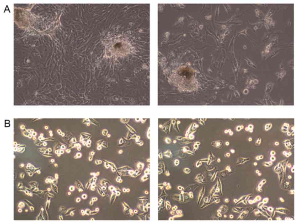
Isolation of prostate epithelial cells
from prostate tissues
Prostate epithelial cells were obtained from
prostate tissue by conventional tissue culture methods. On
observation, cells were identified to be well adhered to the
collagen surface with a good growth state (Fig. 1A). The prostate epithelial cells
exhibited epithelium-like morphology and enhanced cell viability,
overspreading the bottom of the flask at the 16th day. Subsequent
to the formation of a cell monolayer, cells were passaged with
Keratinocyte-SFM medium (Fig. 1B).
The first generation of cells was used for further experiment.
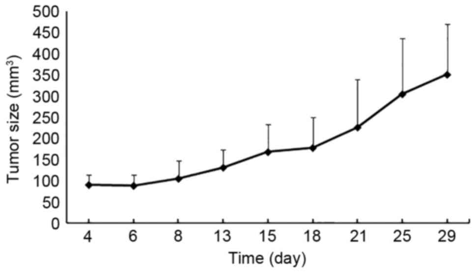
Establishment of a prostate cancer
model in mice
At 4 days after the inoculation of LNCaP cells into
nude mice, the formation of tumors started to be observed. At the
end of the experiment (day 29), the mean size of the tumors was 350
mm3 (Fig. 2). The mice
were then sacrificed, and blood and tumor tissues were collected
for further assays. Thus, a mouse model for prostate cancer was
established.
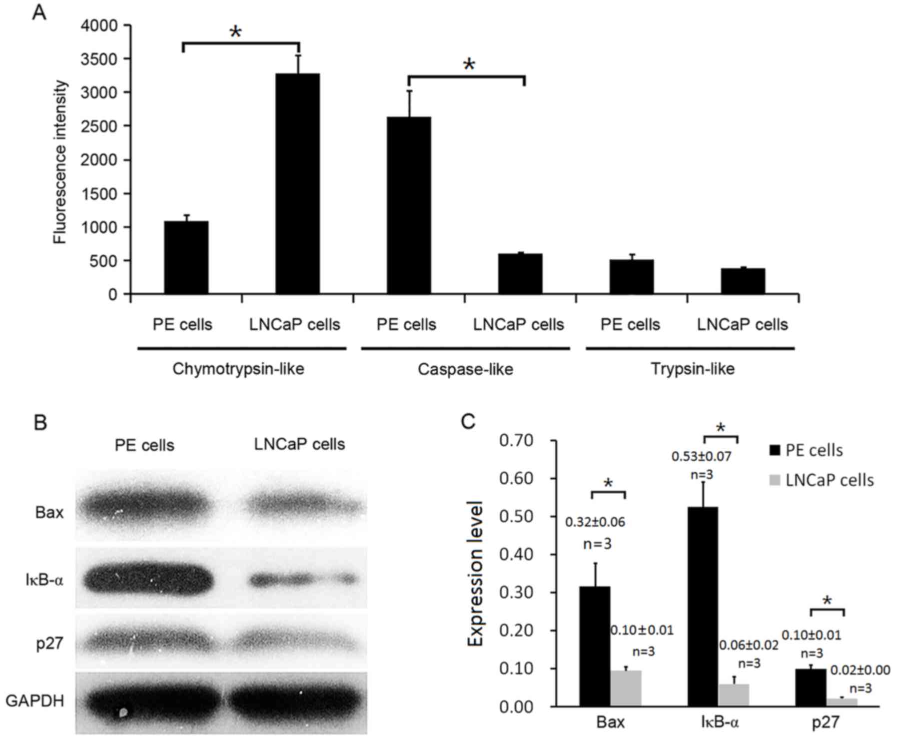
Proteasomal chymotrypsin-like activity
of LNCaP cells is elevated, whereas Bax, IκB-α and p27 protein
levels are decreased
Previous reports have demonstrated an elevated
proteasome level in patients with myeloid hematopoietic
malignancies (18–20), solid tumors (19) and autoimmune diseases (21). To determine the proteasomal activity
variation at acellular level, the proteasomal chymotrypsin-like,
caspase-like and trypsin-like activities were measured in LNCaP
prostate cancer and normal epithelial prostate cells four times.
The proteasomal chymotrypsin-like activity was elevated by ~70% in
LNCaP cells compared with prostate epithelial cells (3,286±259.01
vs. 1,080±100.13; P<0.01). Caspase-like activity was decreased
in LNCaP cells (P<0.01), whereas trypsin-like proteasomal
activity was not significantly altered (Fig. 3A). The expression levels of three of
the most important proteasomal target proteins, Bax, IκB-α, and p27
(22,23), were then assessed by western blotting.
The results indicated all three of the proteins were markedly
reduced in LNCaP cells when compared with PE cells (P<0.01;
Fig. 3B and C), which was consistent
with a previous study (14).
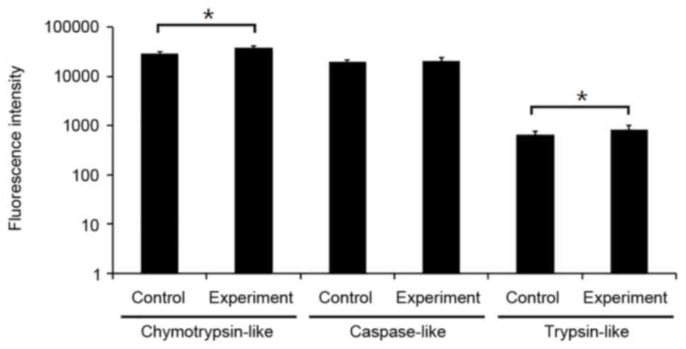
Chymotrypsin-like activity is
increased in xenografts compared with normal mouse prostate tissue,
accompanied by a reduced Bax, IκB-α and p27 protein level
In order to clarify whether the chymotrypsin-like
proteasomal activity was also elevated in vivo, the serum of
the mice with or without LNCaP prostate cancer cell xenografts was
collected for proteasome activity assays. The chymotrypsin-like
activity of the serum in tumor bearing mice was increased by 23%
(37,344.67±2,719.64 vs. 28,845.87±1,880.47; P<0.01) compared
with the control mice, which was in accord with the in vitro
results. In addition, the trypsin-like activity was elevated by 21%
(824.53±164.87 vs. 648±97.5, P<0.01; Fig. 4) in the experimental mice, whereas the
caspase-like activity of the two groups was at a similar level
(Fig. 4). Furthermore, the protein
levels of Bax, IκB-α and p27 were analyzed in three pairs of tumor
bearing and normal mice using western blot analysis. Decreased
levels of Bax, IκB-α and p27 protein were observed in the mouse
tumor tissue compared with the prostate tissue from the control
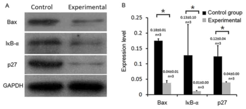
mice (P<0.01; Fig. 5A and B).
Discussion
Prostate cancer is a worldwide concern with a rising
incidence and mortality rate (3,24,25); PSA has been used as a biomarker for
prostate cancer since the 1980s, although its use remains
controversial due to its lack of specificity. Previous studies have
reported that increased chymotrypsin-like proteasomal activity is
associated with various types of tumor, including acute myeloid
leukemia (26) and melanoma (13). In the present study, the
chymotrypsin-like proteasomal activity was significantly elevated
(P<0.01) in prostate cancer cells and tumor-bearing mice
compared with normal epithelial prostate cells and control mice,
suggesting that chymotrypsin-like proteasomal activity may be a
candidate biomarker to supplement PSA in the diagnosis of prostate
cancer. As the inherent limitations of PSA may cause
over-diagnosis, leading to the over-treatment of prostate cancer
and causing psychological distress, loss of bodily function, pain
and suffering for patients (27),
improving the sensitivity and specificity of the detection of
prostate cancer is critical. The chymotrypsin-like proteasomal
activity assay is relatively simple and reproducible, and can be
performed on peripheral blood plasma. Therefore, achymotrypsin-like
activity assay together with PSA detection could potentially
enhance the accuracy of PSA for prostate cancer diagnosis,
ultimately reducing the pain and burden for patients.
Although previous studies (18,19,26) have
demonstrated that plasma proteasomes may act as a biomarker in
several types of tumor, the origin of the proteasomal activity is
has not been identified. Deng et al (28) hypothesized that the aggressiveness of
prostate cancer could be a mechanism; highly malignant cells escape
more frequently into the circulation system, and the
chymotrypsin-like proteasomal activity in the serum could become
elevated with the increase of these cells and their tumor-specific
products. This hypothesis is in accord with the increased
chymotrypsin-like activity in the LnCaP cell medium and the
tumor-bearing mice serum in the present study. In addition, the
caspase-like and trypsin-like activity at both the cell culture and
serum level were altered, as the caspase-like activity was
significantly decreased in the cell experiments, whereas the
trypsin-like activity was elevated in the tumor-bearing mouse
serum, implying that the activity of the proteasomal pathway maybe
altered during, or as a result of, tumorigenesis and
progression.
In the present study, the expression of proteasome
target proteins was also assessed, including p27, which functions
in cell cycle progression; Bax, which is associated with apoptosis;
and IκB-α, which is part of the nuclear factor (NF)-κB pathway
(29). The expression levels of all
three substrates were decreased in prostate cancer cells and
xenograft tumor tissue in the present study. The elevated
chymotrypsin-like activity may have induced the degradation of
these tumor suppressor proteins, ultimately leading to a greater
resistance to apoptosis, and more aggressive cancer behavior. As an
inhibitor of NF-κB, the degradation of IκB-α by the proteasome
facilitates the translocation of NF-κB into the nucleus and thus
promotes cell survival (30),
resulting in the reduced effectiveness of anticancer therapy
(31,32). A relatively low level of p27 is
frequently detected in human tumors, and the degradation of p27 by
the proteasome may result in uncontrolled cell division, ultimately
leading to transformation and tumor development (33). The degradation of Bax has been
identified in aggressive human prostate cancer, which corresponds
with the results of the present study (34). As a Bcl-2 family member, Bax is a
critical molecule upstream of intrinsic cellular apoptosis; its
degradation contributes to maintaining cancer cell survival
(35). It has been reported that the
inhibition of chymotrypsin-like proteasome activity may lead to the
accumulation of IκB-α, Bax and p27 in different types of cancer
cell and tumor models (14), followed
by the induction of cell death. Therefore, proteasome inhibitors
that target chymotrypsin-like activity may be a potential strategy
for prostate cancer treatment.
There are some limitations to the present study.
Firstly, this research detected the proteasomal chymotrypsin-like
activity in cell culture and xenografts, and not in clinical
samples from prostate cancer patients. As a proof of concept, the
present study provided some data to suggest that chymotrypsin-like
activity is a potential candidate biomarker for prostate cancer,
and may build a foundation for future study. Further work to
validate the data of the present study for patients with prostate
cancer will be required. Secondly, proteasomal chymotrypsin-like
activity is not a specific marker for prostate cancer; it is likely
to be increased in various types of carcinoma (36–38).
Therefore, elevated chymotrypsin-like activity cannot independently
indicate the occurrence of prostate cancer; however, it may improve
the accuracy of prostate cancer diagnosis when used in conjunction
with increased PSA level, thus decreasing the chances of
over-diagnosis and over-treatment for patients with suspected
prostate cancer.
Ma et al (26)
reported that the chymotrypsin-like activity in plasma may provide
a powerful biomarker for the risk stratification of acute myeloid
leukemia and advanced-stage myelodysplastic syndrome, which
provides a novel perspective on the application of
chymotrypsin-like activity as a cancer biomarker. Further studies
will be required to verify whether this serological test may serve
as a prognostic factor to detect disease progression in patients
with prostate cancer.
Acknowledgements
Not applicable.
Funding
The present study was supported by Guangzhou General
Science and Technology Project of Health and Family Planning (no.
20161A011011) to Xinghua Wei.
Availability of data and materials
The authors confirm that all data underlying the
findings are fully available without restriction.
Authors' contributions
Conceived and designed the experiments: PT.
Performed the experiments: XW and WZ. Analyzed the data: KX and PF.
Contributed reagents/materials/analysis tools: PT. Contributed to
the writing of the study: XW and PT.
Ethics approval and consent to
participate
The use of human tissue was approved by the Research
Ethics Committee of Guangzhou First People's Hospital, Guangzhou
Medical University (Guangdong, China), and written informed consent
from the patient was acquired prior to the use of the tissue in
research.
Consent for publication
Written informed consent from the patient was
acquired prior to the use of the tissue in research.
Competing interests
The authors declare that they have no competing
interests.
References
|
1
|
Saini S: PSA and beyond: Alternative
prostate cancer biomarkers. Cell Oncol (Dordr). 39:97–106. 2016.
View Article : Google Scholar : PubMed/NCBI
|
|
2
|
Siegel RL, Miller KD and Jemal A: Cancer
statistics, 2015. CA Cancer J Clin. 65:5–29. 2015. View Article : Google Scholar : PubMed/NCBI
|
|
3
|
Lei T, Mao WM, Yang HJ, Chen XZ, Lei TH,
Wang XH, Ying Q, Chen WQ and Zhang SW: Study on cancer incidence
through the cancer registry program in 11 cities and counties,
China. Zhonghua Liu Xing Bing Xue Za Zhi. 30:1165–1170. 2009.(In
Chinese). PubMed/NCBI
|
|
4
|
Fong MK, Hare R and Jarkowski A: A new era
for castrate resistant prostate cancer: A treatment review and
update. J Oncol Pharm Pract. 18:343–354. 2012. View Article : Google Scholar : PubMed/NCBI
|
|
5
|
Rodrigues DN, Butler LM, Estelles DL and
de Bono JS: Molecular pathology and prostate cancer therapeutics:
From biology to bedside. J Pathol. 232:178–184. 2014. View Article : Google Scholar : PubMed/NCBI
|
|
6
|
Lilja H: Testing new PSA subforms to
enhance the accuracy of predicting cancer risk and disease outcome
in prostate cancer. Clin Chem. 54:1248–1249. 2008. View Article : Google Scholar : PubMed/NCBI
|
|
7
|
Lilja H, Ulmert D and Vickers AJ:
Prostate-specific antigen and prostate cancer: Prediction,
detection and monitoring. Nat Rev Cancer. 8:268–278. 2008.
View Article : Google Scholar : PubMed/NCBI
|
|
8
|
Romero Otero J, Garcia Gomez B, Campos
Juanatey F and Touijer KA: Prostate cancer biomarkers: An update.
Urol Oncol. 32:252–260. 2014. View Article : Google Scholar : PubMed/NCBI
|
|
9
|
Cary KC and Cooperberg MR: Biomarkers in
prostate cancer surveillance and screening: Past, present, and
future. Ther Adv Urol. 5:318–329. 2013. View Article : Google Scholar : PubMed/NCBI
|
|
10
|
Prensner JR, Rubin MA, Wei JT and
Chinnaiyan AM: Beyond PSA: The next generation of prostate cancer
biomarkers. Sci Transl Med. 4:127rv32012. View Article : Google Scholar : PubMed/NCBI
|
|
11
|
Voorhees PM, Dees EC, O'Neil B and
Orlowski RZ: The proteasome as a target for cancer therapy. Clin
Cancer Res. 9:6316–6325. 2003.PubMed/NCBI
|
|
12
|
Ciechanover A: The ubiquitin-proteasome
pathway: On protein death and cell life. EMBO J. 17:7151–7160.
1998. View Article : Google Scholar : PubMed/NCBI
|
|
13
|
Stoebner PE, Lavabre-Bertrand T, Henry L,
Guiraud I, Carillo S, Dandurand M, Joujoux JM, Bureau JP and
Meunier L: High plasma proteasome levels are detected in patients
with metastatic malignant melanoma. Br J Dermatol. 152:948–953.
2005. View Article : Google Scholar : PubMed/NCBI
|
|
14
|
Yang H, Zhou P, Huang H, Chen D, Ma N, Cui
QC, Shen S, Dong W, Zhang X, Lian W, et al: Shikonin exerts
antitumor activity via proteasome inhibition and cell death
induction in vitro and in vivo. Int J Cancer. 124:2450–2459. 2009.
View Article : Google Scholar : PubMed/NCBI
|
|
15
|
Piantanelli L, Zaia A, Rossolini G,
Piantanelli A, Basso A and Anisimov VN: Long-live euthymic
BALB/c-nu mice. I. Survival study suggests body weight as a life
span predictor. Mech Ageing Dev. 122:463–475. 2001. View Article : Google Scholar : PubMed/NCBI
|
|
16
|
An B, Goldfarb RH, Siman R and Dou QP:
Novel dipeptidyl proteasome inhibitors overcome Bcl-2 protective
function and selectively accumulate the cyclin-dependent kinase
inhibitor p27 and induce apoptosis in transformed, but not normal,
human fibroblasts. Cell Death Differ. 5:1062–1075. 1998. View Article : Google Scholar : PubMed/NCBI
|
|
17
|
Harmelin A, Danon T, Kela I and Brenner O:
Biopsy of the mouse prostate. Lab Anim. 39:215–220. 2005.
View Article : Google Scholar : PubMed/NCBI
|
|
18
|
Wada M, Kosaka M, Saito S, Sano T, Tanaka
K and Ichihara A: Serum concentration and localization in tumor
cells of proteasomes in patients with hematologic malignancy and
their pathophysiologic significance. J Lab Clin Med. 121:215–223.
1993.PubMed/NCBI
|
|
19
|
Lavabre-Bertrand T, Henry L, Carillo S,
Guiraud I, Ouali A, Dutaud D, Aubry L, Rossi JF and Bureau JP:
Plasma proteasome level is a potential marker in patients with
solid tumors and hemopoietic malignancies. Cancer. 92:2493–2500.
2001. View Article : Google Scholar : PubMed/NCBI
|
|
20
|
Dutaud D, Aubry L, Henry L, Levieux D,
Hendil KB, Kuehn L, Bureau JP and Ouali A: Development and
evaluation of a sandwich ELISA for quantification of the 20S
proteasome in human plasma. J Immunol Methods. 260:183–193. 2002.
View Article : Google Scholar : PubMed/NCBI
|
|
21
|
Egerer K, Kuckelkorn U, Rudolph PE,
Rückert JC, Dörner T, Burmester GR, Kloetzel PM and Feist E:
Circulating proteasomes are markers of cell damage and immunologic
activity in autoimmune diseases. J Rheumatol. 29:2045–2052.
2002.PubMed/NCBI
|
|
22
|
Goldberg AL: Functions of the proteasome:
The lysis at the end of the tunnel. Science. 268:522–523. 1995.
View Article : Google Scholar : PubMed/NCBI
|
|
23
|
Dou QP and Li B: Proteasome inhibitors as
potential novel anticancer agents. Drug Resist Updat. 2:215–223.
1999. View Article : Google Scholar : PubMed/NCBI
|
|
24
|
Siegel R, Ma J, Zou Z and Jemal A: Cancer
statistics, 2014. CA Cancer J Clin. 64:9–29. 2014. View Article : Google Scholar : PubMed/NCBI
|
|
25
|
Heidenreich A, Bastian PJ, Bellmunt J,
Bolla M, Joniau S, van der Kwast T, Mason M, Matveev V, Wiegel T,
Zattoni F, et al: EAU guidelines on prostate cancer. Part.
1:Screening, diagnosis and local treatment with curative
intent–update 2013. Eur Urol 65: 124–137. 2014.
|
|
26
|
Ma W, Kantarjian H, Bekele B, Donahue AC,
Zhang X, Zhang ZJ, O'Brien S, Estey E, Estrov Z, Cortes J, et al:
Proteasome enzymatic activities in plasma as risk stratification of
patients with acute myeloid leukemia and advanced-stage
myelodysplastic syndrome. Clin Cancer Res. 15:3820–3826. 2009.
View Article : Google Scholar : PubMed/NCBI
|
|
27
|
Fowler FJ Jr, Barry MJ, Walker-Corkery B,
Caubet JF, Bates DW, Lee JM, Hauser A and McNaughton-Collins M: The
impact of a suspicious prostate biopsy on patients' psychological,
socio-behavioral, and medical care outcomes. J Gen Intern Med.
21:715–721. 2006. View Article : Google Scholar : PubMed/NCBI
|
|
28
|
Deng X, Zhou P, Wei X, Uhlman M, Lin Y,
Lin X, Wu S, Diao P, Xie H, Liu J, et al: Plasma proteasomal
chymotrypsin-like activity correlates with prostate cancer
progression. Tumour Biol. 36:4115–4121. 2015. View Article : Google Scholar : PubMed/NCBI
|
|
29
|
Frankland-Searby S and Bhaumik SR: The 26S
proteasome complex: An attractive target for cancer therapy.
Biochim Biophys Acta. 1825:64–76. 2012.PubMed/NCBI
|
|
30
|
Adams J: The proteasome: Structure,
function, and role in the cell. Cancer Treat Rev. 29 Suppl 1:S3–S9.
2003. View Article : Google Scholar
|
|
31
|
Jeremias I, Kupatt C, Baumann B, Herr I,
Wirth T and Debatin KM: Inhibition of nuclear factor kappaB
activation attenuates apoptosis resistance in lymphoid cells.
Blood. 91:4624–4631. 1998.PubMed/NCBI
|
|
32
|
Patel NM, Nozaki S, Shortle NH,
Bhat-Nakshatri P, Newton TR, Rice S, Gelfanov V, Boswell SH, Goulet
RJ Jr, Sledge GW Jr and Nakshatri H: Paclitaxel sensitivity of
breast cancer cells with constitutively active NF-kappaB is
enhanced by IkappaBalpha super-repressor and parthenolide.
Oncogene. 19:4159–4169. 2000. View Article : Google Scholar : PubMed/NCBI
|
|
33
|
Chen D and Dou QP: The
ubiquitin-proteasome system as a prospective molecular target for
cancer treatment and prevention. Curr Protein Pept Sci. 11:459–470.
2010. View Article : Google Scholar : PubMed/NCBI
|
|
34
|
Almond JB and Cohen GM: The proteasome: A
novel target for cancer chemotherapy. Leukemia. 16:433–443. 2002.
View Article : Google Scholar : PubMed/NCBI
|
|
35
|
Li B and Dou QP: Bax degradation by the
ubiquitin/proteasome-dependent pathway: Involvement in tumor
survival and progression. Proc Natl Acad Sci USA. 97:pp. 3850–3855.
2000; View Article : Google Scholar : PubMed/NCBI
|
|
36
|
Kondakova IV, Spirina LV, Koval VD,
Shashova EE, Choinzonov EL, Ivanova EV, Kolomiets LA, Chernyshova
AL, Slonimskaya EM, Usynin EA and Afanasyev SG: Chymotripsin-like
activity and subunit composition of proteasomes in human cancers.
Mol Biol (Mosk). 48:444–451. 2014. View Article : Google Scholar : PubMed/NCBI
|
|
37
|
Oldziej A, Bolkun L, Galar M, Kalita J,
Ostrowska H, Romaniuk W and Kloczko J: Assessment of proteasome
concentration and chymotrypsin-like activity in plasma of patients
with newly diagnosed multiple myeloma. Leuk Res. 38:925–930. 2014.
View Article : Google Scholar : PubMed/NCBI
|
|
38
|
Krawczuk-Rybak M, Leszczynska E,
Malinowska I, Matysiak M and Ostrowska H: Proteasome
chymotrypsin-like activity in plasma as a useful marker for
children with acute lymphoblastic leukemia. Scand J Clin Lab
Invest. 72:67–72. 2012. View Article : Google Scholar : PubMed/NCBI
|