Introduction
Breast cancer is one of the most common cancers and
the leading cause of cancer-associated mortality among women,
causing significant morbidity worldwide (1). Nearly 5.8/100,000 new breast cancer
cases are diagnosed in developed countries and >10.5 new breast
cancer cases per 100,000 individuals occur worldwide each year
(1). In the early stages of the
disease, breast cancer, as in case of a number of other cancers,
tends to spread asymptomatically throughout the body and at the
point of diagnosis there is an increased likelihood of
identification of metastatic lesions (2). Conventional therapies, including
surgery, radiotherapy and chemotherapy, have served roles in
treatment of breast cancer, but there remain numerous problems in
tumor therapy. For example, cyclophosphamide, methotrexate and
5-fluorouracil chemotherapy for breast cancer have various side
effects (3,4).
Despite progress in anticancer therapeutics, there
are few efficient drugs with low toxicity available to treat
cancer. Plants have been previously used in cancer therapy
(5). Traditional medical phytotherapy
has been used for cancer treatment for several millennia in Asia,
including in China, Japan and Thailand, and is currently being used
for cancer prevention and therapy worldwide (6–8).
Throughout the centuries, certain plant extracts have been tested
for antitumor potential (9). Plants
are a source of a number of effective anticancer agents in current
use, including taxanes, irinotecan, topotecan, vincristine and
vinblastine (10–12). Plant products such as these can aid in
discovery and development of novel anticancer drugs. In addition,
plant products demonstrate fewer side effects compared with
chemical drugs. There has been an increasing interest in
identifying and isolating natural compounds from medicinal plants
with an aim to develop novel anticancer drugs (13).
Ailanthus altissima is an example of a plant
that has been used in tumor therapy (14). A. altissima, which grows in
Northeast and Central China, can also be present in Europe and the
USA and has been used as a traditional Chinese medicine for a
number of years (15). The bark of
A. altissima has been used for the treatment of ascariasis,
diarrhea, spermatorrhea, bleeding and gastrointestinal diseases in
China (16). The antitumor effect
that has enabled the use of this plant in the treatment of colonic,
cervical, and rectal cancer has been previously described (17). In the present study, ailanthone was
successfully isolated from A. altissima. Ailanthone has a
wide spectrum of biological activities, including anti-allergic,
anti-malarial, anti-HIV, anti-inflammatory, anti-ulcer and
anti-microbial activities (18,19).
Ailanthone has been demonstrated to exhibit in vitro
growth-inhibitory effects against several cancer cell lines,
including HepG2, Hep3B, R-HepG2, HeLa and A549 cells (18,20).
Rosati (20) demonstrated that
ailanthone could induce mitochondrial membrane depolarization and
caspase-3 activation in Jurkat cancer cells. However, the antitumor
activity in breast cancer treatment and the underlying molecular
mechanism remain to be elucidated. In the present study, MCF-7
cells were used to evaluate the antitumor activity of ailanthone
and the underlying molecular mechanism.
Materials and methods
Plant material and extraction
The bark of A. altissima (catalog no.,
NMC-2012-2) was purchased from Anguo Zhong Sheng Medicinal Material
Co., Ltd. (Anguoshi, China). The bark of A. altissima was
powdered, a 10-fold volume of methanol was added to the powdered
sample and allowed to soak for 12 h. Hot reflux was performed two
times serially, each time using a rotary evaporator with 95%
ethanol at 80°C for 2 h, filtered, and the filtrate was merged. The
combined filtrates were concentrated in vacuo, and then
treated with saturated salt solution at concentration of 20%. It
was extracted with petroleum ether and dichloromethane for 2 h, and
dried in vacuo at 40°C respectively. The extract with was separated
with 50 times the amount of 200–300 mesh silica gel column (Qingdao
Haiyang Chemical Co., Ltd., Qingdao, China). The mobile phase was a
solution of dichloromethane and methanol in ratios of 50:1, 30:1,
15:1, 10:1, 5:1, and 500 ml was collected using each ratio. A total
of 211 fractions were collected, and similar fractions were
combined on the basis of TLC to obtain 35 major fractions.
Following further purification by crystallization, ailanthone was
purified as previously described (21).
Cell preparation
Breast cancer MCF-7 cells were purchased from
Shanghai Institute of Biochemistry and Cell Biology (Shanghai,
China), and grown in Dulbecco's Modified Eagle's medium (DMEM,
Gibco; Thermo Fisher Scientific, Inc.). DMEM was supplemented with
100 µg/ml streptomycin, penicillin (Sigma-Aldrich; Merck KGaA,
Darmstadt, Germany) and 10% (v/v) FBS (Gibco; Thermo Fisher
Scientific, Inc.). Cells were cultured at 37°C and 5%
CO2 in a humidified environment.
Cell viability assay
Cell viability was assessed by MTT colorimetric
assay (22). The cells were treated
with various concentrations (0.5, 1.0, 2.0, 4.0 and 8.0 µg/ml) of
ailanthone for 24, 48 and 72 h. At the end of the treatment, 50 µl
MTT (5 mg/ml) was added to each well (1×10⁵ cells/ml)
and the samples were incubated for an additional 1 h. The culture
medium was removed and dissolved by adding 100 µl dimethyl
sulfoxide. The absorbance was measured at a wavelength of 490
nm.
Cell apoptosis
The level of cell apoptosis was determined by flow
cytometry with annexin V (BD Biosciences, Franklin Lakes, NJ, USA)
labeling and propidium iodide (PI) exclusion staining. MCF-7 cells
were treated with 0.5, 1.0, 2.0, 4.0 and 8.0 µg/ml ailanthone for
48 h, collected, washed with PBS and suspended in binding buffer
(BD Biosciences). Subsequently, the cells (1×10⁵
cells/ml) were stained with 10 µl annexin V-fluorescein FITC and 5
µl PI, incubated in dark at room temperature for 15 min according
to the manufacturer's protocol and subjected to flow cytometry
using a flow cytometer (ModFit 6.0, BD Biosciences, USA). Cells
undergoing apoptosis were stained with annexin V and treated as
described above.
Cell cycle distribution
Cells were seeded at a density of
1×10⁶/ml in 6-well microplates and treated with 0.5,
1.0, 2.0, 4.0 and 8.0 µg/ml ailanthone for 48 h. A total of
1×10⁵ cells were collected for each treatment, washed
with PBS, trypsinized with 0.25% trypsin and harvested by
centrifugation for 5 min at 70 × g at room temperature. Cells were
re-suspended with 0.5 ml PBS, fixed overnight with cold 70% ethanol
at −20°C and stained with PI solution containing 10 µg/ml RNase A
and 50 µg/ml PI for 60 min at room temperature. Cells were analyzed
by flow cytometry, as previously described (23).
Reverse transcription-polymerase chain
reaction (RT-PCR) analysis
MCF-7 cells were treated by ailanthone (0.5, 1.0,
2.0, 4.0 and 8.0 µg/ml), and following extraction of total cellular
RNA from the MCF-7 cells using TRIzol reagent (Thermo Fisher
Scientific, Inc., Waltham, MA, USA), 2 µg of total RNA was reverse
transcribed to generate cDNA using the PrimeScript RT reagent kit
(Takara Bio, Inc.). The following thermocycling conditions were
used for RT-PCR: Initial denaturation at 94°C for 30 sec, followed
by 30 cycles of 94°C for 30 sec, 56°C for 30 sec and 72°C for 45
sec, and the final extension at 72°C for 8 min. The PCR products
were resolved in 2% agarose gels and visualized by staining with
ethidium bromide (24). To
semiquantify the PCR products, the bands representing the amplified
products were analyzed by Quantity One analysis software (Bio-Rad
Laboratories, Inc., Hercules, CA, USA). The relative level of the
target mRNA expression was defined as the ratio of the absorbance
of the target band to that of the β-actin band. The following
primer sequences were used for PCR: (Takara RNA PCR Kit AMV,
Ver.3.0) Caspase-3 (442 bp), forward
5′-ATCTCGGTCTGGTACAGATGTCGAT-3′ and reverse
5′-TGAATTTCGCCAAGAATAATACCA-3′; Bcl-associated X (Bax; 258 bp),
forward 5′-CACCAGCTCTGAGCAGATCA-3′, reverse
5′-ATGTCAGCTGCCACTCGGA-3′; B-cell lymphoma 2 (Bcl-2; 383 bp),
forward 5′-TACGAGTGGGATGCGGGAGATGT-3′, reverse
5′-CCACCGAACTCAAAGAAGGC-3′; GAPDH (135 bp), forward
5′-CAATGACCCCTTCATTGACC-3′, reverse 5′-TGGAAGATGGTGATGGGATT-3′.
Western blot analysis
MCF-7 cells were treated by ailanthone (0.5, 1.0,
2.0, 4.0 and 8.0 µg/ml), and lysed in lysis buffer [50 mmol/l
Tris-HCl (pH 8.0), 150 mmol/l NaCl, 0.5% NP40, 0.5% sodium
deoxycholate, 0.1% SDS with protease inhibitor]. Determination of
total protein content was performed using a BCA assay (Beijing
Solarbio Science and Technology, Co., Ltd., Beijing China). The
proteins were separated by 12% SDS-PAGE and transferred to
polyvinylidene difluoride membranes. The membranes were blocked
with 5% milk in Tris-buffered saline-0.05% Tween-20 (TBST) for 3 h
at room temperature. Following washing in TBST, the membranes were
incubated for 2 h at room temperature with 1:1,000 dilutions of
rabbit monoclonal antibodies for BCL-2, (cat no. 2872; Cell
Signaling Technology, Inc., Danvers, MA, USA), Bax (cat no. 2772;
Cell Signaling Technology, Inc.) and Caspase-3, (cat no. 9662; Cell
Signaling Technology, Inc.), and a mouse monoclonal antibody for
β-actin (ab8226; Abcam, Cambridge, UK). Following washing in TBST,
the blots were incubated with horseradish peroxidase-conjugated
goat anti-rabbit immunoglobulin G secondary antibody (rabbit
monoclonal antibody; mouse monoclonal antibody, 1:3,000; Abcam) for
1 h at room temperature. The signal was detected using an ECL
Western Blotting Substrate kit (Applygen Technologies, Inc.,
Beijing, China), and exposed to X-ray film at room temperature.
Protein expression levels were quantitated using Quantity-one
software (v 4.62; Bio-Rad Laboratories, Inc.).
Statistical analysis
SPSS software (version 16; SPSS, Inc., Chicago, IL,
USA) was used for statistical analysis of the data. Data are
presented as the mean ± standard deviation. One-way analysis of
variance followed by Dunnett's test was used for statistical
analysis. P<0.05 was considered to indicate a statistically
significant difference.
Results
Inhibitory effect of ailanthone on
proliferation of tumor cells
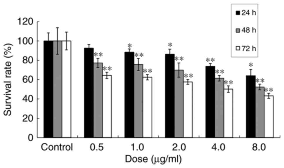
The effect of ailanthone on viability of MCF-7 cells
was measured by MTT assay. Compared with the untreated control, the
viability of MCF-7 cells treated with 0.5, 1.0, 2.0, 4.0 or 8.0
µg/ml of ailanthone was 92.62, 88.46, 86.36, 73.74 and 64.05%,
respectively, following 24 h of treatment; 77.27, 75.65, 69.89,
61.40 and 52.40%, respectively, following 48 h of treatment; and
64.36, 62.48, 57.64, 50.24 and 43.24%, respectively, following 72 h
of treatment (Fig. 1). The data
indicated that ailanthone inhibits MCF-7 cell proliferation.
Effects of ailanthone on apoptosis of
MCF-7 cells
Following 48 h of treatment with ailanthone, 22.28,
27.99, 35.88, 49.77 and 75.51% of cells were apoptotic at doses of
0.5, 1.0, 2.0, 4.0 and 8.0 µg/ml, respectively (Fig. 2). In total, 2.13% of control cells
were apoptotic at 48 h, which is significantly different to all
treatment groups (P<0.01).
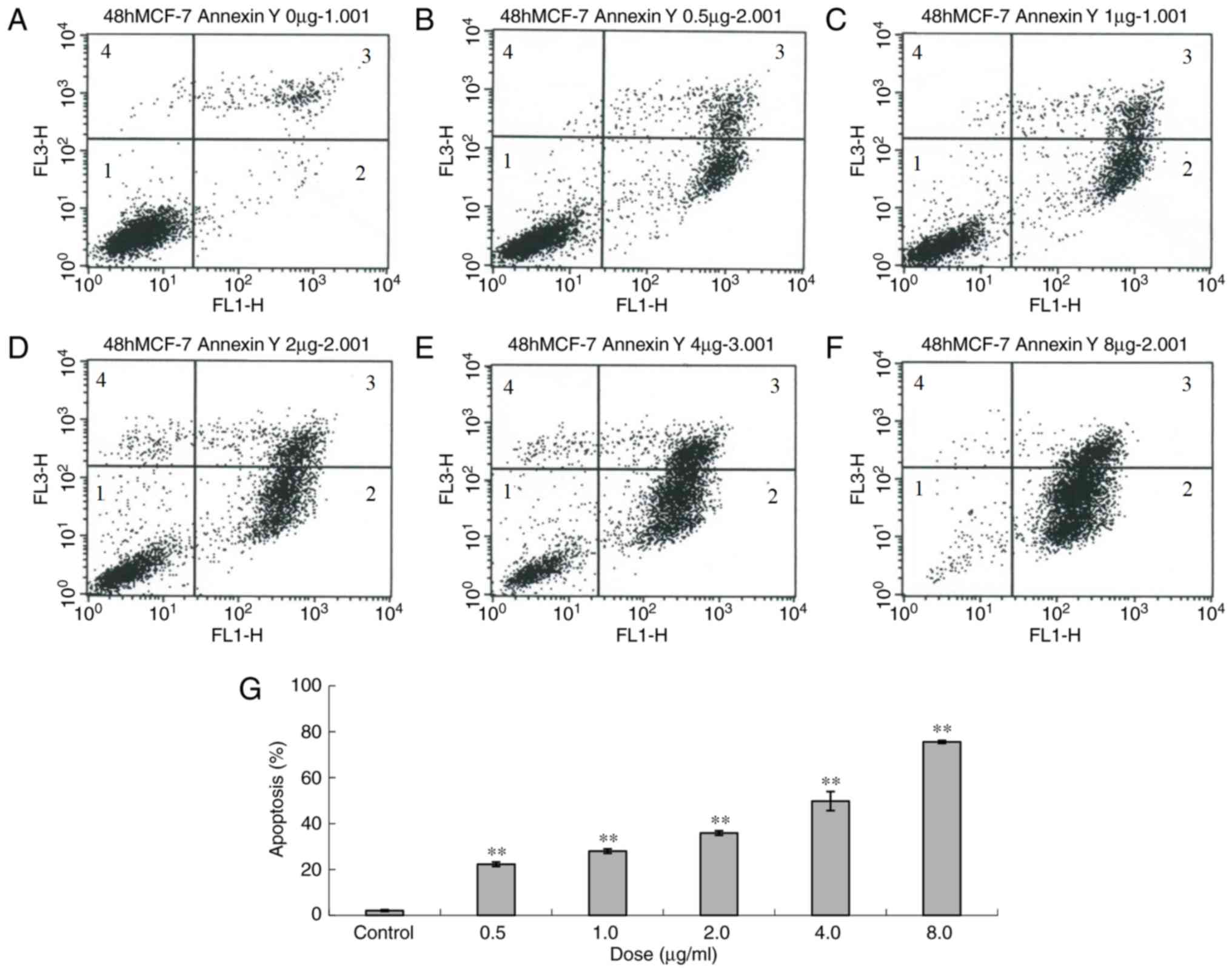 | Figure 2.Apoptosis of MCF-7 cells following
treatment with different doses of ailanthone (0.5, 1, 2, 4 and 8
µg/ml) for 48 h, measured by flow cytometry. (A) Control (0 µg/ml)
and (B) 0.5 µg/ml, (C) 1.0 µg/ml, (D) 2.0 µg/ml, (E) 4.0 µg/ml and
(F) 8.0 µg/ml ailanthone. (G) Histogram of apoptosis of MCF-7
cells. Data are presented as the mean ± standard deviation (n=3).
**P<0.01 vs. the control and treatment groups. 1, normal cells;
2, early apoptosis; 3, late apoptosis; 4, dead cells. |
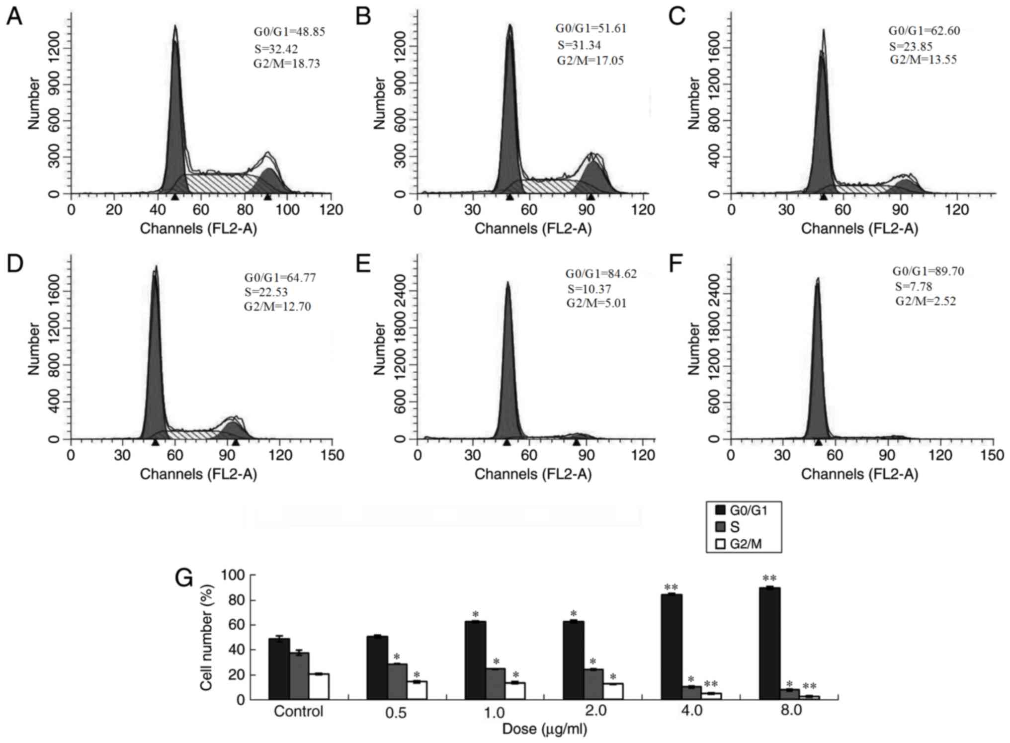
Effects of ailanthone on cell cycle of
MCF-7 cells
Following treatment with 0.5, 1.0, 2.0, 4.0 and 8.0
µg/ml ailanthone for 48 h, all treatment groups exhibited
statistically significant differences compared with the control.
Following treatment with ailanthone, the proportion of cells in the
G0/G1 phase increased and percentage of cells
in S and G2/M phases decreased significantly compared
with the control group (Fig. 3).
Alteration of expression levels of
Bcl-2 and Bax mRNA
Following 48 h of treatment with different doses of
ailanthone, RT-qPCR demonstrated that in the MCF-7 cells,
expression of the Bax and caspase-3 genes increased, whereas
expression levels of the Bcl-2 gene decreased. Doses of 1.0, 2.0,
4.0 and 8.0 µg/ml of ailanthone resulted in significantly altered
expression, compared with the control group (Fig. 4).
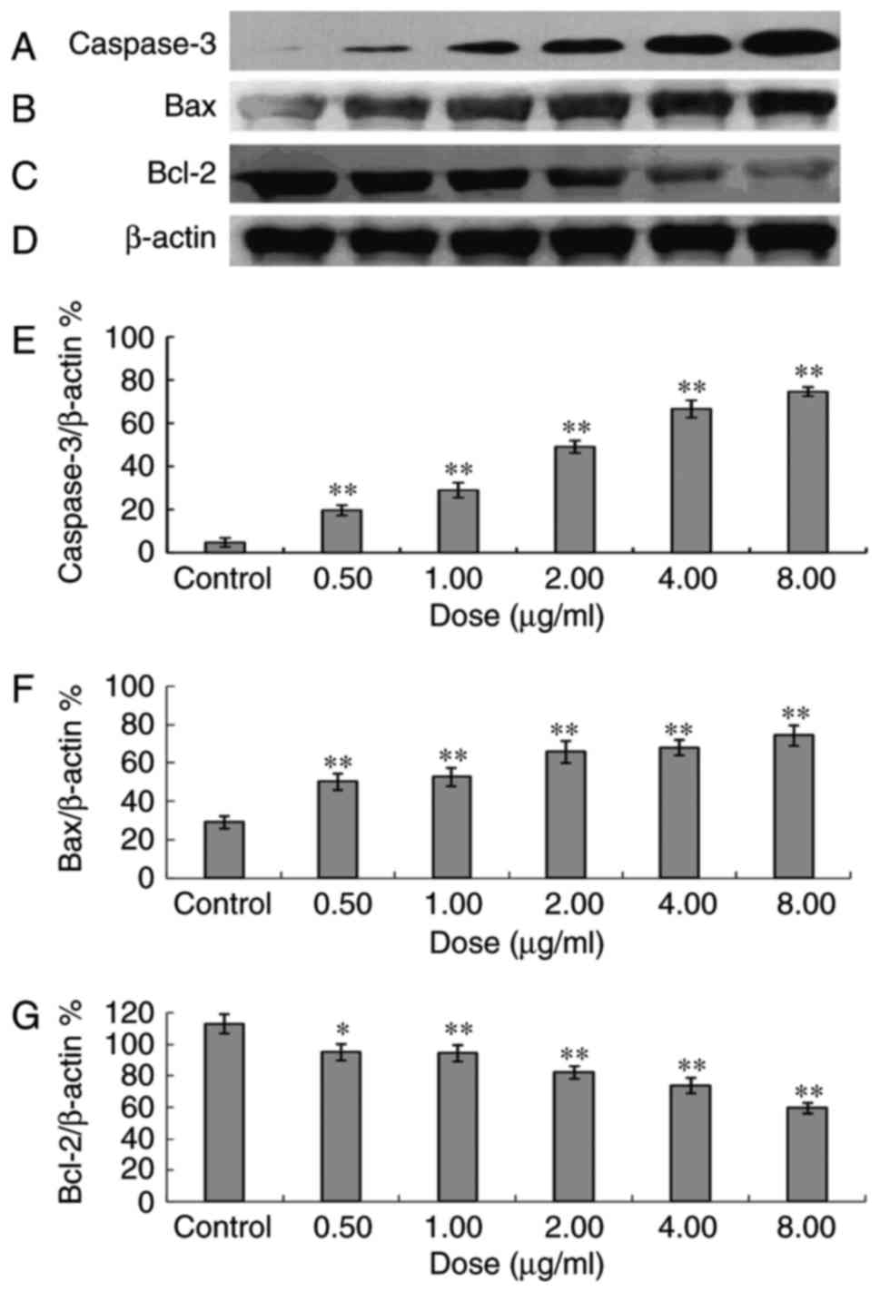
Effects of ailanthone on protein
expression levels by western blotting
Following 48 h of treatment with 0.5, 1.0, 2.0, 4.0
or 8.0 µg/ml ailanthone, it was observed that ailanthone promoted
the expression of Bax and caspase-3 proteins, whereas the
expression of Bcl-2 protein was inhibited. The inhibitory effect on
Bcl-2 protein expression increased with the increase of the doses
(P<0.05). The Bax/Bcl-2 ratios were 0.27, 0.53, 0.56, 0.80, 0.93
and 1.25 at dosages of 0, 0.5, 1.0, 2.0, 4.0 and 8.0 µg/ml,
respectively, and thus increased following treatment with
ailanthone in a dose-dependent manner (Fig. 5).
Discussion
Breast cancer is the primary cause of mortality
among women in the world, according to statistics from 2009
(2). Currently, certain cytotoxic
drugs are used for the treatment of breast cancer, including
daunorubicin, doxorubicin, cisplatin and bleomycin (25). However, these agents are costly and
have been demonstrated to induce several side effects, including
emesis, anemia, myelosuppression and cellular resistance (3). Therefore, it is necessary to identify
alternative drugs or therapies to minimize side effects (26). As a result, natural medicines that
cause fewer side effects have been attracting substantial attention
(27). Previous research has
demonstrated that natural antitumor drugs may serve important roles
in the future. A phytochemical study demonstrated the presence of
quassinoids in A. altissima and these compounds have been
previously demonstrated to exhibit potent antitumor properties
(28). To the best of our knowledge,
the present study is the first to report that ailanthone isolated
from A. altissimahas inhibits proliferation of MCF-7
cells.
The majority of drugs achieve antitumor effects by
inducing apoptosis in tumor cells (29). Cell cycle analysis is used to
determine the distribution of cells in different phases of the cell
cycle and enables investigation of tumor proliferation as opposed
to apoptosis. There are three cell cycle regulation points in
G1, S and G2 phases, which can modulate cell
cycle progression. Induction of tumor cell cycle arrest in
G0/G1 phase is a target for the development
of antitumor therapy (30). Certain
molecules, including tumor protein p53, serve a role in cell cycle
inhibition and induction of apoptosis; cells may be arrested in the
G1 phase and apoptosis may be induced by p53 (31). In the present study, the results of
flow cytometry revealed that, following treatment with ailanthone,
apoptosis in MCF-7 cells increased. Analysis of the cell cycle by
flow cytometry demonstrated an increase in the number of MCF-7
cells in G0/G1 phase following treatment with
ailanthone and a decrease in the number of cells in S phase,
indicating that the cells were arrested in the
G0/G1 phase by so that the cells could not
enter S phase or perform DNA synthesis, thus inhibiting
proliferation. This checkpoint may be involved in the effects of
ailanthone on the cell cycle of MCF-7 cells (32), but this potential mechanism remains to
be further investigated. Caspases are mediators of apoptosis, of
which caspase-3 are frequently activated death protease that
catalyzes specific cleavage of numerous cellular proteins (33). Tumor inhibition and apoptosis, as well
as the expression of caspase-3, Bcl-2 and Bax were determined by
flow cytometry. The results demonstrated that ailanthone can induce
apoptosis in tumor cells. Apoptosis is an active cell suicide
process that is regulated by p53 (34). The effect of Bcl-2 depends on the
ratio of its expression with Bax; this ratio determines whether
cells undergo apoptosis or survival upon signal stimulation
(35). Excessive Bax expression in
cells promotes apoptosis, whereas excessive Bcl-2 expression
promotes survival (36).
Western blot analysis was used to detect levels of
Bax and Bcl-2 protein expression; it was demonstrated that
following treatment of MCF-7 cells with 0.5, 1.0, 2.0, 4.0 or 8.0
µg/ml ailanthone for 48 h, Bax expression increased, whereas that
of Bcl-2 decreased markedly. The results indicated that the
mechanism underlying ailanthone-induced MCF-7 cell apoptosis may be
associated with the adjusting of the Bax and Bcl-2 family proteins.
Experimental results demonstrated that ailanthone exhibited an
inhibitory effect on cellular proliferation and induced apoptosis.
The promotion of Bax and the inhibition of Bcl-2 proteins may
further enhance the antitumor effect.
Genetic abnormalities in the phosphatidylinositol
3-kinase (PI3K)/RAC serine/threonine-protein kinase (AKT) signaling
pathway are frequently observed in human tumors; previous studies
indicate that this pathway is involved in the development of
multiple cancer types (37). The role
of the PI3K/AKT pathway and its potential as a therapeutic target
for tumor treatment has been investigated in preclinical studies
into a number of tumor types, including lung, breast and renal
cancer, neuroblastoma and glioblastoma. The results of these
studies indicate that the PI3K/AKT signaling pathway and those
downstream of it are potential targets for therapeutic intervention
(38–41). The PI3K/AKT pathway serves a role in
apoptosis, cell cycle progression and tumorigenesis; therefore, we
hypothesize that ailanthone-induced apoptosis may also involve the
PI3K/AKT pathway, demonstrating that the ailanthone treatment of
Huh7 cells resulted in a decrease in the expression of PI3K and AKT
phosphorylation at threonine-408 and serine-473.
Treatment of MCF-7 cells with ailanthone resulted in
cell apoptosis. In the present study, the antitumor effect of
ailanthone indicated that this compound may be beneficial for the
treatment of breast cancer. Further investigation is required to
identify the mechanism underlying the antitumor activity.
In conclusion, the present study demonstrated that
ailanthone, isolated from A. altissima, exhibited an
inhibitory effect on MCF-7 cells and promoted cell apoptosis by
upregulating Bax protein and mRNA. Ailanthone inhibited the protein
and mRNA expression of Bcl-2, indicating that is has potential
antitumor activity. Ailanthone may be a novel phytomedicine for
tumor therapy.
Acknowledgements
The authors would like to thank Professor Jun-Qing
Liang (Shijiazhuang Yiling Pharmaceutical Co., Ltd., Hebei, China)
for providing necessary facilities to perform experiments in the
present study. The present study was supported by the National
Natural Science Foundation of China (grant no., 81302664 and
81703001) and the Hebei Medical University Development Project
(2016-kyfz111). We are grateful for the key discipline construction
project of the Universities in Hebei (ZD 2017003); Chengde medical
college high level talent research startup fund (201705) and Key
Discipline Construction Projects of Higher Schools in Hebei. The
authors also wish to extend thanks for the financial support of
Syngenta Ltd. (2017-Hebei Medical University-Syngenta-04).
Competing interests
The authors declare that they have no competing
interests.
References
|
1
|
Khoobchandani M, Ojeswi BK, Sharma B and
Srivastava MM: Chenopodium album prevents progression of cell
growth and enhances cell toxicity in human breast cancer cell
lines. Oxid Med Cell Longev. 2:160–165. 2009. View Article : Google Scholar : PubMed/NCBI
|
|
2
|
Tokgun O, Akca H, Mammadov R, Aykurt C and
Deniz G: Convolvulus galaticus, crocus antalyensis, and lilium
candidum extracts show their antitumor activity through induction
of p53-mediated apoptosis on human breast cancer cell line MCF-7
Cells. J Med Food. 11:1000–1005. 2012. View Article : Google Scholar
|
|
3
|
Binkley JM, Harris SR, Levangie PK, Pearl
M, Guglielmino J, Kraus V and Rowden D: Patient perspectives on
breast cancer treatment side effects and the prospective
surveillance model for physical rehabilitation for women with
breast cancer. Cancer. 8:2207–2216. 2012. View Article : Google Scholar
|
|
4
|
Sitzia J and Huggins L: Side effects of
cyclophosphamide, methotrexate, and 5-fluorouracil (CMF)
chemotherapy for breast cancer. Cancer Pract. 6:13–21. 1998.
View Article : Google Scholar : PubMed/NCBI
|
|
5
|
da Rocha AB, Lopes RM and Schwartsmann G:
Natural products in anticancer therapy. Curr Opin Pharmacol.
1:364–369. 2001. View Article : Google Scholar : PubMed/NCBI
|
|
6
|
Efferth T, Li PC, Konkimalla VS and Kaina
B: From traditional Chinese medicine to rational cancer therapy.
Trends Mol Med. 13:353–361. 2007. View Article : Google Scholar : PubMed/NCBI
|
|
7
|
Olaku O and White JD: Herbal therapy use
by cancer patients: A literature review on case reports. Eur J
Cancer. 47:508–514. 2011. View Article : Google Scholar : PubMed/NCBI
|
|
8
|
Dennis T, Fanous M and Mousa S: Natural
products for chemopreventive and adjunctive therapy in oncologic
disease. Nutr Cancer. 61:587–597. 2009. View Article : Google Scholar : PubMed/NCBI
|
|
9
|
Ho JW, Leung YK and Chan CP: Herbal
medicine in the treatment of cancer. Curr Med Chem Anticancer
Agents. 2:209–214. 2002. View Article : Google Scholar : PubMed/NCBI
|
|
10
|
Pezzuto JM: Plant-derived anticancer
agents. Biochem Pharmacol. 53:121–133. 1997. View Article : Google Scholar : PubMed/NCBI
|
|
11
|
Kinghorn AD, Farnsworth NR, Doel Soejarto
D, Cordell GA, Pezzuto JM, Udeani GO, Wani MC, Wall ME, Navarro HA,
Kramer RA, et al: Novel strategies for the discovery of
plant-derived anticancer agents. Pure Appl Chem. 71:611–618. 1999.
View Article : Google Scholar
|
|
12
|
Lee KH: Anticancer drug design based on
plant-derived natural products. J Biomed Sci. 6:236–250. 1999.
View Article : Google Scholar : PubMed/NCBI
|
|
13
|
Bishayee A: Editorial: Current advances in
cancer prevention and treatment by natural products. Curr Pharm
Biotechnol. 13:115–116. 2012. View Article : Google Scholar : PubMed/NCBI
|
|
14
|
Efferth T, Li PC, Konkimalla VS and Kaina
B: From traditional Chinese edicine to rational cancer therapy.
Trends Mol Med. 13:353–361. 2007. View Article : Google Scholar : PubMed/NCBI
|
|
15
|
De Feo V, De Martino L, Quaranta E and
Pizza C: Isolation of phytotoxic compounds from tree-of-heaven
(Ailanthus altissima swingle). J Agric Food Chem. 51:1177–1180.
2003. View Article : Google Scholar : PubMed/NCBI
|
|
16
|
Rahman S, Fukamiya N, Ohno N, Tokuda H,
Nishino H, Tagahara K, Lee KH and Okano M: Inhibitory effects of
quassinoid derivatives on Epstein-Barr virus early antigen
activation. Chem Pharm Bull (Tokyo). 45:675–677. 1997. View Article : Google Scholar : PubMed/NCBI
|
|
17
|
Wang Y, Wang WJ, Su C, Zhang DM, Xu LP, He
RR, Wang L, Zhang J, Zhang XQ and Ye WC: Cytotoxic quassinoids from
Ailanthus altissima. Bioorg Med Chem Lett. 23:654–657. 2013.
View Article : Google Scholar : PubMed/NCBI
|
|
18
|
Okunade AL, Bikoff RE, Casper SJ, Oksman
A, Goldberg DE and Lewis WH: Antiplasmodial activity of extracts
and quassinoids isolated from seedlings of Ailanthus altissima
(Simaroubaceae). Phytother Res. 17:675–677. 2003. View Article : Google Scholar : PubMed/NCBI
|
|
19
|
Kundu P and Laskar S: A brief resume on
the genus Ailanthus: chemical and pharmacological aspects.
Phytochem Rev. 9:379–412. 2010. View Article : Google Scholar
|
|
20
|
Rosati A, Quarantam E, Ammirante M, Turco
MC, Leone A and De Feo V: Quassinoids can induce mitochondrial
membrane depolarisation and caspase 3 activation in human cells.
Cell Death Differ. 11 Suppl 2:S216–S218. 2004. View Article : Google Scholar : PubMed/NCBI
|
|
21
|
Casinovi CG, Ceccherelli P, Grandolini G
and Bellavita V: On the structure of ailanthone. Tetrahedron Lett.
5:3991–3997. 1964. View Article : Google Scholar
|
|
22
|
Bishayee A, Háznagy Radnai E, Mbimba T,
Sipos P, Morazzoni P, Darvesh AS, Bhatia D and Hohmann J:
Anthocyaninrich black current extract suppresses the growth of
human hepatocellular carcinoma cells. Nat Pred Commun. 5:1613–1618.
2010.
|
|
23
|
Kim MJ, Kim YJ, Park HJ, Chung JH, Leem KH
and Kim HK: Apoptotic effect of red wine polyphenols on human colon
cancer SNU-C4cells. Food Chem Toxicol. 44:898–902. 2006. View Article : Google Scholar : PubMed/NCBI
|
|
24
|
Lu YJ, Xu Q, Chen L, Zuo Y, Liu S, Hu Y,
Li X, Li Y and Zhao X: Expression of semaphorin 6D and its receptor
plexin-A1 in gastric cancer and their association with tumor
angiogenesis. Oncology. 12:3967–3974. 2016.
|
|
25
|
Moysich KB, Beehler GP, Zirpoli G, Choi JY
and Baker JA: Use of common medications and breast cancer risk.
Cancer Epidemiol Biomarkers Prev. 7:1564–1595. 2008. View Article : Google Scholar
|
|
26
|
Kim DW, Hong GH, Lee HH, Choi SH, Chun BG,
Won CK, Hwang IK and Won MH: Effect of colloidal silver against the
cytotoxicity of hydrogen peroxide and naphthazarin on primary
cultured cortical astrocytes. Int J Neurosci. 117:387–400. 2007.
View Article : Google Scholar : PubMed/NCBI
|
|
27
|
Duan JA, Su SL and Qian DW: Approaches and
advances in the resources chemistry of Chinese medicinal material.
Chin J Nat Med. 7:333–340. 2009. View Article : Google Scholar
|
|
28
|
Zhuo Z, Hu J, Yang X, Chen M, Lei X, Deng
L, Yao N, Peng Q, Chen Z, Ye W and Zhang D: Ailanthone inhibits
Huh7 cancer cell growth via cell cycle arrest and apoptosis in
vitro and in vivo. Sci Rep. 5:161852015. View Article : Google Scholar : PubMed/NCBI
|
|
29
|
James BR and Griffith TS: Activation of
systemic antitumor immunity via TRAIL-induced apoptosis.
OncoImmunology. 17:1178–1180. 2012. View Article : Google Scholar
|
|
30
|
Chen M, Xu XYX D, et al: Inhibiting Bcl-2
gene expression enhance radiosensitivity of non-small cell lung
cancer NCI-H460 cells. China Oncology. 20:641–5. 2010.
|
|
31
|
Bai X, Che F, Li J, Ma Y, Zhou Y, Zhai J
and Meng L: Effects of adenovirus-mediated p16 and p53 genes
transfer on apoptosis and cell cycle of lung carcinoma cells.
Zhonghua Bing Li Xue Za Zhi. 29:354–358. 2000.(In Chinese).
PubMed/NCBI
|
|
32
|
Zhang Z, Leonard SS, Huang C, Vallyathan
V, Castranova V and Shi X: Role of reactive oxygen species and
MAPKs in vanadate-induced G(2)/M phase arrest. Free Radic Biol Med.
34:1333–1042. 2003. View Article : Google Scholar : PubMed/NCBI
|
|
33
|
Porter AG and Jänicke RU: Emerging roles
of caspase-3 in apoptosis. Cell Death Differ. 6:99–104. 1999.
View Article : Google Scholar : PubMed/NCBI
|
|
34
|
Alvarez S, Drane P, Meiller A, Bras M,
Deguin-Chambon V, Bouvard V and May E: A comprehens ive s tudy of
p53 transcriptional activity in thymus and spleen of gamma
irradiated mouse: High sens itivity of genes involved in the two
main apoptotic pathways. Int J Radiat Biol. 82:761–770. 2006.
View Article : Google Scholar : PubMed/NCBI
|
|
35
|
Raisova M, Hossini AM, Eberle J, Riebeling
C, Wieder T, Sturm I, Daniel PT, Orfanos CE and Geilen CC: The
Bax/Bcl-2 ratio determines the susceptibility of human melanoma
cells to CD95/Fas-mediated apoptosis. J Invest Dermatol.
117:333–340. 2001. View Article : Google Scholar : PubMed/NCBI
|
|
36
|
Samarghandian S, Nezhad MA and Mohammadi
G: Role of caspases, Bax and Bcl-2 in chrysin-induced apoptosis in
the A549 human lung adenocarcinoma epithelial cells. Anticancer
Agents Med Chem. 14:901–909. 2014. View Article : Google Scholar : PubMed/NCBI
|
|
37
|
Wong KK, Engelman JA and Cantley LC:
Targeting the PI3K signaling pathway in cancer. Curr Opin Genet
Dev. 20:87–90. 2010. View Article : Google Scholar : PubMed/NCBI
|
|
38
|
Rao E, Jiang C, Ji M, Huang X, Iqbal J,
Lenz G, Wright G, Staudt LM, Zhao Y, McKeithan TW, et al: The
miRNA-17~92 cluster mediates chemoresistance and enhances tumor
growth in mantle cell lymphoma via PI3K/AKT pathway activation.
Leukemia. 26:1064–1072. 2012. View Article : Google Scholar : PubMed/NCBI
|
|
39
|
Markman B, Dienstmann R and Tabernero J:
Targeting the PI3K/Akt/mTOR pathway-beyond rapalogs. Oncotarget.
1:530–543. 2010.PubMed/NCBI
|
|
40
|
Neri LM, Cani A, Martelli AM, Simioni C,
Junghanss C, Tabellini G, Ricci F, Tazzari PL, Pagliaro P, McCubrey
JA and Capitani S: Targeting the PI3K/Akt/mTOR signaling pathway in
B-precursor acute lymphoblastic leukemia and its therapeutic
potential. Leukemia. 28:739–748. 2014. View Article : Google Scholar : PubMed/NCBI
|
|
41
|
Slomovitz BM and Coleman RL: The
PI3K/AKT/mTOR pathway as a therapeutic target in endometrial
cancer. Clin Cancer Res. 18:5856–5864. 2012. View Article : Google Scholar : PubMed/NCBI
|