Introduction
Gastric cancer (GC) is one of the most common
malignant tumors globally (1–4). In China, GC is the second leading cause
of cancer-associated mortality (5).
In spite of the decreasing incidence and mortality rate of GC among
the Chinese population, 723,100/951,600 novel cases of GC resulted
in mortality in 2012 (6). The
treatment protocols available include surgery, chemotherapy,
radiotherapy and molecular targeted therapy (7,8).
Nevertheless, the median survival remains <12 months and the
5-year survival rate is ~25% as a result of tumor recurrence and
metastasis (9). Therefore,
identifying new prognostic biomarkers and therapeutic targets for
GC is of high importance.
Nectin-4 is a member of the Nectin family of four
Ca2+-independent immunoglobulin-like cell adhesion
molecules and is involved in several functional processes,
including cell adhesion, movement, proliferation, differentiation,
polarization and survival (10–13).
Nectin-4 is expressed specifically in the embryo and placenta,
whereas Nectin-1-3 are identified in adult tissues (14,15).
Several studies have indicated that Nectin-4 is overexpressed in a
variety of tumors and tumor cell lines (16–19). In
human pancreatic cancer, Nectin-4 induced tumor proliferation,
angiogenesis and indicated poor prognosis (20). High expression of Nectin-4 in
hepatocellular carcinoma was associated with poor prognosis and was
an independent prognostic marker for patients with HCC (21). Takano et al (22) reported that Nectin-4 was implicated in
cell proliferation and growth of tumors via the Rac1 signaling
pathway in human lung adenocarcinoma cells. In a different study,
Nectin-4 regulated Rac-1 activity by activating the
phosphoinositide 3-kinase (PI3K)/protein kinase B (AKT) signaling
pathway to mediate cell proliferation and migration (23). However, to the best of our knowledge,
the expression of Nectin-4, as well as its prognostic significance,
has not been investigated in GC.
In the present study, Nectin-4 mRNA and protein
expression were determined in GC tissues and adjacent non-tumorous
tissues using the reverse transcription-quantitative polymerase
chain reaction (RT-qPCR) and immunohistochemistry (IHC), its
association with clinical parameters as well as its prognostic
significance in patients with GC were determined. The results
indicate that Nectin-4 may be a predictive biomarker for poor
prognosis in patients with GC.
Materials and methods
Tissue samples
A total of 20 pairs of fresh-frozen GC tissues and
surrounding non-cancerous tissues were collected for RT-qPCR assay
from the Department of Pathology at the Affiliated Hospital of
Nantong University between January 2015 to June 2015. Additionally,
between January 2010 and January 2015, 303 formalin-fixed
paraffin-embedded tissues, including 212 GC tumor samples and 91
adjacent normal tissues, were collected for immunohistochemistry
from the Affiliated Hospital of Nantong University. Two
pathologists (from the Affiliated Hospital of Nantong University)
blinded to each other checked the grade [Tumor Node Metastasis
(TNM) classification of malignant tumors, 7th edition, Union for
International Cancer Control] (24)
and histological type of all cases. The age range was between 22
and 84 years (median age, 55.3 years), and the samples were from 72
females and 140 males. The patients did not receive any prior
treatment for their cancer. The Human Research Ethics Committee of
Nantong University Affiliated Hospital approved the present study
and all clinical methods applied, and all patients provided written
informed consent prior to participation in this clinical trial and
research.
RT-qPCR
Trizol® reagent (Thermo Fisher
Scientific, Inc., Waltham, MA, USA) was used according to the
manufacturer's protocol to extract total RNA from the samples, and
RNA was reverse-transcribed into cDNA for amplification, using 5×
Buffer (1 µg), RT Enzyme Mix I (2 µl), Oligo dT Prime (0.5 µl),
randon 6mers (2 µl) and total RNA (1 µg) and a temperature protocol
of 37°C for 15 min, 85°C for 5 sec and 4°C for 5 min. Nectin-4
primers used in qPCR were as follows: Forward primer
5′-CAAAATCTGTGGCACATTGG-3′ and reverse primer
5′-GCTGACATGGCAGACGTAGA-3′. For qPCR, SYBR®Premix Ex
Taq™ reagent (Takara Biotechnology Co., Ltd., Dalian, China) was
used, and thermo cycling conditions as follows: 95°C for 30 sec,
95°C for 5 sec and 60°C for 30 sec maintained for 40 cycles. As an
internal control, β-actin was amplified using a forward primer of
5′-AGAGCCTCGCCTTTGCCGATCC-3′ and a reverse primer of
5′-CTGGGCCTCGTCGCCCACATA-3′. For quantification (25), StepOnePlus™ PCR (Applied Biosystems;
Thermo Fisher Scientific, Inc.) was used. All procedures were
repeated three times.
Tissue microarray (TMA) construction
and IHC analysis
TMAs consisted of formalin-fixed GC tumor tissues
(n=212) and paired normal tissues (n=91). TMAs were generated in
the Department of Clinical Pathology of Nantong University Hospital
(Jiangsu, China) using the Tissue Microarray System Quick-Ray
(UT06; Unitma Co., Ltd., Seongnam, South Korea). Each core of the
paraffin-recipient blocks was 2 mm in diameter and samples taken
from paraffin-embedded tissue sections were arrayed in them. TMA
specimens were sliced into 4-µm-thick sections and located on
Superfrost glass microscope slides. IHC was performed as described
previously (26). Subsequently, an
antibody against Nectin-4 (1:100; cat no. Ab192033; Abcam,
Cambridge, MA, USA) was incubated with the slides at 4°C overnight.
Following 3 washes with PBS, the slides were incubated with
fluorescein isothiocyanate-conjugated goat anti-rabbit IgG heavy
and light chain (1:200; cat. no. ab97050; Abcam) secondary antibody
at 37°C for 1 h. The slides were then stained with 0.05%
diaminobenzidine for 3 min at room temperature at a dilution of
1:20, and the nuclei were counterstained with 2% haemotoxylin (for
5–10 min at room temperature) for color rendering. The stained
samples were visualized under a light microscope, and the
percentage of Nectin-4-positive cells, and cell staining intensity
(as described below) was calculated.
TMA-IHC data evaluation
IHC staining was analyzed by two independent
pathologists, who had no knowledge of the present investigation or
the clinicopathological data of each specimen. To evaluate the
protein expression, the staining intensity was graded between 0 and
3, as well as the percentage of cells stained (0–100). The staining
intensity was designated negative (0), weakly positive (1), moderately positive (2) or strongly positive (3). The final score was calculated by
multiplying the percentage of Nectin-4-positive cells and the
staining intensity, with potential results ranging between 0 and
300. The score was subsequently categorized into low expression and
high expression based on a threshold value determined by the X-file
software of the score (Rimm Laboratory at Yale University;
http://www.tissuearray.org/rimmlab)
as previously described (24). The
threshold value for Nectin-4 was 70, and the final scores were
classified into two groups: Low expression (≤70) and high
expression (>70).
Statistical analysis
Differences between Nectin-4 mRNA expression in
fresh GC and matching tumor-adjacent tissues were analyzed using
the Wilcoxon non-parametric signed-rank test. A Pearson
χ2 test was applied to evaluate the associations of
Nectin-4 expression with clinicopathological variables of patients
with GC. The univariate Cox's regression model was used to
determine factors with prognostic significance, and a multivariate
Cox's regression model was used for further evaluation. P<0.05
was considered to indicate a statistically significant difference.
All statistical analyses were performed using SPSS software
(version 20.0; IBM Corp., Armonk, NY, USA).
Results
Evaluation of Nectin-4 mRNA expression
in GC using RT-qPCR
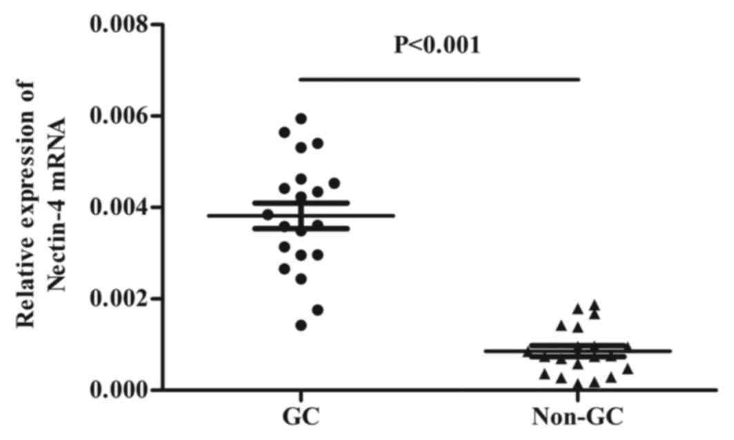
RT-qPCR was employed to determine the relative
expression of Nectin-4 mRNA which was isolated from 20 GC tissues
and matched tumor-adjacent normal tissue. It was identified that
Nectin-4 mRNA expression was significantly higher in GC samples
(0.0038±0.0012) compared with the paired adjacent non-tumor samples
(0.0008±0.0005) (P<0.001; Fig.
1).
Nectin-4 expression in GC tissues by
IHC
To validate the results acquired from RT-qPCR,
TMA-IHC was employed to evaluate Nectin-4 protein expression on 212
GC tissues and 91 matched peritumoral tissues. In GC tissues,
positive staining was mainly observed in the cell membrane and
cytoplasm. High expression levels of Nectin-4 were detected in
60.4% (128/212) of GC tumors compared with only 15.4% (14/91) of
matched adjacent normal tissue samples (χ2=51.76;
P<0.001), which was consistent with the current RT-qPCR results
(Fig. 2).
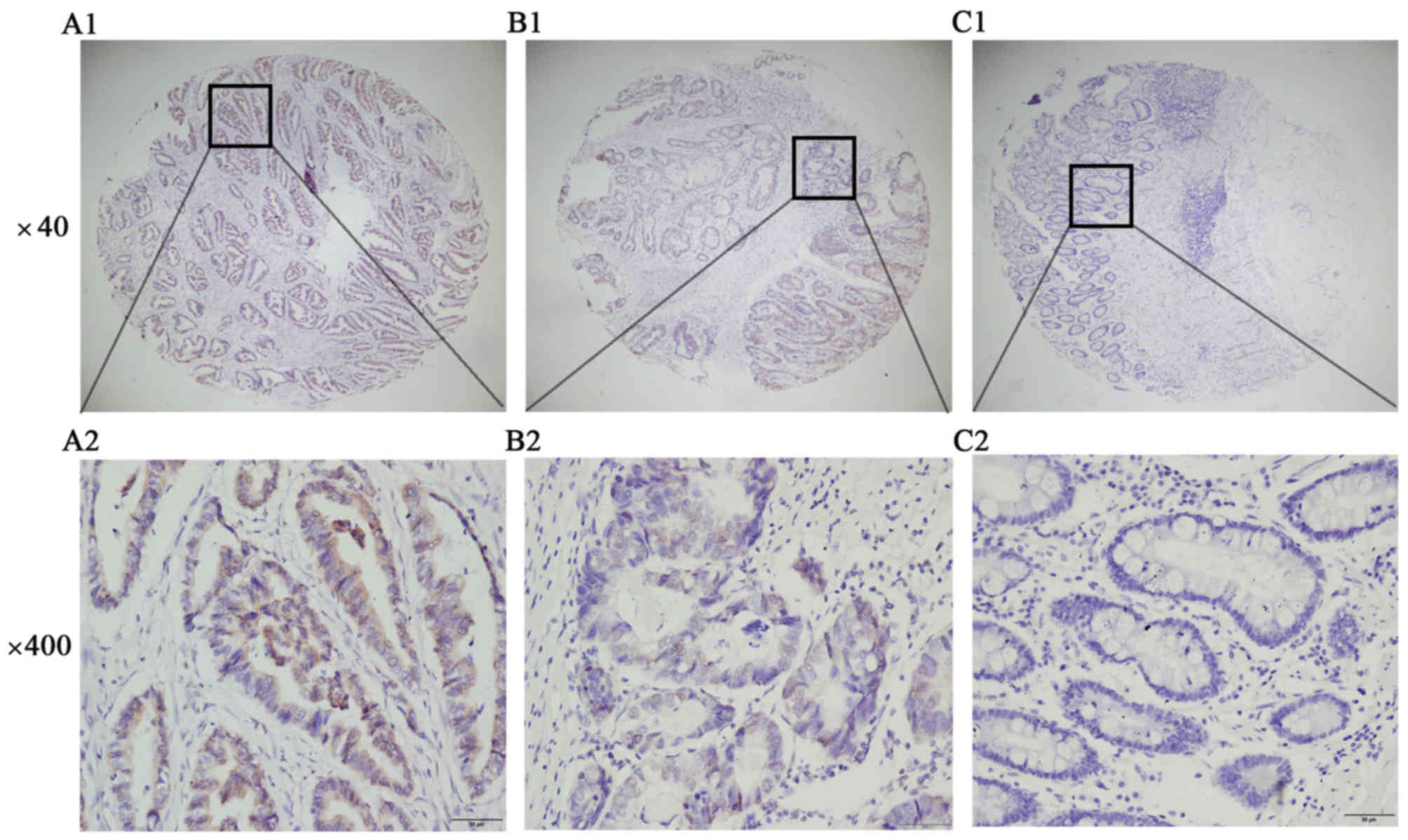 | Figure 2.Representative Nectin-4 protein
expression in GC and matched, peritumoral tissues. (A1, 2) Strong
positive IHC staining of Nectin-4 in GC. (B1, 2) Positive
expression of Nectin-4 in GC. (C1, 2) Peritumoral tissues, negative
expression of Nectin-4 in GC. Magnification, ×40 (A1, B1, C1), ×400
(A2, B2, C2). |
Association between Nectin-4 protein
expression and clinicopathological parameters of patients with
GC
The association between high Nectin-4 expression and
different types of clinicopathological parameters of patients with
GC is indicated in Table I. It was
identified that high expression of Nectin-4 was notably associated
with differentiation (P=0.004), primary tumor (P=0.001), lymph node
metastasis (P<0.001) and TNM stage (P<0.001). However, no
association was observed between Nectin-4 and other clinical
features, including sex, age, tumor location, tumor size and
preoperative carcinoembryonic antigen level (27) (Table I),
distant metastasis (M) occurred in only 10 cases, therefore its
association with Nectin-4 was not evaluated.
 | Table I.Association of Nectin-4 expression in
tumorous tissues with clinicopathological characteristics in
patients with gastric cancer. |
Table I.
Association of Nectin-4 expression in
tumorous tissues with clinicopathological characteristics in
patients with gastric cancer.
|
|
| Nectin-4 |
|---|
|
|
|
|
|---|
| Clinicopathological
characteristic | n | Low or no
expression, n (%) | High expression, n
(%) | Pearson
χ2 | P-value |
|---|
| Total | 212 | 84 (39.62) | 128 (60.38) |
|
|
| Sex |
|
|
| 1.803 | 0.179 |
|
Male | 140 | 60 (42.86) | 80 (57.14) |
|
|
|
Female | 72 | 24 (33.33) | 48 (66.67) |
|
|
| Age at diagnosis,
years |
|
|
| 0.016 | 0.898 |
|
≤60 | 92 | 36 (39.13) | 56 (60.87) |
|
|
|
>60 | 120 | 48 (40.00) | 72 (60.00) |
|
|
| Tumor size, cm |
|
|
| 3.441 | 0.064 |
|
<3 | 63 | 31 (49.21) | 32 (50.70) |
|
|
| ≥3 | 149 | 53 (35.57) | 96 (65.30) |
|
|
| Location |
|
|
| 1.816 | 0.403 |
|
Lower | 140 | 58 (41.43) | 82 (58.57) |
|
|
|
Middle | 45 | 14 (31.11) | 31 (68.89) |
|
|
|
Upper | 27 | 12 (44.44) | 15 (55.56) |
|
|
|
Differentiation |
|
|
| 8.408 | 0.004a |
| Low
grade | 126 | 40 (31.75) | 86 (68.25) |
|
|
| Middle
and high grade | 70 | 37 (52.86) | 33 (47.14) |
|
|
|
Other | 16 | 7 | 9 |
|
|
| Primary tumor |
|
|
| 14.826 | 0.001a |
| T1 | 51 | 27 (52.94) | 24 (47.06) |
|
|
| T2 | 41 | 23 (56.10) | 18 (43.90) |
|
|
|
T3+T4 | 120 | 34 (28.33) | 86 (71.67) |
|
|
| Lymph node
metastasis |
|
|
| 18.544 |
<0.001a |
|
N0 | 103 | 55 (53.40) | 48 (46.60) |
|
|
|
N1 | 31 | 12 (38.71) | 19 (61.29) |
|
|
|
N2+N3 | 78 | 17 (21.79) | 61 (78.21) |
|
|
| Stage grouping with
TNM |
|
|
| 32.749 |
<0.001a |
| Stage
I | 76 | 43 (56.58) | 33 (43.42) |
|
|
| Stage
II | 42 | 24 (57.14) | 18 (42.86) |
|
|
| Stage
III+IV | 94 | 17 (18.09) | 77 (81.91) |
|
|
| Preoperative CEA,
ng/ml |
|
|
| 0.421 | 0.517 |
| ≤5 | 159 | 65 (40.88) | 94 (59.12) |
|
|
|
>5 | 53 | 19 (35.85) | 34 (64.15) |
|
|
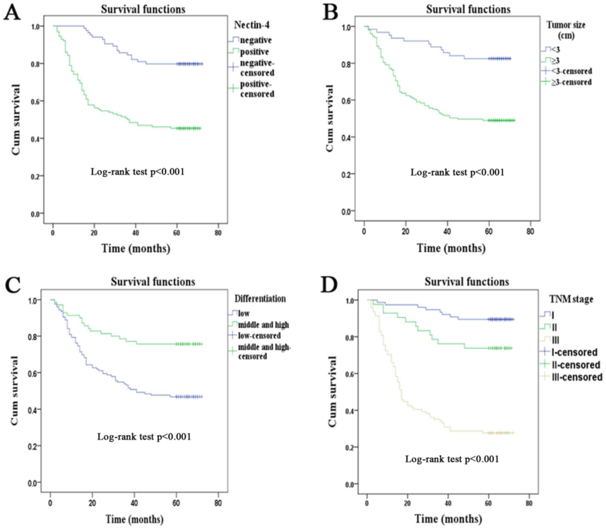
Survival analysis
A univariate analysis was used to investigate all
relevant features, and it was identified that high Nectin-4
expression (P<0.001), along with tumor size (P<0.001), tumor
differentiation (P<0.001), T (P<0.001), N (P<0.001) and
tumor TNM stage (P<0.001), was markedly associated with the
survival of patients (Table II).
Multivariate regression analysis was subsequently employed to
further confirm that Nectin-4 expression (P=0.002) and tumor TNM
stage (P<0.001) were independent prognostic indicators in GC
(Table II). Kaplan-Meier survival
curves revealed that patients with GC with high Nectin-4
expression, large tumor size (≥3 cm), low-differentiated tumors and
advanced TNM stage had a significantly poorer prognosis
(P<0.001; Fig. 3).
| Table II.Univariate and multivariate analysis
of prognostic factors of 5-year overall survival in patients with
gastric cancer. |
Table II.
Univariate and multivariate analysis
of prognostic factors of 5-year overall survival in patients with
gastric cancer.
|
| Univariate
analysis | Multivariate
analysis |
|---|
|
|
|
|
|---|
| Characteristic | HR | P-value | 95% CI | HR | P-value | 95% CI |
|---|
| Nectin-4
expression | 3.815 |
<0.001a | 2.243–6.490 | 2.402 | 0.002a | 1.364–4.232 |
| High
vs. low and none |
|
|
|
|
|
|
| Sex | 0.825 | 0.387 | 0.533–1.276 |
|
|
|
| Male
vs. female |
|
|
|
|
|
|
| Age, years | 1.033 | 0.881 | 0.675–1.580 |
|
|
|
| ≤60 vs.
>60 |
|
|
|
|
|
|
|
Location | 0.814 | 0.141 | 0.619–1.070 |
|
|
|
| Lower vs. middle
vs. upper |
|
|
|
|
|
|
| Tumor
size, cm | 3.810 |
<0.001a | 2.023–7.179 | 1.638 | 0.151 | 0.835–3.214 |
| <3
vs. ≥3 |
|
|
|
|
|
|
|
Differentiation | 0.375 |
<0.001a | 0.220–0.640 | 0.749 | 0.323 | 0.422–1.329 |
| Low vs.
middle and high grade vs. others |
|
|
|
|
|
|
| Primary tumor | 2.768 |
<0.001a | 1.913–4.006 |
|
|
|
| T1 vs.
T2 vs. T3+T4 |
|
|
|
|
|
|
| Lymph node
metastasis | 2.224 |
<0.001a | 1.850–2.674 |
|
|
|
|
N0 vs.
N1 vs. N2+N3 |
|
|
|
|
|
|
| TNM stage | 3.660 |
<0.001a | 2.589–5.172 | 2.442 |
<0.001a | 1.651–3.612 |
| I vs.
II vs. III+IV |
|
|
|
|
|
|
| Preoperative CEA,
ng/ml | 1.203 | 0.441 | 0.752–1.925 |
|
|
|
| ≤5 vs.
>5 |
|
|
|
|
|
|
Discussion
In the present study, RT-qPCR analysis was used to
compare the mRNA expression of Nectin-4 in GC tissues with matched
normal tissues. The result identified a marked increase in tumor
tissues, which as consistent with previous studies (19–23).
TMA-IHC was subsequently performed to further validate this result,
and it was identified that Nectin-4 protein was similarly highly
expressed in GC tumorous tissues when compared with adjacent
non-tumorous tissues. High protein expression of Nectin-4 in GC was
associated with certain pathological and clinical features
including differentiation degree, primary tumor, lymph node
metastasis and TNM stage. Univariate analysis was employed to
investigate the association between clinicopathological
characteristics including Nectin-4 and overall survival of patients
with GC. The result revealed that Nectin-4 expression, tumor size,
tumor differentiation, T, N and tumor TNM stage had a negative
association with the overall 5-year survival rate in GC.
Multivariate analysis further indicated that Nectin-4 expression as
well as TNM stage independently predicted adverse outcomes of
patients with GC. Kaplan-Meier analysis provided a visual
representation that the lifetime of patients with low Nectin-4
expression was notably longer than that of patients with high
expression.
As aforementioned, previous in vitro and
in vivo evidence suggested the function of Nectin-4 in a
number of tumors as a carcinogenic factor, which was consistent
with results of the present study (19,22,23). In
gallbladder carcinoma, ectopic expression of Nectin-4 had been
demonstrated to be associated with cell proliferation, movement
ability and anchorage-independent growth in in vitro
experiments and a mouse model (23).
Takano et al (22) reported
that Nectin-4 was highly associated with unfavorable clinical
outcomes in non-small cell lung cancer, and increased expression of
Nectin-4 was able to promote the formation of lamellipodia and
increase the invasive ability of cancer cells through the Rac1
signaling pathway, and the proliferative ability of tumor cells
silenced with siRNA of Nectin-4 was significantly decreased.
Additionally, nectin-4, together with cancer antigen 125, may help
to make a distinction between benign gynecological diseases and
ovarian cancer (19).
Rac1 signaling is involved in in tumor growth and
progression of numerous types of cancer, and a number of functions
in the progression of cancer, including proliferation,
differentiation, migration, invasion, survival and cancer
metastasis (28–30). Rac1 signaling is necessary for the
extension of protrusions, including the formation of lamellipodia,
which is crucial for cell-cell adherence (31,32).
Cell-cell adhesive processes are important for cancer progression
(33). Nectin family members are
homophilic and heterophilic cell adhesion molecules that bind
afadin scaffold molecule, an actin filament (F-actin)-binding
protein through their cytoplasmic tails and associate with the
actin cytoskeleton (33,34). Nectins serve critical functions in
cellular activities including movement, differentiation,
polarization and the entry of viruses, in cooperation with other
cell adhesion molecules and cell-surface membrane receptors
(10,13,35,36).
Nectin-4 belongs to the family of cell adhesion molecules that
regulate the formation of adherence functions, and Nectin-4 is
different from the other the Nectin family members owing to its
distinct distribution, in that it is expressed specifically in the
embryo and placenta as opposed to adult tisues (14,15).
Overexpression of Nectin-4 has been identified to be an essential
contributor to cell proliferation and highly malignant tumor
phenotypes and is known to serve a function as a significant
mediator of the Rac1 signaling pathway (22). Activation of Rac1 signaling enables
Nectin-4 to enhance the lamellipodia formation, cellular
proliferation and migration of the cell (37). Previous studies have also demonstrated
that the Rac1 signaling pathway is mainly modulated by the PI3K/AKT
and mitogen-activated protein kinase/extracellular-signal-regulated
kinase (ERK) kinase (MEK)/ERK signaling pathways in several types
of human tumor (38–40). Zhang et al (23) also demonstrated that Rac1 was a
downstream target of the PI3K/AKT pathway and demonstrated that
this pathway cooperated with Nectin-4 and Rac1 to mediate cell
proliferation and migration during tumor progression; furthermore,
no association was identified between Nectin-4 and the MEK/ERK
signaling pathway (23).
Collectively, these studies suggested that Nectin-4 exerted
oncogenic properties and may be a candidate for targeted therapy in
certain types of human cancer.
There are several limitations of the present study.
The expression of Nectin-4 in patients with GC was studied
retrospectively, therefore whether Nectin-4 is the main promoting
factor of GC progression or a consequence of GC development remains
uncertain, and additional study is required to resolve the precise
nature of the association. Furthermore, TMA-IHC was employed to
evaluate Nectin-4 protein expression. The results were subjective
and semiquantitative, therefore additional methods are required to
validate the expression level of Nectin-4 in cancer tissues and
cells. Finally, in vitro and in vivo studies were not
performed to identify the underlying molecular mechanisms of
Nectin-4 expression in GC.
The results of the present study suggested that
expression of Nectin-4 was higher in GC tissues and was
significantly associated with poorer outcome. Therefore, Nectin-4
was able to serve as a novel marker, and is a promising target to
evaluate the prognosis of patients with GC. The results of the
present study enrich current knowledge of Nectin-4 in the
occurrence and development of GC. Further study should be carried
out to explore the underlying molecular mechanism of action of this
molecule in GC development. In conclusion, the present study
provides rationale to further investigation into the involvement of
Nectin-4 in GC development in addition to Nectin-4 as a potential
prognostic marker and therapeutic target in GC.
Funding
The present study was supported by the National
Natural Science Foundations for Young Scientists of China (grant
no. 81502053).
Availability of data and materials
The datasets generated and analyzed in the present
study are included in this published article.
Authors' contributions
YZ, YL and QN conceived and designed the
experiments. JZ, QS and HH performed the experiments. QS and WY
performed statistical analysis and data interpretation. YZ wrote
the manuscript.
Ethics and consent to participate
The Human Research Ethics Committee of Nantong
University Affiliated Hospital approved the present study and all
clinical methods applied, and all patients provided written
informed consent prior to participation in this clinical trial and
research.
Consent for publication
All patients provided consent for publication.
Competing interests
The authors declare that they have no competing
interests.
References
|
1
|
Hartgrink HH, Jansen EP, van Grieken NC
and van de Velde CJ: Gastric cancer. Lancet. 374:477–490. 2009.
View Article : Google Scholar : PubMed/NCBI
|
|
2
|
Jemal A, Bray F, Center MM, Ferlay J, Ward
E and Forman D: Global cancer statistics. CA Cancer J Clin.
61:69–90. 2011. View Article : Google Scholar : PubMed/NCBI
|
|
3
|
Goggins WB and Wong GK: Poor survival for
US Pacific Islander cancer patients: Evidence from the
surveillance, epidemiology, and end results database: 1991 to 2004.
J Clin Oncol. 25:5738–5741. 2007. View Article : Google Scholar : PubMed/NCBI
|
|
4
|
Jemal A, Siegel R, Ward E, Hao Y, Xu J,
Murray T and Thun MJ: Cancer statistics, 2008. CA Cancer J Clin.
58:71–96. 2008. View Article : Google Scholar : PubMed/NCBI
|
|
5
|
Lin Y, Ueda J, Kikuchi S, Totsuka Y, Wei
WQ, Qiao YL and Inoue M: Comparative epidemiology of gastric cancer
between Japan and China. World J Gastroenterol. 17:4421–4428. 2011.
View Article : Google Scholar : PubMed/NCBI
|
|
6
|
Torre LA, Bray F, Siegel RL, Ferlay J,
Lortet-Tieulent J and Jemal A: Global cancer statistics, 2012. CA
Cancer J Clin. 65:87–108. 2015. View Article : Google Scholar : PubMed/NCBI
|
|
7
|
Choi YY, Noh SH and Cheong JH: Evolution
of gastric cancer treatment: From the golden age of surgery to an
era of precision medicine. Yonsei Med J. 56:1177–1185. 2015.
View Article : Google Scholar : PubMed/NCBI
|
|
8
|
Peng P, Wu W, Zhao J, Song S, Wang X, Jia
D, Shao M, Zhang M, Li L, Wang L, et al: Decreased expression of
Calpain-9 predicts unfavorable prognosis in patients with gastric
cancer. Sci Rep. 6:296042016. View Article : Google Scholar : PubMed/NCBI
|
|
9
|
Garrido M, Fonseca PJ, Vieitez JM, Frunza
M and Lacave AJ: Challenges in first line chemotherapy and targeted
therapy in advanced gastric cancer. Expert Rev Anticancer Ther.
14:887–900. 2014. View Article : Google Scholar : PubMed/NCBI
|
|
10
|
Takai Y, Ikeda W, Ogita H and Rikitake Y:
The immunoglobulin-like cell adhesion molecule nectin and its
associated protein afadin. Annu Rev Cell Dev Biol. 24:309–342.
2008. View Article : Google Scholar : PubMed/NCBI
|
|
11
|
Yasumi M, Shimizu K, Honda T, Takeuchi M
and Takai Y: Role of each immunoglobulin-like loop of nectin for
its cell-cell adhesion activity. Biochem Biophys Res Commun.
302:61–66. 2003. View Article : Google Scholar : PubMed/NCBI
|
|
12
|
Nakanishi H and Takai Y: Roles of nectins
in cell adhesion, migration and polarization. Biol Chem.
385:885–892. 2004. View Article : Google Scholar : PubMed/NCBI
|
|
13
|
Takai Y, Miyoshi J, Ikeda W and Ogita H:
Nectins and nectin-like molecules: Roles in contact inhibition of
cell movement and proliferation. Nat Rev Mol Cell Biol. 9:603–615.
2008. View
Article : Google Scholar : PubMed/NCBI
|
|
14
|
Reymond N, Fabre S, Lecocq E, Adelaïde J,
Dubreuil P and Lopez M: Nectin4/PRR4, a new afadin-associated
member of the nectin family that trans-interacts with nectin1/PRR1
through V domain interaction. J Biol Chem. 276:43205–43215. 2001.
View Article : Google Scholar : PubMed/NCBI
|
|
15
|
Fabre S, Reymond N, Cocchi F, Menotti L,
Dubreuil P, Campadelli-Fiume G and Lopez M: Prominent role of the
Ig-like V domain in trans-interactions of nectins. Nectin3 and
nectin 4 bind to the predicted C-C'-C'-D beta-strands of the
nectin1 V domain. J Biol Chem. 277:27006–27013. 2002. View Article : Google Scholar : PubMed/NCBI
|
|
16
|
Fabre-Lafay S, Monville F, Garrido-Urbani
S, Berruyer-Pouyet C, Ginestier C, Reymond N, Finetti P, Sauvan R,
Adélaïde J, Geneix J, et al: Nectin-4 is a new histological and
serological tumor associated marker for breast cancer. BMC Cancer.
7:732007. View Article : Google Scholar : PubMed/NCBI
|
|
17
|
Fabre-Lafay S, Garrido-Urbani S, Reymond
N, Gonçalves A, Dubreuil P and Lopez M: Nectin-4, a new serological
breast cancer marker, is a substrate for tumor necrosis
factor-alpha-converting enzyme (TACE)/ADAM-17. J Biol Chem.
280:19543–19550. 2005. View Article : Google Scholar : PubMed/NCBI
|
|
18
|
Athanassiadou AM, Patsouris E, Tsipis A,
Gonidi M and Athanassiadou P: The significance of Survivin and
Nectin-4 expression in the prognosis of breast carcinoma. Folia
Histochem Cytobiol. 49:26–33. 2011. View Article : Google Scholar : PubMed/NCBI
|
|
19
|
Derycke MS, Pambuccian SE, Gilks CB,
Kalloger SE, Ghidouche A, Lopez M, Bliss RL, Geller MA, Argenta PA,
Harrington KM and Skubitz AP: Nectin 4 overexpression in ovarian
cancer tissues and serum: Potential role as a serum biomarker. Am J
Clin Pathol. 134:835–845. 2010. View Article : Google Scholar : PubMed/NCBI
|
|
20
|
Nishiwada S, Sho M, Yasuda S, Shimada K,
Yamato I, Akahori T, Kinoshita S, Nagai M, Konishi N and Nakajima
Y: Nectin-4 expression contributes to tumor proliferation,
angiogenesis and patient prognosis in human pancreatic cancer. J
Exp Clin Cancer Res. 34:302015. View Article : Google Scholar : PubMed/NCBI
|
|
21
|
Ma J, Sheng Z, Lv Y, Liu W, Yao Q, Pan T,
Xu Z, Zhang C and Xu G: Expression and clinical significance of
Nectin-4 in hepatocellular carcinoma. Onco Targets Ther. 9:183–190.
2016.PubMed/NCBI
|
|
22
|
Takano A, Ishikawa N, Nishino R, Masuda K,
Yasui W, Inai K, Nishimura H, Ito H, Nakayama H, Miyagi Y, et al:
Identification of nectin-4 oncoprotein as a diagnostic and
therapeutic target for lung cancer. Cancer Res. 69:6694–6703. 2009.
View Article : Google Scholar : PubMed/NCBI
|
|
23
|
Zhang Y, Liu S, Wang L, Wu Y, Hao J, Wang
Z, Lu W, Wang XA, Zhang F, Cao Y, et al: A novel PI3K/AKT signaling
axis mediates Nectin-4-induced gallbladder cancer cell
proliferation, metastasis and tumor growth. Cancer Lett.
375:179–189. 2016. View Article : Google Scholar : PubMed/NCBI
|
|
24
|
Jun KH, Lee JS, Kim JH, Kim JJ, Chin HM
and Park SM: The rationality of N3 classification in the 7th
edition of the International Union Against Cancer TNM staging
system for gastric adenocarcinomas: A case-control study. Int J
Surg. 12:893–896. 2014. View Article : Google Scholar : PubMed/NCBI
|
|
25
|
Livak KJ and Schmittgen TD: Analysis of
relative gene expression data using real-time quantitative PCR and
the 2(-Delta Delta C(T)) method. Methods. 25:402–408. 2001.
View Article : Google Scholar : PubMed/NCBI
|
|
26
|
Ni S, Xu L, Huang J, Feng J, Zhu H, Wang G
and Wang X: Increased ZO-1 expression predicts valuable prognosis
in non-small cell lung cancer. Int J Clin Exp Pathol. 6:2887–2895.
2013.PubMed/NCBI
|
|
27
|
Qin HF, Qu LL, Liu H, Wang SS and Gao HJ:
Serum CEA level change and its significance before and after
Gefitinib therapy on patients with advanced non-small cell lung
cancer. Asian Pac J Cancer Prev. 14:4205–4208. 2013. View Article : Google Scholar : PubMed/NCBI
|
|
28
|
Wang Y, Chen Y, Wu M, Lan T, Wu Y, Li Y
and Qian H: Type II cyclic guanosine monophosphate-dependent
protein kinase inhibits Rac1 activation in gastric cancer cells.
Oncol Lett. 10:502–508. 2015. View Article : Google Scholar : PubMed/NCBI
|
|
29
|
Zhu G, Fan Z, Ding M, Zhang H, Mu L, Ding
Y, Zhang Y, Jia B, Chen L, Chang Z and Wu W: An EGFR/PI3K/AKT axis
promotes accumulation of the Rac1-GEF Tiam1 that is critical in
EGFR-driven tumorigenesis. Oncogene. 34:5971–5982. 2015. View Article : Google Scholar : PubMed/NCBI
|
|
30
|
Bousquet E, Calvayrac O, Mazières J,
Lajoie-Mazenc I, Boubekeur N, Favre G and Pradines A: RhoB loss
induces Rac1-dependent mesenchymal cell invasion in lung cells
through PP2A inhibition. Oncogene. 35:1760–1769. 2016. View Article : Google Scholar : PubMed/NCBI
|
|
31
|
Jaffe AB and Hall A: Rho GTPases in
transformation and metastasis. Adv Cancer Res. 84:57–80. 2002.
View Article : Google Scholar : PubMed/NCBI
|
|
32
|
Takai Y, Sasaki T and Matozaki T: Small
GTP-binding proteins. Physiol Rev. 81:153–208. 2001. View Article : Google Scholar : PubMed/NCBI
|
|
33
|
Samanta D and Almo SC: Nectin family of
cell-adhesion molecules: Structural and molecular aspects of
function and specificity. Cell Mol Life Sci. 72:645–658. 2015.
View Article : Google Scholar : PubMed/NCBI
|
|
34
|
Sakisaka T, Ikeda W, Ogita H, Fujita N and
Takai Y: The roles of nectins in cell adhesions: Cooperation with
other cell adhesion molecules and growth factor receptors. Curr
Opin Cell Biol. 19:593–602. 2007. View Article : Google Scholar : PubMed/NCBI
|
|
35
|
Rikitake Y, Mandai K and Takai Y: The role
of nectins in different types of cell-cell adhesion. J Cell Sci.
125:3713–3722. 2012. View Article : Google Scholar : PubMed/NCBI
|
|
36
|
Fuchs A and Colonna M: The role of NK cell
recognition of nectin and nectin-like proteins in tumor
immunosurveillance. Semin Cancer Biol. 16:359–366. 2006. View Article : Google Scholar : PubMed/NCBI
|
|
37
|
Fortugno P, Josselin E, Tsiakas K, Agolini
E, Cestra G, Teson M, Santer R, Castiglia D, Novelli G,
Dallapiccola B, et al: Nectin-4 mutations causing ectodermal
dysplasia with syndactyly perturb the rac1 pathway and the kinetics
of adherens junction formation. J Invest Dermatol. 134:2146–2153.
2014. View Article : Google Scholar : PubMed/NCBI
|
|
38
|
Henderson V, Smith B, Burton LJ, Randle D,
Morris M and Odero-Marah VA: Snail promotes cell migration through
PI3K/AKT-dependent Rac1 activation as well as PI3K/AKT-independent
pathways during prostate cancer progression. Cell Adh Migr.
9:255–264. 2015. View Article : Google Scholar : PubMed/NCBI
|
|
39
|
Vial E, Sahai E and Marshall CJ: ERK-MAPK
signaling coordinately regulates activity of Rac1 and RhoA for
tumor cell motility. Cancer Cell. 4:67–79. 2003. View Article : Google Scholar : PubMed/NCBI
|
|
40
|
Ray RM, Vaidya RJ and Johnson LR: MEK/ERK
regulates adherens junctions and migration through Rac1. Cell Motil
Cytoskeleton. 64:143–156. 2007. View Article : Google Scholar : PubMed/NCBI
|