Introduction
Hepatocellular carcinoma (HCC) is the most fatal
cancer worldwide (1). Known causes of
liver cancer include hepatitis B or C infection, alcohol-induced
cirrhosis (2) and chronic diseases,
including obesity and diabetes, which lead to liver fibrosis
(3). HCC is characterized by mixed
cell types, strong resistance to chemotherapy and local (rather
than distant) metastasis, all of which present challenges to
effective treatment.
Transforming growth factor-β (TGF-β) has been
suggested as a useful biomarker for fibrosis, cirrhosis and
epithelial-mesenchymal transition (EMT) in patients with HCC
(4). EMT is a predisposing factor for
the development of HCC (5) and has
been shown to be induced by several cytokines, including
interleukins, TGF-β and interferons (6).
TGF-β induces differentiation of embryonic stem
cells, somatic stem cells and cancer stem cells (7). In addition, TGF-β induces
differentiation of quiescent hepatic stellate cells into activated
cells in the liver (8), fibrosis
through extracellular matrix (ECM) deposition and HCC (9). TGF-β binds several proteins, activates
several signaling pathways and is involved in HCC progression
(10). TGF-β enhances tumor
initiating ability through EMT of cancer stem cells (11). Tumor initiating ability is a known
characteristic of cancer stem cells (12).
Cancer stem cells are characterized by resistance to
anticancer drugs (13), high sphere
forming efficacy (14), EMT (15) and specific protein expression that
promotes transformation (16). HCC
stem cells express cluster of differentiation (CD)90, CD133 and
CD24 (17), and retain side
population (SP) characteristics, as well as resistance to
anticancer drugs (18). In addition,
liver cancer typically exhibits strong resistance to anticancer
drugs, since detoxification is one of its functions (19).
The present study focused on the effect of TGF-β on
cancer stem cells in vitro. The proliferation, fraction of
SP cells and expression of relevant signaling molecules were
assessed following TGF-β and SB431542 treatment using Huh-7 and
Huh-Bat cells as a model for HCC.
Materials and methods
Cell culture
Huh-7 and Huh-Bat [Huh-7 transfected with human
Na+-taurocholate co-transporting polypeptide (bile acid
transporter) cDNA] cell lines were obtained from the Korean Cell
Line Bank (Seoul, Korea). Cells were cultured in Dulbecco's
modified Eagle's medium (DMEM; Thermo Fisher Scientific, Inc.,
Waltham, MA, USA) containing 10% fetal bovine serum (FBS; Thermo
Fisher Scientific, Inc.). For adherent cultures, 5×10⁵
cells were seeded onto tissue culture dishes (BD Biosciences, San
Jose, CA, USA). All cultures were maintained at 37°C in a
humidified 5% CO2 atmosphere.
Cell survival rate following SB431542
and TGF-β treatment
Cells were cultured (5×10⁵ cells) in DMEM
containing 10% FBS at 37°C. After 24 h, the cells were washed twice
with PBS, and fresh medium was added. Cells were treated for 72 h
at 37°C with 0.01% dimethyl sulfoxide (vehicle control), 1 µM
SB431542 (TGF-β inhibitor; Sigma-Aldrich; Merck KGaA, Darmstadt,
Germany), 1 ng/ml TGF-β (LC Laboratories, Woburn, MA, USA) or 1 µM
SB431542 plus 1 ng/ml TGF-β. Following treatment for 72 h, cells
were harvested for subsequent analysis. Cell survival rates were
estimated by the number of viable cells, counted by positive
staining with 0.4% trypan blue dye (Sigma-Aldrich; Merck KGaA) in a
Neubauer chamber.
Cell cycle analysis
To determine the effect of treatment on cell cycle
progression, at 72 h following treatment, cell samples were
harvested and fixed in 70% ethanol for 25 min. Subsequent to
washing in PBS, samples were treated with 100 µg/ml RNaseA for 90
min at 37°C, and stained with 10 µg/ml propidium iodide (PI). Flow
cytometry was performed in triplicate for each experiment using a
FACSCalibur (BD Biosciences).
SP cell analysis
SP cell analysis was performed as reported
previously (20). Briefly, cells were
detached, transferred to microcentrifuge tubes, washed in PBS and
resuspended at 1×10⁶ cells/ml. Samples were stained with Hoechst
33342 dye (5 µg/ml; Sigma-Aldrich; Merck KGaA) or Hoechst dye and
50–100 µM verapamil (an efflux blocker), in DMEM containing 10% FBS
for 90 min at 37°C. Following staining, samples were centrifuged
for 3 min at 150 × g and 4°C and collected for analysis of the SP
fraction. PI (1 µg/ml) was added prior to analysis to identify and
exclude dead cells. Samples were analyzed using a FACSAria (BD
Biosciences).
Expression of CD133, E-Cadherin,
α-smooth muscle actin (SMA) and vimentin
To determine the phenotype of the cell populations
following TGF-β and/or SB431542 treatment, cells were harvested and
stained for CD133, E-cadherin, α-SMA and vimentin expression.
Briefly, cell samples were washed twice with PBS, then fixed and
permeabilized according to the manufacturer's protocol (BD
Biosciences). Following centrifugation for 3 min at 150 × g and
4°C, cells were washed and incubated for 45 min at 4°C with
anti-human CD133-APC (cat. no. 130-090-826; 1:50; Miltenyi Biotec
Ltd., Bergisch Gladbach, Germany), PE anti-human E-cadherin (cat.
no. 562870; 1:50; BD Biosciences), anti-human α-SMA-fluorescein
isothiocyanate (cat. no. ab8211; 1:50; Abcam, Cambridge, UK) and
anti-human vimentin-Alexa488 (cat. no. 562338; 1:50; BD
Biosciences). Following staining, cells were washed with PBS three
times, and flow cytometry analysis was performed in triplicate
using FACSCalibur (BD Biosciences).
Immunoblotting
Total cell lysates were prepared in 100 µl lysis
buffer (Cell Signaling Technology, Inc., Danvers, MA, USA). Protein
concentrations were measured using Bio-Rad protein assay kit I,
according to the manufacturer's protocol (cat. no. 5000001; Bio-Rad
Laboratories, Inc., Hercules, CA, USA). Cell lysates were separated
using 10% SDS-PAGE, and proteins were electrotransferred onto
Hybond-enhanced chemiluminescence nitrocellulose membranes (GE
Healthcare Life Sciences, Chalfont, UK). Blots were blocked for 1 h
with blocking buffer (5% skim milk in TBS-tween 20) and incubated
overnight at 4°C with mouse anti-phospho-SAPK/JNK (Thr183/Tyr185;
cat. no. 9251; 1:1,000; Cell Signaling Technology, Inc.), rabbit
anti-phospho-c-Jun (ser63; cat. no. 2361; 1:1,000; Cell Signaling
Technology, Inc.), rabbit anti-phospho-smad2 (cat. no. 8828;
1:1,000; Cell Signaling Technology), and mouse anti-β-actin (cat.
no. sc47778; 1:1,000; Santa Cruz Biotechnology, Inc., Dallas, TX,
USA). Blots were washed and incubated for 1 h at room temperature
with peroxidase-conjugated AffiniPure rabbit anti-mouse IgG (cat.
no. 315-005-003; 1:2,500; Jackson ImmunoResearch Laboratories,
Inc., West Grove, PA, USA) or peroxidase-conjugated AffiniPure
mouse anti-rabbit IgG (cat. no. 211-005-109; 1:2,500; Jackson
ImmunoResearch Laboratories, Inc.). Labeled proteins were detected
using an enhanced chemiluminescence detection system (GE Healthcare
Life Sciences).
Statistical analysis
At least three replicate experiments were performed
for all analyses. Data are expressed as the mean ± standard error.
Student's t-tests were used to compare the results of treated and
control cells. P<0.05 was considered to indicate a statistically
significant difference.
Results
TGF-β treatment inhibits proliferation
of Huh-7 and Huh-Bat cells
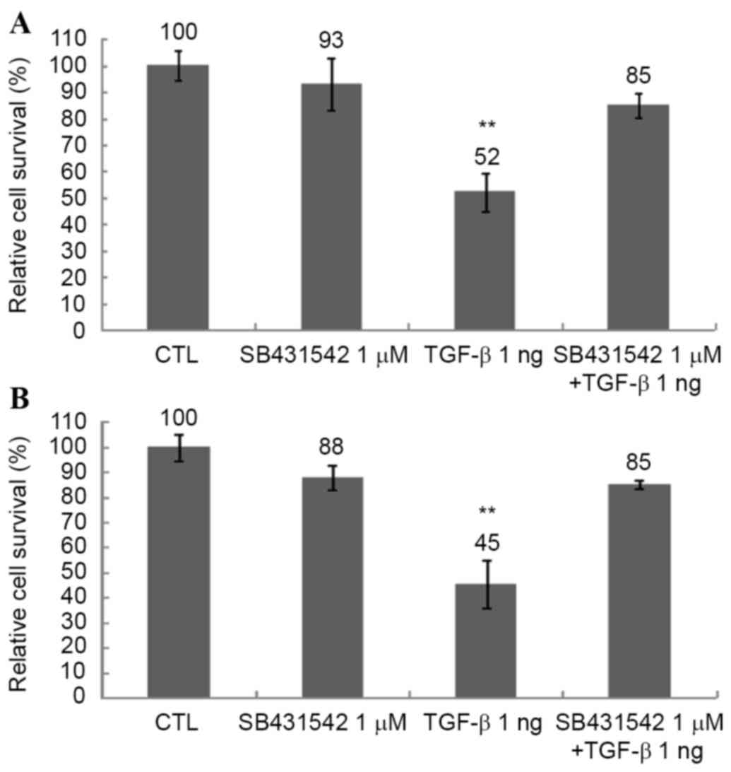
To determine the effect of TGF-β on the survival
rate of Huh-7 and Huh-Bat cells, cultures were treated with 1 µM
SB431542, 1 ng/ml TGF-β or 1 µM SB431542 plus 1 ng/ml TGF-β for 72
h at 37°C. The survival of Huh-7 and Huh-Bat cells was reduced by
TGF-β treatment and this effect was blocked by 1 µM SB431542
(Fig. 1). These results revealed that
TGF-β specifically inhibited the survival of HCC cells in
vitro.
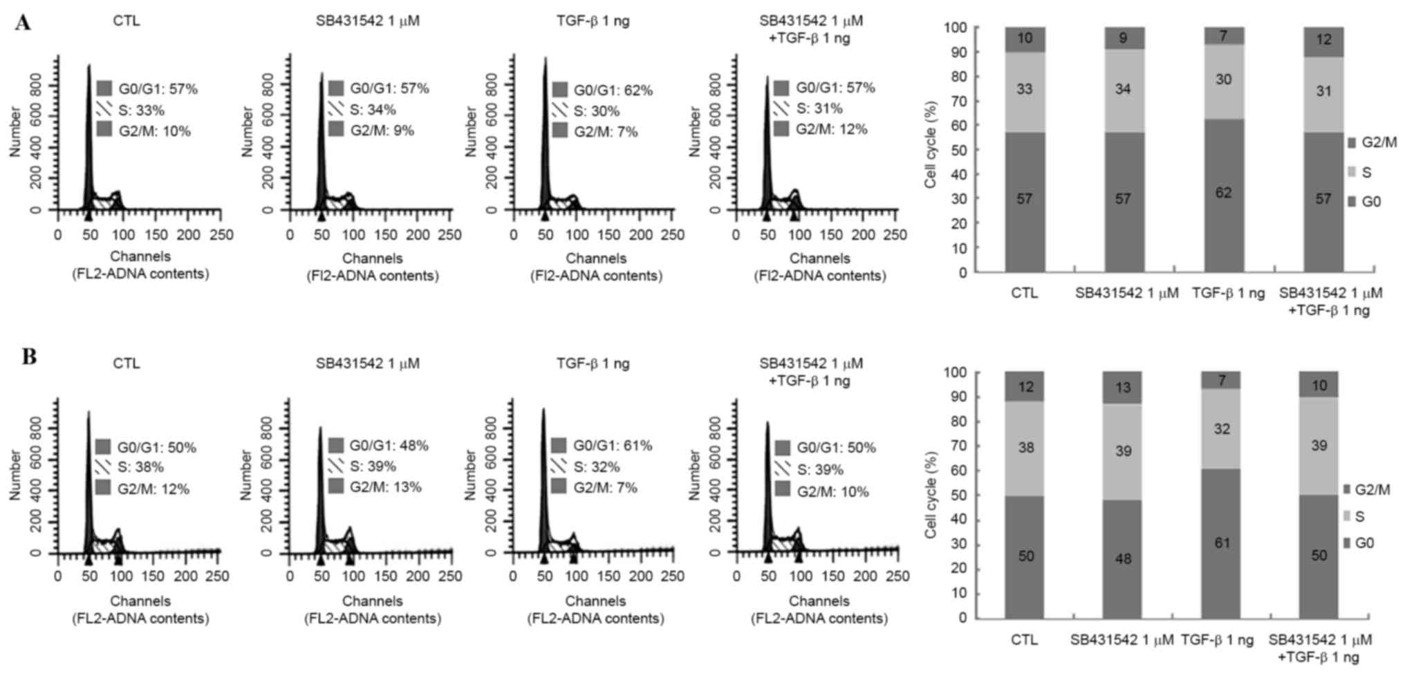
TGF-β treatment induces cell cycle
arrest in Huh-7 and Huh-Bat cells
TGF-β is known to induce cell cycle arrest in
several types of cancer (21). TGF-β
was found to decrease the survival rate of Huh-7 and Huh-Bat cells
after 72 h (Fig. 1). To confirm the
inhibitory effects of TGF-β on cell survival rate, the cell cycle
was analyzed following TGF-β and SB431542 treatment in Huh-7 and
Huh-Bat cells. TGF-β induced a 5% increase of Huh-7 cells in G0/G1
and an 11% increase of Huh-Bat cells in G0/G1 compared with the
control; cell cycle arrest was blocked by SB431542 (Fig. 2). These findings indicated that TGF-β
reduces survival of HCC cells by induction of cell cycle
arrest.
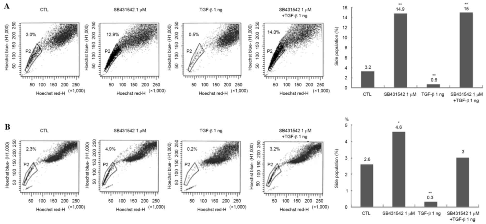
TGF-β treatment reduces the number of
SP cells in Huh-7 and Huh-Bat cultures
To determine whether TGF-β treatment targeted cancer
stem cells in Huh-7 and Huh-Bat cultures, SP cells were analyzed
using Hoechst dye staining following 1 µM SB431542, 1 ng/ml TGF-β
or 1 µM SB431542 plus 1 ng/ml TGF-β treatment for 72 h. TGF-β
treatment reduced the proportion of SP cells, whereas SB431542
treatment increased the proportion of SP cells, and TGF-β plus
SB431542 blocked the effect of TGF-β on SP cells (Fig. 3). These results indicated that the
effect of TGF-β may be specific for cancer stem cells.
Expression of CD133, E-cadherin, α-SMA
and vimentin in Huh-7 and Huh-bat cells following TGF-β
treatment
To confirm the effect of TGF-β on SP cells, the
expression levels of the cancer stem cell markers CD133 and
epithelial mesenchymal transition markers (EMT, E-cadherin, α-SMA
and vimentin) were analyzed. Expression of the cancer stem cell
marker CD133 and EMT markers (E-cadherin, α-SMA and vimentin)
differed depending on cell type. Huh-7 cells expressed high levels
of CD133 and vimentin compared with Huh-Bat cells (Fig. 4). However, following SB431542
treatment, expression of CD133 and vimentin was upregulated in
Huh-Bat cells. By contrast, expression of these markers was similar
in control and TGF-β-treated cells (Fig.
4). The present results revealed that the expression of cancer
stem cell and EMT markers was dependent on cell type, and was
upregulated by blockade of TGF-β signaling.
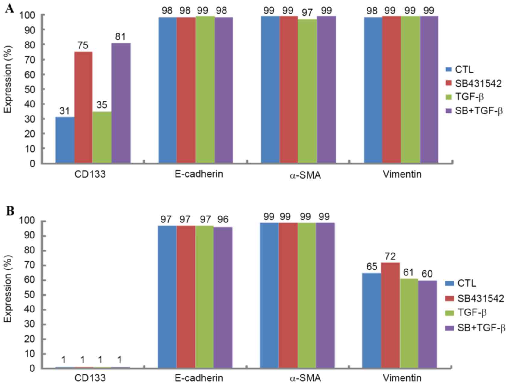 | Figure 4.Expression of CD133, E-cadherin, α-SMA
and vimentin in Huh-7 and Huh-Bat cells following SB431542 and
TGF-β treatment. Huh-7 or Huh-Bat cells (5×10⁵ cells)
were seeded in Dulbecco's modified Eagle's medium containing 10%
fetal bovine serum. Cells were incubated for 72 h with dimethyl
sulfoxide (control), 1 µM SB431542, 1 ng/ml TGF-β or 1 µM SB431542
plus 1 ng/ml TGF-β. Expression of CD133, E-cadherin, α-SMA and
vimentin was determined by fluorescence-activated cell sorting
analysis. (A) Huh-7 cells, (B) Huh-Bat cells. α-SMA, α-smooth
muscle actin; TGF-β, transforming growth factor-β; CTL, control;
CD133, cluster of differentiation 133. |
Expression of p-JNK, p-c-Jun and
p-smad2 in Huh-7 and Huh-Bat cells following TGF-β treatment
The expression of JNK signaling molecules, including
p-SAPK/JNK, p-c-Jun and p-smad2, which are all associated with
protein regulation by TGF-β in Huh-7 and Huh-Bat cells following
SB431542 and TGF-β treatment, was examined. p-SAPK/JNK, p-c-Jun and
p-smad2 were upregulated by TGF-β treatment, which was blocked by
TGF-β plus SB431542 treatment (Fig.
5). These results indicated that TGF-β specifically upregulates
proteins associated with growth inhibition that targeted the SP
subpopulation.
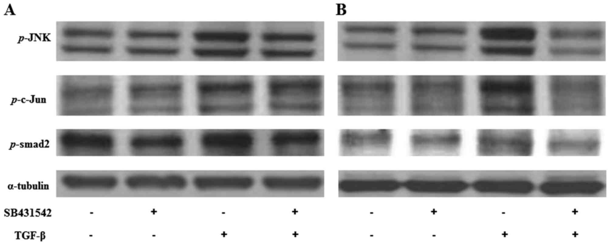 | Figure 5.Expression of p-SAPK/JNK, p-c-Jun, and
p-smad2 in Huh-7 and Huh-Bat cells following SB431542 and TGF-β
treatment. Huh-7 or Huh-Bat cells (5×10⁵ cells) were
seeded in Dulbecco's modified Eagle's medium containing 10% fetal
bovine serum. Cells were incubated for 72 h with DMSO (control), 1
µM SB431542, 1 ng/ml TGF-β or 1 µM SB431542 plus 1 ng/ml TGF-β. (A)
Huh-7 cells. (B) Huh-bat cells. Expression of p-JNK, p-c-Jun and
p-smad2 was detected by immunoblotting. Cells were treated with
distilled water and DMSO (lane 1), 1 µM SB431542 (lane 2), 1 ng/ml
TGF-β (lane 3) or 1 µM SB431542 plus 1 ng/ml TGF-β (lane 4). TGF-β,
transforming growth factor-β; SAPK, stress-activated protein
kinase; JNK, c-Jun N-terminal kinase; DMSO, dimethyl sulfoxide. |
Discussion
TGF-β performs a role in liver fibrosis through
activation of stellate cells and TGF-β promotion of ECM
accumulation. TGF-β also plays an important role in differentiation
of stem cells and has been used as an agent for differentiation of
stem cells. In relevance to cancer, TGF-β has been shown to induce
metastatic ability through EMT or suppress cancer cell
proliferation through cell cycle arrest. In the present study, the
anticancer effects of TGF-β on cancer stem cells were studied in
HCC cell lines. It was hypothesized that TGF-β treatment reduces
the proliferation of cancer stem cells. Therefore, proliferation,
SP cell numbers and expression of cancer stem cell markers, EMT
markers and TGF-β signaling molecules were evaluated following
TGF-β treatment of Huh-7 and Huh-Bat cells.
TGF-β treatment decreased survival cell
(specifically of the SP fraction) and upregulated expression of JNK
signaling molecules. These effects were blocked by the TGF-β
receptor inhibitor SB431542. Furthermore, the effect of TGF-β was
through cell cycle arrest. These results are similar to those of
previous studies investigating the tumor suppressive effects of
TGF-β in liver cancer. Baek et al reported that TGF-β
inhibited cell proliferation through cell cycle arrest (22). Senturk et al demonstrated that
TGF-β induced downregulation of p21 (Cip1) and p15 (Ink4b) and
inhibited cell proliferation through cell cycle arrest in HCC cell
lines (23). In addition, Hashimoto
et al identified that TGF-β inhibited cell proliferation
through G2 arrest in HCC cells (24).
With regard to TGF-β signaling molecules,
TGF-β-induced upregulation of p-JNK, p-c-Jun and p-smad2 expression
was observed. Hu et al reported that upregulated TGF-β/smad
signaling was associated with suppression of hepatocarcinogenesis
(25). Dzieran et al also
reported that TGF-β inhibited proliferation and induced apoptosis
through increased smad3 activity in human HCC cell lines (26). Park et al reported that TGF-β1
induced apoptosis through the activation of p38, JNK and caspases
(27). Suzuki et al reported
that TGF-β regulated autophagy activation, which is involved in
cancer progression through the smad and JNK pathways (28). Kim et al reported that TGF-β
induces apoptosis through cleavage of Bcl-2-associated death
promoter in a smad3-dependent mechanism in FaO HCC cells (29). The present results indicated that
TGF-β inhibited proliferation through the activation of JNK and
smad signaling.
SPs have been proposed as cancer stem cells due to
their tumor initiation ability and drug resistance (18). TGF-β was found to decrease the SP
fraction through inhibition of cell proliferation. Therefore, the
present results demonstrated that TGF-β treatment specifically
targeted cancer stem cells in HCC cell lines. Ehata et al
reported that TGF-β decreased the number of SP cells through
repression of ATP binding cassette subfamily G member 2 (ABCG2)
transcription, which prevented the direct binding of smad2/3 to its
promoter/enhancer in gastric carcinoma cells (30).
TGF-β is a known EMT-inducing agent in several
cancers (31). However, in the
present study, TGF-β did not induce upregulation of the liver
cancer stem cell marker (CD133) and EMT markers (E-cadherin, α-SMA
and vimentin). This may be due to the high level of expression of
EMT markers (E-cadherin, α-SMA and vimentin) in Huh-7 and Huh-bat
cells. Relevant to EMT, Yin et al reported that TGF-β
induced EMT but decreased SP cells and expression of ABCG2 in the
breast cancer MCF7 cell line (32).
In contrast to the present results, You et al reported that
TGF-β upregulated CD133 expression and increased tumor initiation
ability in an HCC cell line (33).
Martin et al reported that TGF-β increased the proportion of
CD133+ cells in liver cancer cell lines (34). Nishimura et al reported that
TGF-β induced increased proliferative capacity and drug resistance
in SP cells in the hepatic tumor cell line K-251 (35). These findings were different from the
present results, and may be due to differences in culture
environment and drug treatment methods.
In conclusion, the present results indicated that
TGF-β may be used to specifically target SP cells through the
induction of JNK signaling. This effect of TGF-β on liver cancer
stem cells indicated that TGF-β requires investigation as a novel
method to treat liver cancer.
Acknowledgements
The present study was supported by grants from the
Seoul National University Hospital [grant no. 0320140200
(2014–1290)] and the ILDONG pharmaceutical corporation [grant no.
0620133000 (2013–1890)].
References
|
1
|
El-Serag HB: Hepatocellular carcinoma. N
Engl J Med. 365:1118–1127. 2011. View Article : Google Scholar : PubMed/NCBI
|
|
2
|
Armengol C, Bartolí R, Sanjurjo L, Serra
I, Amézaga N, Sala M and Sarrias MR: Role of scavenger receptors in
the pathophysiology of chronic liver diseases. Crit Rev Immunol.
33:57–96. 2013.PubMed/NCBI
|
|
3
|
Petta S and Craxi A: Hepatocellular
carcinoma and non-alcoholic fatty liver disease: From a clinical to
a molecular association. Curr Pharm Des. 16:741–752. 2010.
View Article : Google Scholar : PubMed/NCBI
|
|
4
|
Song BC, Chung YH, Kim JA, Choi WB, Suh
DD, Pyo SI, Shin JW, Lee HC, Lee YS and Suh DJ: Transforming growth
factor-beta1 as a useful serologic marker of small hepatocellular
carcinoma. Cancer. 94:175–180. 2002. View Article : Google Scholar : PubMed/NCBI
|
|
5
|
Reichl P, Haider C, Grubinger M and
Mikulits W: TGF-β in epithelial to mesenchymal transition and
metastasis of liver carcinoma. Curr Pharm Des. 18:4135–4147. 2012.
View Article : Google Scholar : PubMed/NCBI
|
|
6
|
Saito T, Yoshida K, Matsumoto K, Saeki K,
Tanaka Y, Ong SM, Sasaki N, Nishimura R and Nakagawa T:
Inflammatory cytokines induce a reduction in E-cadherin expression
and morphological changes in MDCK cells. Res Vet Sci. 96:288–291.
2014. View Article : Google Scholar : PubMed/NCBI
|
|
7
|
Selesniemi K, Reedy M, Gultice A, Guilbert
LJ and Brown TL: Transforming growth factor-beta induces
differentiation of the labyrinthine trophoblast stem cell line
SM10. Stem Cells Dev. 14:697–711. 2005. View Article : Google Scholar : PubMed/NCBI
|
|
8
|
Kim JB, Ann YH, Park SY, Jee HG, Kim HR,
Lee JH, Yu SJ, Lee HS and Kim YJ: Side population in LX2 cells
decreased by transforming growth factor-β. Hepatol Res. 44:229–237.
2014. View Article : Google Scholar : PubMed/NCBI
|
|
9
|
Giannelli G, Mazzocca A, Fransvea E, Lahn
M and Antonaci S: Inhibiting TGF-β signaling in hepatocellular
carcinoma. Biochim Biophys Acta. 1815:214–223. 2011.PubMed/NCBI
|
|
10
|
Meindl-Beinker NM, Matsuzaki K and Dooley
S: TGF-β signaling in onset and progression of hepatocellular
carcinoma. Dig Dis. 30:514–523. 2012. View Article : Google Scholar : PubMed/NCBI
|
|
11
|
Zhang Z, Zhu F, Xiao L, Wang M, Tian R,
Shi C and Qin R: Side population cells in human gallbladder cancer
cell line GBC-SD regulated by TGF-β-induced epithelial-mesenchymal
transition. J Huazhong Univ Sci Technolog Med Sci. 31:749–755.
2011. View Article : Google Scholar : PubMed/NCBI
|
|
12
|
Bonnet D and Dick JE: Human acute myeloid
leukemia is organized as a hierarchy that originates from a
primitive hematopoietic cell. Nat Med. 3:730–737. 1997. View Article : Google Scholar : PubMed/NCBI
|
|
13
|
Reya T, Morrison SJ, Clarke MF and
Weissman IL: Stem cells, cancer, and cancer stem cells. Nature.
414:105–111. 2001. View
Article : Google Scholar : PubMed/NCBI
|
|
14
|
Cioce M, Gherardi S, Viglietto G, Strano
S, Blandino G, Muti P and Ciliberto G: Mammosphere-forming cells
from breast cancer cell lines as a tool for the identification of
CSC-like- and early progenitor-targeting drugs. Cell Cycle.
9:2878–2887. 2010. View Article : Google Scholar : PubMed/NCBI
|
|
15
|
Pinto CA, Widodo E, Waltham M and Thompson
EW: Breast cancer stem cells and epithelial mesenchymal
plasticity-implications for chemoresistance. Cancer Lett.
341:56–62. 2013. View Article : Google Scholar : PubMed/NCBI
|
|
16
|
Kai K, Arima Y, Kamiya T and Saya H:
Breast cancer stem cells. Breast Cancer. 17:80–85. 2010. View Article : Google Scholar : PubMed/NCBI
|
|
17
|
Michishita M, Ezaki S, Ogihara K, Naya Y,
Azakami D, Nakagawa T, Sasaki N, Arai T, Shida T and Takahashi K:
Identification of tumor-initiating cells in a canine hepatocellular
carcinoma cell line. Res Vet Sci. 96:315–322. 2014. View Article : Google Scholar : PubMed/NCBI
|
|
18
|
Chiba T, Kita K, Zheng YW, Yokosuka O,
Saisho H, Iwama A, Nakauchi H and Taniguchi H: Side population
purified from hepatocellular carcinoma cells harbors cancer stem
cell-like properties. Hepatology. 44:240–251. 2006. View Article : Google Scholar : PubMed/NCBI
|
|
19
|
Dietrich CG, Geier A, Wasmuth HE, Matern
S, Gartung C, de Waart DR and Elferink RP: Influence of biliary
cirrhosis on the detoxification and elimination of a food derived
carcinogen. Gut. 53:1850–1855. 2004. View Article : Google Scholar : PubMed/NCBI
|
|
20
|
Goodell MA, Brose K, Paradis G, Conner AS
and Mulligan RC: Isolation and functional properties of murine
hematopoietic stem cells that are replicating in vivo. J Exp Med.
183:1797–1806. 1996. View Article : Google Scholar : PubMed/NCBI
|
|
21
|
Damdinsuren B, Nagano H, Kondo M, Natsag
J, Hanada H, Nakamura M, Wada H, Kato H, Marubashi S, Miyamoto A,
et al: TGF-beta1-induced cell growth arrest and partial
differentiation is related to the suppression of Id1 in human
hepatoma cells. Oncol Rep. 15:401–408. 2006.PubMed/NCBI
|
|
22
|
Baek HJ, Pishvaian MJ, Tang Y, Kim TH,
Yang S, Zouhairi ME, Mendelson J, Shetty K, Kallakury B, Berry DL,
et al: Transforming growth factor-β adaptor, β2-spectrin, modulates
cyclin dependent kinase 4 to reduce development of hepatocellular
cancer. Hepatology. 53:1676–1684. 2011. View Article : Google Scholar : PubMed/NCBI
|
|
23
|
Senturk S, Mumcuoglu M, Gursoy-Yuzugullu
O, Cingoz B, Akcali KC and Ozturk M: Transforming growth
factor-beta induces senescence in hepatocellular carcinoma cells
and inhibits tumor growth. Hepatology. 52:966–974. 2010. View Article : Google Scholar : PubMed/NCBI
|
|
24
|
Hashimoto O, Ueno T, Kimura R, Ohtsubo M,
Nakamura T, Koga H, Torimura T, Uchida S, Yamashita K and Sata M:
Inhibition of proteasome-dependent degradation of Wee1 in
G2-arrested Hep3B cells by TGF beta 1. Mol Carcinog. 36:171–182.
2003. View
Article : Google Scholar : PubMed/NCBI
|
|
25
|
Hu X, Rui W, Wu C, He S, Jiang J, Zhang X
and Yang Y: Compound astragalus and salvia miltiorrhiza extracts
suppress hepatocarcinogenesis by modulating transforming growth
factor-β/Smad signaling. J Gastroenterol Hepatol. 29:1284–1291.
2014. View Article : Google Scholar : PubMed/NCBI
|
|
26
|
Dzieran J, Fabian J, Feng T, Coulouarn C,
Ilkavets I, Kyselova A, Breuhahn K, Dooley S and Meindl-Beinker NM:
Comparative analysis of TGF-β/Smad signaling dependent cytostasis
in human hepatocellular carcinoma cell lines. PLoS One.
8:e722522013. View Article : Google Scholar : PubMed/NCBI
|
|
27
|
Park SS, Eom YW, Kim EH, Lee JH, Min DS,
Kim S, Kim SJ and Choi KS: Involvement of c-Src kinase in the
regulation of TGF-beta1-induced apoptosis. Oncogene. 23:6272–6281.
2004. View Article : Google Scholar : PubMed/NCBI
|
|
28
|
Suzuki HI, Kiyono K and Miyazono K:
Regulation of autophagy by transforming growth factor-β (TGF-β)
signaling. Autophagy. 6:645–647. 2010. View Article : Google Scholar : PubMed/NCBI
|
|
29
|
Kim BC, Mamura M, Choi KS, Calabretta B
and Kim SJ: Transforming growth factor beta 1 induces apoptosis
through cleavage of BAD in a Smad3-dependent mechanism in FaO
hepatoma cells. Mol Cell Biol. 22:1369–1378. 2002. View Article : Google Scholar : PubMed/NCBI
|
|
30
|
Ehata S, Johansson E, Katayama R, Koike S,
Watanabe A, Hoshino Y, Katsuno Y, Komuro A, Koinuma D, Kano MR, et
al: Transforming growth factor-β decreases the cancer-initiating
cell population within diffuse-type gastric carcinoma cells.
Oncogene. 30:1693–1705. 2011. View Article : Google Scholar : PubMed/NCBI
|
|
31
|
Tirino V, Camerlingo R, Bifulco K, Irollo
E, Montella R, Paino F, Sessa G, Carriero MV, Normanno N, Rocco G
and Pirozzi G: TGF-β1 exposure induces epithelial to mesenchymal
transition both in CSCs and non-CSCs of the A549 cell line, leading
to an increase of migration ability in the CD133+ A549 cell
fraction. Cell Death Dis. 4:e6202013. View Article : Google Scholar : PubMed/NCBI
|
|
32
|
Yin L, Castagnino P and Assoian RK: ABCG2
expression and side population abundance regulated by a
transforming growth factor beta-directed epithelial-mesenchymal
transition. Cancer Res. 68:800–807. 2008. View Article : Google Scholar : PubMed/NCBI
|
|
33
|
You H, Ding W and Rountree CB: Epigenetic
regulation of cancer stem cell marker CD133 by transforming growth
factor-beta. Hepatology. 51:1635–1644. 2010. View Article : Google Scholar : PubMed/NCBI
|
|
34
|
Martin M, Ancey PB, Cros MP, Durand G, Le
Calvez-Kelm F, Hernandez-Vargas H and Herceg Z: Dynamic imbalance
between cancer cell subpopulations induced by transforming growth
factor beta (TGF-β) is associated with a DNA methylome switch. BMC
Genomics. 15:4352014. View Article : Google Scholar : PubMed/NCBI
|
|
35
|
Nishimura T, Azuma T, Yokoyama A, Ochiai
H, Saito H and Hibi T: New mechanism of transforming growth
factor-beta signaling in hepatoma: Dramatic up-regulation of tumor
initiating cells and epidermal growth factor receptor expression.
Hepatol Res. 39:501–509. 2009. View Article : Google Scholar : PubMed/NCBI
|